Giant ganglion cyst at the posterior thigh
36
GIANT GANGLION CYST AT THE POSTERIOR THİGH Aslan A1*, Atay T2, Baykal YB2, Kırdemir V2, Konya MN1, Sofu H3, Baydar ML4 1. 2. 3. 4.
Afyonkarahisar State Hospital, Departmants of Orthopaedics and Traumatology. Afyonkarahisar. Turkey Süleyman Demirel Üniversity, Medical Faculty, Departmants of Orthopaedics and Traumatology. Isparta. Turkey. Suluova State Hospital, Departmants of Orthopaedics and Traumatology. Amasya. Turkey TBMM, Ankara, Turkey
Correspondence Dr. Ahmed Aslan. Afyonkarahisar State Hospital, Departmants of Orthopaedics and Traumatology. Afyonkarahisar. Turkey Email:
[email protected] Aslan A, Atay T, Baykal YB, Kırdemir V, Konya MN, Sofu H, Baydar ML. Giant ganglion cyst at the posterior thigh. Diag Ther Stud. 2013; 2(3): 36 - 40. ABSTRACT Ganglion is a cystic formation originating from the synovial herniation and is located on the joint capsule or tendon sheath. It is generally seen in the wrist joint and more common in female gender. We present the clinical and radiological findings of a giant ganglion with an atypical location at the posterior compartment of a patient’s right thigh.Sixty-six year-old female patient was consulted to our department with a history of increasing pain and swelling at her right thigh during the last eight years. Magnetic resonance imaging (MRI) resulted in cystic mass in between the biceps femoris and semitendinosus muscle groups. The giant cystic mass excised totally with its surrounding capsule. Recurrence was not diagnosed in yearly follow-up. Giant and atypically located ganglion cysts may be seen in thigh, and thus it should be kept in mind in the differential diagnosis of the soft tissue lesions in this anatomical location. Key words: Ganglion cyst, atypical localization, surgical treatment, magnetic resonance Imaging INTRODUCTION Ganglion is a cystic formation originating from the synovial herniation and is located on the joint capsule or tendon sheath. It is generally seen in the wrist joint and more common in female gender. Although the etiology is controversial, repeated micro-trauma is believed to be an important factor 1 - 2. Trauma may be a direct hit, a crush injury, sprain or as a result of overuse syndrome of an extremity. The anatomic location or the tissue of origin may be an articular, capsular, tendinous or bony structure. Clinical features are characterized by mild pain, local swelling (except the bony type) and palpable mass. It may be diagnosed as a Baker’s cyst at the knee joint and less commonly Diagnostic and Therapeutic Study 2013; 2(3): 36 - 40.
36
Giant ganglion cyst at the posterior thigh
37
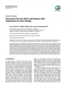
around the ankle joint in the lower extremity associated with osteoarthritis or rheumatoid arthritis, as well as the shoulder joint 2 - 6. We present the clinical and radiological findings of a giant ganglion with an atypical location at the posterior compartment of a patient’s right thigh. CASE REPORT Sixty-six year-old female patient was consulted to our department with a history of increasing pain and swelling at her right thigh during the last eight years. She had no clinical history of trauma. Roentgenograms and laboratory test did not reveal any pathological finding. Magnetic resonance imaging (MRI) resulted in a 10x14x21 cmsized cystic mass in between the biceps femoris and semitendinosus muscle groups accompanied by a 7x5 cm-sized solid soft tissue mass at the superoanterior part (Figure 1). There was no osseos destruction.
Figure 1. Ganglion cyst: Lesion located that is at the posterior side of right thigh, including septations proximally, bi-lobulated with a sharp rim, hyperintense in T2A sagital sections, hypointense in T1A coronal sections, 10x14x21 cm-sized, cystic in nature. It is located as an intermuscular mass which caused a propulsion on neighboring muscles. Demarcation of the lesion from the muscle planes is clear. It is also medial to the neurovascular structures and significant neighboring to them is present. Incisional biopsy of this atypical cyst was performed. Following capsular tissue dissection around the cystic mass, 750 cc yellow-colored, clear, muscinous type fluid was drained and sent to pathology department in optimal conditions (Figure 2a and 2b). Then, Diagnostic and Therapeutic Study 2013; 2(3): 36 - 40.
37
Giant ganglion cyst at the posterior thigh
38
the giant cystic mass excised totally with its surrounding capsule. Compressive bandage was applied post-operatively. Pathological evaluation was reported as ganglion cyst. Recurrence was not diagnosed in yearly follow-up.
Figure 2a. Clinical appearance of the lesion.
Figure 2b. Drained fluid from the lesion.
Diagnostic and Therapeutic Study 2013; 2(3): 36 - 40.
38
Giant ganglion cyst at the posterior thigh
39
DISCUSSION Ganglion cysts are generally diagnosed in women at third to fifth decades of life. The etiology is controversial. Repeated micro-trauma is believed to be an important factor. Our case did not have any history of trauma. Ganglion cysts generally originate from joint capsule or tendon sheath. Although most common location is the wrist joint; proximal tibiofibular joint, posterior compartment of the knee joint (as Baker’s cyst), ankle joint, and shoulder joint may less commonly be the anatomical locations 5 - 6. Yılmaz et al evaluated 1620 knee joint MRIs and diagnosed 23 ganglions associated with the ligaments or tendons of the knee joint 7. Ganglion cyst located at the posterior thigh was very rarely reported in the literature. Tuberculosis, rheumatoid tenosynovitis, lipoma, fibroma, osteoma, sarcoma and aneurismal lesions are included in the differential diagnosis of the ganglia. Glossy, yellowish, viscous, and gelatinous fluid in fine-needle aspiration is diagnostic for ganglion cyst. MRI is a very useful tool which helps to understand anatomical features of any soft tissue tumor and gives very useful information according to signal characteristics of the lesion. Lobular, multi-septated collections which cover the entire lesion and are characterized with hyperintense signal on T2-weighted and gradient-echo images are specific for ganglion cysts. Excision is the definitive treatment in symptomatic cases. In our case, total excision following biopsy was performed. Recurrence rates up to 40% due to inadequate excision were reported in the literature 6. Recurrence was not diagnosed in our patient in yearly follow-up. Atypically located ganglia may lead to severe complications because of being in close relationship with neurovascular structures. Vayvada et al reported an 11x3x2 cm-sized giant ganglion cyst of the quadriceps femoris tendon which was palpable at the anterolateral hip region and leading to chronic pain 8. Wu et al reported a case of 51 year-old patient with a ganglion cyst originating from intermuscular septum and leading to severe compression syndrome on siatic nerve 9. In another study, Gül et al diagnosed drop foot due to a ganglion cyst 10. Compression on iliopsoas muscle caused by a ganglion cyst originating from the capsule of the hip joint was described by Akman et al 11. In a 76 year-old female patient with massive edema of the lower extremity, Gong et al diagnosed a giant cyst as the etiology of femoral vein occlusion [12]. According to a case report published by Belhan et al, congenital ganglion cyst was diagnosed in the right arm of an eight month-old baby 13. Garg et al reported a 24x10x12 cm-sized giant ganglion originating from semimembranosus muscle 14. In conclusion, giant and atypically located ganglion cysts may be seen in thigh, and thus it should be kept in mind in the differential diagnosis of the soft tissue lesions in this anatomical location. Careful dissection during surgery is crucial for total excision of the cyst with its entire capsule and because of possible close relationship with neurovascular structures. ACKNOWLEDGEMENT We thank to Prof.N.Hürriyet Aydoğan, Dr.Emre Yaman.
Diagnostic and Therapeutic Study 2013; 2(3): 36 - 40.
39
Giant ganglion cyst at the posterior thigh
40
COMPETING INTERESTS The authors declare that they have no competing interests. REFERENCES 1. Barnes WE, Larsen RD, Posch JL. Review of ganglia of the hand and wrist with analysis of surgical treatment. Plast Reconst Surg. 1964; 34: 570 - 8. 2. Cook TD. Ganglion of the Hip. Surgery. 1952; 32: 129 - 31. 3. Thommasen HV, Johnston S, Thommasen A. Management Of The Occasional Wrist Ganglion. Can J Rural Med. 2006; 11: 51 - 2. 4. Angelides AC. Ganglions of the hand and wrist. In: Green DP, Hotchkiss RN, Pederson WC, Eds. Operative Hand Surgery. 4th Ed, Volumes 1. New York: Churchill Livingston, 1998; 2157 - 71. 5. Garcia-Alvarez F, Garcia-Pequerul JM, Avila JL, Sainz JM, Castiella T:Ganglion Cysts Associated With Cruciate Ligaments Of The Knee: A Possible Cause Of Recurrent Knee Pain. Acta Orthop Belg. 2000; 66: 490 - 4. 6. Kim YJ, Chae SU, Choi BS, Kim JY, Jo HJ. Intramuscular ganglion of the quadriceps femoris. Knee Surg Relat Res. 2013; 25: 40 - 2. 7. Yilmaz T, Genç B, Argin M, Memiş A, Arkun R. Ganglion cysts of the knee originating from tendons and ligament]. Tani Girisim Radyol. 2004; 10: 246 - 51. 8. Vayvada H, Tayfur V, Menderes A, Yilmaz M, Barutcu A. Giant ganglion cyst of the quadriceps femoris tendon. Knee Surg Sports Traumatol Arthrosc. 2003; 11: 260 - 2. 9. Wu KW, Hu MH, Huang SC, Kuo KN, Yang SH. Giant ganglionic cyst of the hip as a rare cause of sciatica. J Neurosurg Spine. 2011; 14: 484 - 7. 10. Gül M, Özkaya U, Parmaksızoğlu A, Sökücü S, Kabukçuoğlu Y. Drop foot case caused by a ganglion cyst. The Medical Bulletin of Sisli Etfal Hospital. 2008; 42: 25- 7. 11. Akman Ş, Gür B,Sülün T,Aksoy B. A case of a ganglion cyst originating from the hip joint and surgical outcome. Acta Orthop Traumatol Turc. 2002; 36: 76 - 8. 12. Gong W, Ge F, Chen L.A giant ganglion cyst of hip joint causing lower limb edema. Saudi Med J. 2010; 31: 569 - 71. 13. Nicholson LT, Freedman HL.Intramuscular dissection of a large ganglion cyst into the gastrocnemius muscle. Orthopedics. 2012; 35: e1122 - 4. 14. Garg S, Al-Jabri T, Mutnal S, Moftah F. A giant ganglion cyst of the semimembranosus tendon: a case report. Cases J. 2009; 2: 830.
Diagnostic and Therapeutic Study 2013; 2(3): 36 - 40.
40