encephalography (EEG)2,3,10,14; electrocorticography (ECoG) from either subdural7 or epidural5 locations; field potentials, measured by electrodes monitoring ...
CHAPTER 23
Brain-Machine Interface: Restoring Neurological Function through Bioengineering Jeffrey G. Ojemann, M.D., Eric C. Leuthardt, and Kai J. Miller
R
ecent advances in computing and neuroscience have made realistic the idea of a machine controlled directly from brain signals. Such devices are known as a brain computer interface (BCI).15 Advances in sensory prosthetics show promise (e.g., cochlear implant or visual cortex stimulation) allowing for restorative options. Essentially, a BCI is a machine that can decode human intent from brain signal alone to create a new communication channel for patients with severe motor impairments.16 For such clinical success to occur for BCI, several components must be present (Fig. 23.1). In particular, there must be signal acquisition (e.g., what are you recording), signal processing, device output (including user learning of the device, whether it be a computer or a prosthetic, and device-signal interaction), and an operating protocol (e.g., how do you turn it on). These classes of devices could have enormous impact on patients with severe motor impairment, including those with spinal cord injury, stoke, neuromuscular disorders, and amputations. These patients currently have very few options for any substantive intervention in altering their level of function, although progress in amputee prosthetics is impressive recently. The neurosurgical issues surrounding BCI have been recently reviewed6 and I will outline herein the key features of a clinically relevant system. BCIs require a signal that arises from the user’s intent and that can be linked to some element of the outside world. The signal need not be a direct recording of a linked neural activity (e.g., motor cortex neurons to drive a motor device) because any signal that can ultimately be manipulated with accuracy will be adequate. Nevertheless, these measures are usually performed with electrodes. Recordings can be either invasive (implanted beneath the skin) or noninvasive (externally acquired). The most common BCI signals are electroencephalography (EEG)2,3,10,14; electrocorticography (ECoG) from either subdural7 or epidural5 locations; field potentials, measured by electrodes monitoring brain activity from within the parenchyma1; and “single units,” activity measured by microelectrodes monitoring individual neuron action potenCopyright © 2007 by The Congress of Neurological Surgeons 0148-703/07/5401-0134
134
tial firing.4,13 EEG is, of course, the most accessible, and, as such, the most extensively studied. Common signals used from EEG include the sensorimotor (beta) rhythms that change with motor activity, and the P300 response to novel stimuli.2 Intraparenchymal measures have been successful in achieving some degree of control, but gliosis and the invasive nature have limited both long-term application and any human experimental model. ECoG is commonly recorded during invasive monitoring for seizure localizations. In recovered patients, following our Institutional Review Board approved protocol, we have studied the signals that can be used as a BCI platform (5,7,8a). The use of ECoG as an experimental platform was based on the insights garnered from previous EEG-based BCIs and the associated understanding of sensorimotor rhythms.17 Whereas lower frequencies of mu (8 –12 Hz) and beta (18 –26 Hz) rhythms are thought to be produced by thalamocortical circuits,8,11 the higher frequencies, or gamma (30 Hz) rhythms, are thought to be produced by smaller cortical assemblies. When looking beyond the common 60-Hz artifacts, we have found that very focal and separable signals can be found at 70 Hz and above. In fact, the ECoG spectra overlap between rest and motor activity up to as high as 50 Hz (the point of intersection in the frequency domain, denoted Jo), but above Jo, a highly separable signal can be achieved (Fig. 23.2). We denote this frequency band the -band, distinct from gamma or high-gamma as it is functionally defined as the range in which behaviorally mediated changes in the ECoG signal are most clearly delineated. BCIs based on EEG oscillations have focused exclusively on mu and beta rhythms because gamma and -rhythms are inconspicuous at the scalp.9 In contrast, -band signals have been successfully used to run BCI devices.5,7 If separable signals can be identified and used, then the overall efficacy of a BCI system can be greatly improved. The use of -band signals allows for different motor signals (e.g., first and third finger) to be used independently. Twodimensional control has been demonstrated using this approach.12 The specific device output will depend on the patient’s particular circumstances (Fig. 23.1). Restoring limb function Clinical Neurosurgery • Volume 54, 2007
Clinical Neurosurgery • Volume 54, 2007
FIGURE 23.1. Components of a BCI. A brain signal must be recorded, processed, and then converted into a device control (from, Leuthardt EC, Schalk G, Moran DW, Ojemann JG: The emerging world of motor neuroprosthetics: A neurosurgical perspective. Neurosurgery 59:1–14, 20066).
is a conceptually straightforward goal, and bioprosthetics that translate brain signal to arm and/or leg function would have obvious implications, although even accurate control of a wheelchair would have enormous implications. For locked-in patients, a communication device would be of highest priority. The accuracy of control is a special consideration. In some situations, a low error rate is much more important than speed (e.g., failure to stop a wheelchair, inability to ask for help, misdirecting a prosthetic limb, etc.), whereas some communication devices may have a larger tolerance for error, in which case, speed becomes more important. Current BCI systems remain too slow in their flow of information to go beyond yes/no responses or very basic word processing. Accuracy may improve with user learning. Advances in speed and accuracy will be critical for future applications. In summary, advances in signal identification, computing, and device development make the promise of a motor neuroprosthetic very bright for near-term implementation. Neurosurgeons will be at the forefront of device development and, especially, implementation.6 We will need to bring to the discussion important issues surrounding safety, practicality, and risk/benefit discussions, as with any invasive procedure. © 2007 The Congress of Neurological Surgeons
Brain-Machine Interface
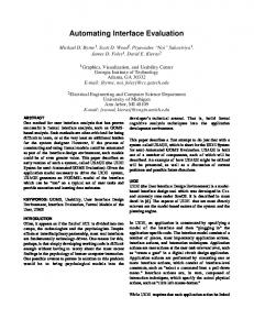
FIGURE 23.2. Spectral power in the ECoG signal for movement and for rest. With motor tasks, the signal is actually lower than compared with rest for lower frequencies. The crossover point, Jo, is typically approximately 30 to 40 Hz, within the classic gamma band. Beyond the range of Jo is functionally defined as the -band. -band signals provide a reliable and spatially focal signal ideal for a BCI source signal.
REFERENCES 1. Andersen RA, Burdick JW, Musallam S, Pesaran B, Cham JG: Cognitive neural prosthetics. Trends Cogn Sci 8:486 – 493, 2004. 2. Donchin E, Spencer KM, Wijesinghe R: The mental prosthesis: Assessing the speed of a P300-based brain-computer interface. IEEE Trans Rehabil Eng 8:174 –179, 2000. 3. Elbert T, Rockstroh B, Lutzenberger W, Birbaumer N: Biofeedback of slow cortical potentials. Electroencephalogr Clin Neurophysiol 48: 293–301, 1980. 4. Kennedy PR, Bakay RA: Restoration of neural output from a paralyzed patient by a direct brain connection. Neuroreport 9:1707–1711, 1998. 5. Leuthardt EC, Miller KJ, Schalk G, Rao RN, Ojemann JG: Electrocorticography-based brain computer interface—The Seattle experience. IEEE Trans Neural Syst Rehabil Eng 14:194 –198, 2006. 6. Leuthardt EC, Schalk G, Moran DW, Ojemann JG: The emerging world of motor neuroprosthetics: A neurosurgical perspective. Neurosurgery 59:1–14, 2006. 7. Leuthardt EC, Schalk G, Wolpaw JR, Ojemann JG, Moran DW: A brain-computer interface using electrocorticographic signals in humans. J Neural Eng 1:63–71, 2004. 8. Levine SP, Huggins JE, BeMent SL, Kushwaha RK, Schuh LA, Passaro EA, Rohde MM, Ross DA: Identification of electrocorticogram patterns as the basis for a direct brain interface. J Clin Neurophysiol 16:439 – 447, 1999. 8a. Miller KJ, Leuthardt EC, Schalk G, Rao RP, Anderson NR, Moran DW, Miller JW, Ojemann JG: Special changes in cortical surface potentials during motor movement. Neurosci 27:2424 –2432, 2007. 9. Pfurtscheller G, Cooper R: Frequency dependence of the transmission of the EEG from cortex to scalp. Electroencephalogr Clin Neurophysiol 38:93–96, 1975. 10. Pfurtscheller G, Flotzinger D, Kalcher J: Brain-computer interface—A new communication device for handicapped persons. J Microcomp App 16:293–299, 1993. 11. Pfurtscheller G, Graimann B, Huggins JE, Levine SP, Schuh LA: Spatiotemporal patterns of beta desynchronization and gamma synchronization in corticographic data during self-paced movement. Clin Neurophysiol 114:1226 –1236, 2003. 12. Schalk G, Leuthardt EC, Moran D, Ojemann J, Wolpaw JR: Twodimensional cursor control using electrocorticographic signals in hu-
135
Ojemann et al.
mans. Presented at the Society for Neuroscience Annual Meeting, San Diego, October 23, 2004. 13. Serruya MD, Hatsopoulos NG, Paninski L, Fellows MR, Donoghue JP: Instant neural control of a movement signal. Nature 416:141– 142, 2002. 14. Vidal JJ: Real-time detection of brain events in EEG. IEEE Proceedings Special Issue on Biological Signal Processing and Analysis 65:633– 664, 1977. 15. Wolpaw JR, Birbaumer N, Heetderks WJ, McFarland DJ, Peckham PH,
136
Clinical Neurosurgery • Volume 54, 2007
Schalk G, Donchin E, Quatrano LA, Robinson CJ, Vaughan TM: Brain-computer interface technology: A review of the first international meeting. IEEE Trans Rehabil Eng 8:164 –173, 2000. 16. Wolpaw JR, Birbaumer N, McFarland DJ, Pfurtscheller G, Vaughan TM: Brain-computer interfaces for communication and control. Clin Neurophysiol 113:767–791, 2002. 17. Wolpaw JR, McFarland DJ, Vaughan TM, Schalk G: The Wadsworth Center brain-computer interface (BCI) research and development program. IEEE Trans Neural Syst Rehabil Eng 11:204 –207, 2003.
© 2007 The Congress of Neurological Surgeons