الفيزياء
2012 نيسان23-22 اجلامعة املستنصرية/املؤمتر العلمي التخصصي احلادي والعشرون لكلية الرتبية
Brain Tumor Area Calculation using Morphological Operations Mohammed Y. Kamil(1), Amel H. Abbas (2) Department of Physics, College of Sciences, AL–Mustansiriyah University. (1) E-mail:
[email protected] (2) E-mail:
[email protected]
Abstract: Brain tumor is serious and life-threatening because it found in a specific area inside the skull. Computed Tomography (CT scan) which be directed into intracranial cavity produces a complete image of the brain. This image is visually examined by the physician for diagnosis of brain tumor. This study provides a computer aided method for calculating the area of the tumor with high accuracy is better than technique within CT scan device. This method determines the extracted the position and shape tumor based on morphological operations (dilation and erosion), enhancement filters and thresholding. Then, automatically calculate of tumor area for the area of interest Keywords: medical image, brain tumor, morphological processing. دطاب ِطادح ورَ اٌذِاغ تاضرخذاَ اٌعٍُّاخ اٌّىرفىٌىجُح ِذّذ َىضف واًِ أًِ دطُٓ عثاش اٌجاِعح اٌّطرٕظرَح,َ وٍُح اٌعٍى,لطُ اٌفُسَاء :اٌّطرخٍض )CT scan( ٟثٛ اٌؾبعلٟش اٌّمطؼل٠ٛ اٌزظل. ِٕطملخ ِؾلذدح داخلً اٌغّغّلخٟاعذ فٛز٠ ٗٔبح أل١ذد ٌٍؾِٙٚ ش١سَ اٌذِبؽ خطٚ لت١ب ِلٓ لجلً اٌطج٠س ثظلشٛلزُ فؾلض ٘لزٖ اٌظل٠ .سح وبٍِلخ ٌٍلذِبؽٕٛلزظ ِٕلٗ طل٠ ف داخً اٌغّغّخ٠ٛ رغٌٝٗ إٙ١عْٛ رٛى٠ . اٌذِبؽٟسَ فٌٛض ا١ٌزشخ لبصٙ داخلً عٟدح فلٛعلٌّٛلخ ا١ٕلخ أفؼلً ِلٓ اٌزم١ٌسَ ثذلخ ػبٌٛرش ٌؾغبة ِغبؽخ اٛ١مخ ثّغبػذح اٌىّج٠فش ؽشٛ٘زٖ اٌذساعخ ر )لخ٠اٌزؼشٚ لخ (رّلذد١عٌٛٛسفٌّٛلبد ا١ٍّ أعلبط اٌؼٝسَ ػٍلٛ لىً اٌلٚ للغِٛ ملخ اعلزخالص٠ رؾذد ٘لزٖ اٌطش.خ١اال ؼخ اٌّمطؼ .َ إٌّطمخ راد األ٘زّبٟسَ فٌٛ ٌّغبؽخ اٟ صُ اٌؾغبة اٌزٍمبئ.ت١اٌزؼزٚ ٓ١ِش ؾبد اٌزؾغٚ 1. Introduction Image scanner devices such as computed tomography (CT), magnetic resonance imaging (MRI) or positron emission tomography (PET) are nowadays a standard instrument for diagnosis. Among these devices, CT-scanners are today widely used in radiotherapy departments all over the world it has several advantages. The main advantages of a CTscanner are to obtain physical information, like patient anatomy, size, shape, and in homogeneities; the other is to obtain the electron density from the different anatomical structures of the patient for the radiotherapy treatment planning [1]. One of the most important image processing branches is image segmentation which is used as a tool in medical image analysis. In image segmentation the level to which the subdivision of an image into its constituent regions or objects is carried depends on the problem being solved. In other words, when the object of focus is separated, image segmentation should stop [2]. The main goal of segmentation is to divide an image into parts having a strong correlation with areas of interest in the image. Segmentation can be primarily classified as complete and partial. Complete segmentation results in a set of disjoint regions corresponding solely to input image objects. While in partial segmentation resultant regions do not correspond directly with input image [3]. Image segmentation is often treated as a pattern recognition problem since segmentation requires classification of pixels [4]. A brain tumor is a mass of cells that have grown and multiplied uncontrollable i.e. a brain tumor is an uncontrolled growth of solid mass formed by undesired cells, either normally found in the different part of the brain, such as glial cells, neurons, lymphatic tissue, blood vessels, pituitary and pineal gland, skull, or spread from cancers mainly located in other organs [5]. There are different type of brain tumor they are i) Gliomas, ii) Medulloblastoma, iii) Lymphoma, iv) Meningioma, v) Craniopharyngioma, vi) Pituitary adenoma [6]. )1( اجمللذ/عذد خاص
356
جملة كلية الرتبية
الفيزياء
2012 نيسان23-22 اجلامعة املستنصرية/املؤمتر العلمي التخصصي احلادي والعشرون لكلية الرتبية
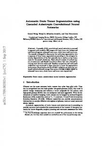
Different brain tumor detection algorithms have been developed in the past years. K. Somasundaram and T. Kalaiselvi [7] have proposed a technique for automatic detection of brain tumor using maxima transform. S. Patil, et al. [8] have discussed various techniques which need to consider during the preprocessing of MRI and CT scan. Their techniques are based on preprocessing using median filter and using morphological operations. S. Roy et al. [9] have proposed a preprocessing technique to remove skull or non-brain tissues from brain MRI based on global threshold and computational geometry like Convex Hull. Anam Mustaqeem et al. [10] have also given a tumor detection algorithm using watershed and thresholding based segmentation. B. K. Saptalakar et al. [11] have proposed another tumor detection method based on watershed segmentation. 2. Morphological Processing The word morphology signifies the study of form or structure. In image processing, we use mathematical morphology as a means to identify and extract meaningful image descriptors based on properties of form or shape within the image. Key areas of application are segmentation together with the automated counting and inspection. Morphology encompasses a powerful and important of methods which can be precisely treated mathematically within the framework of set theory. While this set-theoretic framework does offer the advantages associated with mathematical rigour, it is not readily accessible to the less mathematically trained reader and the central ideas and uses of morphology can be much more easily grasped through a practical and intuitive discussion. [12] The principle two morphological operations are dilation and erosion. Dilation allows objects to expand, thus potentially filling in small holes and connecting disjoint objects. Erosion shrinks objects by etching away (eroding) their boundaries. These operations can be customized for an application by the proper selection of the structuring element, which determines exactly how the objects will be dilated or eroded [12]. Morphological operations are the logical transformation based on comparison of pixel neighbourhood with a specified pattern which is known as structure element. Open is a combination of two morphological operation i.e. erosion followed by dilation. The mechanism of dilation and erosion operate in a very similar way to the convolution Kernels employed in spatial filtering. The structuring element slides over the image so that its center pixel successively lies on top of each foreground or background pixel as appropriate. The new value of each image pixel, then depends on the values of the pixels in the neighborhoods defined by the structuring element. Fig. (1) shows the results of dilation and erosion on a simple binary image. The foreground pixels are shaded and the background pixels are clear. In the diagram demonstrating dilation, the newly created foreground pixels are shaded darker to differentiate them from the original foreground pixels. Note that whenever the structuring element goes over the boundary of the image, we only consider that part of the neighborhood that lies within the boundary of the image [13, 14]. 3. Proposed Methodology The proposed methodology can be divided into ten steps. Step 1: input image from ST-scan device, RGB image, size 1099×650 and JPEG format. Step 2: image crop to remove the unwanted portions from the input image like personal information of patients. Step 3: convert RGB image to gray level image. Step 4: morphological gradient using difference of gray-scale dilation from gray-scale erosion with flat 9×9 structuring element. Step 5: Enhancement the image by using Contrast-limited adaptive histogram equalization method. Step 6: Using filter region of interest (ROI) in an image by Specify polygonal in order to select the bounded region of the tumor. Step 7: Subtract original image from final image. Step 8: Convert image to binary image, based on threshold. Step 9: Using Morphologically closing image (dilation followed by erosion). )1( اجمللذ/عذد خاص
357
جملة كلية الرتبية
الفيزياء
2012 نيسان23-22 اجلامعة املستنصرية/املؤمتر العلمي التخصصي احلادي والعشرون لكلية الرتبية
Step 10: calculated the area of all the remaining portions including the tumor by measure properties of image regions.
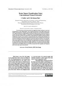
Figure (1): the results of dilation and erosion on binary image [12]. 4. Results and Discussion The CT-scan image are collected for this study from the Al-Kindy Teaching Hospital in Baghdad. The proposed method is applied on different types of tumor CT scan images. The shape and area of the tumor is various. Test example for abnormal case to patient name: Sanaa Daowd, Age: 1973, Date: 23-3-2015. Figures (2-7) shown the progress of the proposed algorithm (step by step) from the original image to area calculate of brain tumor. We introduce algorithm approaches forِ CT scan images and investigate its implemented to the detection of region of interest. The masses are assumed to be distinctive regions that are relatively brighter than the surrounding background.
Figure (2): CT-scan image of brain tumor
)1( اجمللذ/عذد خاص
358
جملة كلية الرتبية
الفيزياء
2012 نيسان23-22 اجلامعة املستنصرية/املؤمتر العلمي التخصصي احلادي والعشرون لكلية الرتبية
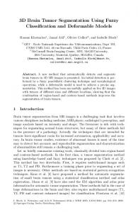
Figure (3): after cropping for gray image.
Figure (4): after using the morphological gradient.
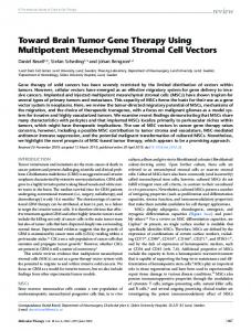
Figure (5): after using Enhancement.
Figure (6): using region of interest.
Figure (7): Extracted area calculation for brain tumor. )1( اجمللذ/عذد خاص
359
جملة كلية الرتبية
الفيزياء
2012 نيسان23-22 اجلامعة املستنصرية/املؤمتر العلمي التخصصي احلادي والعشرون لكلية الرتبية
5. Conclusions In this study, we present a preprocessing and segmentation to a region of interest that are available for area calculation for brain tumor. Processing of brain tumor CT scan images are more interesting and difficult process than MRI images. The proposed algorithm results showed the ease of physician in the selection of the affected area after automatically highlighted, also the accuracy of the calculate of the tumor area; better than the method of CT scan device, which takes calculated only distance (straight line), this accuracy in tumors that are of irregular shape. References [1] Shum, H.Y., Chan, S.C. and S.B. Kang, Image-Based Rendering, Springer ScienceBusiness Media, LLC, Spring Street, New York, USA, 2007. [2] R.C. Gonzalez, R.E. Woods and S.L.Eddins, Digital image processing using MATLAB, Second edition, Gatesmark Publishing;, USA, 2009. [3] M. Sonka, V. Hlavac and R. Boyle, Image processing, analysis, and machine vision, Third edition, Thomson, USA, 2008. [4] Issac N. Bankman, Handbook of medical image processing and analysis, Second edition, Academic press, USA, 2008. [5] “The Essential Guide to Brain Tumors” National Brain tumor society. http://www.braintumor.org/patients-family-friends/about-braintumors/ publications/essentialguide.pdf [6] Roger J. Packer, Henry S. Friedman, Larry E. Kun, and Gregory N. Fuller “Tumors of The Brain Stem, Cerebellum, Fourth Ventricl”, Page 171-192. http://soc-neuroonc.org/levin/Levin_ch06_p171-192.pdf [7] K. Somasundaram and T. Kalaiselvi, “Automatic Detection of Brain Tumor from MRI Scans using Maxima Transform”, National Conference on Image Processing, Vol. 1, (2010). [8] S. Patil and V. R. Udupi, “Preprocessing to be considered forMR and CT Images Containing tumors”, IOSR Journal of Electrical and Electronics Engineering, Vol. 1, issue 4, July–August (2012). [9] S. Roy, K. Chatterjee, I. K. Maitra and S. K. Bandyopadhyay, “Artefact Removal from MRI of Brain Images”, International Refereed Journal of Engineering and Science (IRJES), Vol. 2, issue 3, March (2013). [10] Anam Mustaqeem, Ali Javed and Tehseen Fatima, “An Efficient Brain Tumor Detection Algorithm using Watershed & Thresholding Based Segmentation”, International Journal Image, Graphics and Signal Processing, Vol.4, No.10, September (2012). [11] B. K. Saptalakar and H. Rajeshwari, “Segmentation based Detection of Brain Tumor”, International Journal of Computer and Electronic Research, vol. 2, issue 1, February (2013). [12] Ch.Solomon and T.Breckon, Fundamentals of Digital Image Processing: A Practical Approach, John Wiley & Sons, Ltd., 2011 [13] E. Scott Umbaugh, Digital Image Processing and Analysis: Human and Computer Vision Applications with CVIP Tools, Taylor & Francis Group, 2nd edition, 2011. [14] A. Pahsa,”morphological image processing with fuzzy logic” Journal of Aeronautics and Space Technologies, Vol. 2 Issue 3, p27, (2006).
)1( اجمللذ/عذد خاص
360
جملة كلية الرتبية