Articles in PresS. J Neurophysiol (June 2, 2004). 10.1152/jn.00351.2004
Brief trains of action potentials enhance pyramidal neuron excitability via endocannabinoid-mediated suppression of inhibition
Dale A. Fortin, Joseph Trettel & Eric S. Levine Department of Pharmacology, University of Connecticut Health Center, Farmington, CT 06030 Running Title - Action potentials enhance pyramidal neuron excitability
Corresponding Author: Eric S. Levine Dept. of Pharmacology, MC-6125 University of Connecticut Health Center 263 Farmington Ave. Farmington, CT 06030 Telephone: Fax: Email:
(860) 679-2145 (860) 679-3693
[email protected]
1 Copyright © 2004 by the American Physiological Society.
Abstract Depolarization-induced suppression of inhibition (DSI) is a form of retrograde signaling at GABAergic synapses that is initiated by the calcium- and depolarization-dependent release of endocannabinoids from postsynaptic neurons. In the neocortex, pyramidal neurons (PNs) appear to use DSI as a mechanism for regulating somatic inhibition from a subpopulation of GABAergic inputs that express the type 1 cannabinoid receptor. Although postsynaptic control of afferent inhibition may directly influence the integrative properties of neocortical PNs, little is known regarding the patterns of activity that evoke endocannabinoid release and the impact such disinhibition may have on the excitability of PNs. Here we provide the first systematic survey of AP-induced DSI in the neocortex. The magnitude and time course of DSI was directly related to the number and frequency of postsynaptic APs and was enhanced in the presence of the cholinergic receptor agonist carbachol. This AP-induced DSI was mediated by endocannabinoids, as it was prevented by the cannabinoid receptor antagonist AM251 and potentiated by the endocannabinoid transport inhibitor AM404. We also explored the consequences of neocortical DSI on PN excitability by examining the responsiveness of PNs to evoked synaptic stimulation. We found that endocannabinoid-mediated DSI markedly increased PN responsiveness to excitatory synaptic inputs and promoted AP discharge with a time course that paralleled DSI expression. Taken together, our data suggest a role for endocannabinoids in regulating the output of cortical PNs.
Keywords: cannabinoid, cortex, patch clamp, GABA, depolarization-induced suppression of inhibition
2
Introduction The integration and processing of information by the neocortex is dependent on the activity of pyramidal neurons (PNs), the principal cells of the cortex. In turn, the output of PNs is highly dependent on synaptic interactions with local-circuit interneurons that synthesize and release GABA. These inhibitory interneurons are quite diverse in their morphological, neurochemical, and electrophysiological phenotype (Cauli et al. 1997; Gupta et al. 2000; Kawaguchi 2001; Kawaguchi and Kubota 1997; Thomson et al. 1996). In addition, distinct subclasses of interneurons selectively innervate specific functional domains of the PN membrane. For example, basket cells provide potent inhibition to the soma and proximal dendrites, whereas other types of interneurons form synaptic contacts exclusively on the axon initial segment or onto specific apical dendritic domains (Buhl et al. 1994; Tamas et al. 1997). Soma-targeting interneurons are of particular interest as they maintain high firing rates in vivo and are ideally positioned to constrain PN firing and regulate synchronous activity (Cobb et al. 1995; Miles et al. 1996; reviewed in Paulsen and Moser 1998). Thus, the integrative properties of PNs may depend on their ability to selectively modulate the strength of these somatic inhibitory inputs. A candidate mechanism for regulating perisomatic inhibition is depolarization-induced suppression of inhibition (DSI), a form of short-term plasticity at GABAergic synapses originally described in cerebellum and hippocampus (Llano et al. 1991; Pitler and Alger 1992).
Depolarization-induced suppression of inhibition is mediated by fast retrograde signaling. In general, postsynaptic depolarization results in the synthesis and release of an endocannabinoid that acts presynaptically to suppress GABA release, as shown in the hippocampus, cerebellum, and neocortex (Kreitzer and Regehr 2001a; Maejima et al. 2001; Ohno-Shosaku et al. 2001; Trettel and Levine 2003; Wilson and Nicoll 2001). The two best characterized endocannabinoids, arachidonylethanolamine and 2-arachidonylglycerol, inhibit neurotransmitter release by activating the Gi/o-coupled type-1 cannabinoid receptor (CB1R, reviewed in Di Marzo et al. 1998). In the neocortex, CB1R expression is primarily restricted to a subclass of interneurons that co-express the neuropeptide cholecystokinin (CCK, Marsicano and Lutz 1999) and that selectively innervate the soma and basal dendrites of PNs (Kawaguchi 2001; Kawaguchi and Kubota 1997; Wang et al. 2002). Consistent with these anatomical and physiological studies, we recently demonstrated that endocannabinoid-mediated DSI in the neocortex selectively targets perisomatic inhibitory inputs (Trettel et al. 2004).
3
The range of stimuli that trigger neocortical DSI and its impact on the regulation of PN output are not well characterized. We have recently shown that a single train of postsynaptic APs is sufficient to induce endocannabinoid-mediated DSI (Trettel et al. 2004). In the present studies, we examined the relationship between the level of postsynaptic activity and DSI expression. To begin to explore the consequences of neocortical DSI on PN firing, we examined the responsiveness of PNs to excitatory synaptic input during AP-induced DSI.
Materials and Methods Slice Preparation Swiss CD-1 mice (P12 - 20; Charles River, Wilmington, MA) were sacrificed according to guidelines approved by University of Connecticut Health Center Animal Care Committee. Brains were harvested and immersed in “cutting and incubating” solution composed of (in mM): 125 NaCl, 2.5 KCl, 1.25 NaH2PO4, 25 NaHCO3, 0.5 CaCl2, 4 MgCl2, 4 MgSO4, 4 lactic acid, 2 pyruvic acid, 20 glucose, 0.4 ascorbic acid, and 0.25 kynurenic acid oxygenated with 95% O2/ 5% CO2 (pH 7.3, 310 ± 5 mmol.kg-1). Transverse slices (300 µm) containing somatosensory or auditory cortex (Paxinos & Franklin, 2001) were cut with a vibratome (Microslicer, Dosaka EM, Kyoto, Japan). Slices were incubated for 30 - 40 min at room temperature before being transferred to a heated recording chamber (32 - 33°C) fixed to the stage of an upright microscope (Olympus BX51WI, Olympus America Inc., Melville, NY) fitted with a x40 water immersion objective lens (0.8 NA). The recording chamber was continuously perfused with oxygenated bath solution containing (in mM): 125 NaCl, 2.5 KCl, 1.25 NaH2PO4, 25 NaHCO3, 2 CaCl2, 2 MgCl2, 17.5 glucose (pH 7.3, 315 ± 4 mmol.kg-1).
Electrophysiology Recordings were obtained from layer 2/3 pyramidal neurons (PNs) that were visually identified using infrared DIC video microscopy. All recorded neurons responded to a 500 ms depolarizing current injection with spike frequency adaptation typical of PNs (Connors and Gutnick 1990; McCormick et al. 1985). Spontaneous inhibitory postsynaptic potentials (sIPSPs) were recorded under whole-cell current clamp configuration with patch electrodes made from borosilicate glass (3 - 5 MΩ ) filled with internal solution containing (in mM): 130 KCl, 0.10 CaCl2, 10 HEPES, 1 EGTA, 4 Na2-ATP, 0.3 Na-GTP, and 10 di-tris phosphocreatine, pH 7.3 with KOH (285 ± 2 mmol.kg-1). The ECl was -1.3 mV based on the Nernst equation. Resting membrane potentials were measured immediately upon breaking into whole-cell mode (range = -56 to -72 mV) and
4
were not corrected for liquid junction potential. The membrane potential was maintained near -70 mV by small hyperpolarizing or depolarizing DC injections.
In order to isolate GABA-mediated sIPSPs, excitatory synaptic responses were blocked by addition of the non-NMDA receptor antagonist 6,7-dinitroquinoxaline-2,3-dione (DNQX; 10 µM) and the NMDA receptor antagonist (RS)-3-(2-carboxypiperazin-4-yl)-propyl-1-phosphonic acid (CPP; 3 µM) to the perfusate. The remaining synaptic potentials were upward, depolarizing potentials (see example sweeps in Figure 1 A) and were completely blocked by the GABAA antagonist bicuculline methiodide (BIC; 20 µM; n = 10; p < 0.001), indicating that these sIPSPs represent an outward Cl- flux through GABAA receptors. For experiments involving mixed evoked postsynaptic potentials (PSPs), a concentric bipolar tungsten electrode (WPI, Sarasota, FL) was positioned 100 - 150 µm lateral to the patched neuron. Extracellular stimuli consisted of individual square-wave current pulses (duration = 50 - 150 µs, amplitude = 50 - 200 µA). KCl was replaced with K-gluconate in the internal pipette solution in order to obtain an ECl of -70 mV. For whole-cell voltage-clamp recordings of evoked excitatory postsynaptic currents (EPSCs) the internal solution consisted of 117 mM CsCH3O3S, 8 mM CsCl, 10 mM HEPES, 1 mM EGTA, 0.1 mM CaCl2, 1.5 mM MgCl2, 4 mM Na2-ATP, 0.5 mM Na-GTP, and 5 mM QX-314 (pH 7.3, 295 ± 5 mmol.kg-1). Excitatory currents were evoked using a bipolar tungsten electrode placed 100200 µm lateral to the recorded neuron. Evoked PSPs and sIPSPs were filtered at 2.9 kHz and digitized at ≥6 kHz using a HEKA EPC9 amplifier (Heka Elektronic, Darmstadt, Germany) connected to a 16-bit A/D converter (PCI-16, Instrutech Corp., Port Washington, NY). Neurons were rejected from analysis if (1) the holding current increased by more than 50 pA in order to maintain a Vm of -70 mV or -55 mV for spontaneous or evoked experiments, respectively; (2) the input resistance (Ri) changed by >15% during the course of an experiment; or (3) Ri fell below 100 MΩ. All drugs were delivered by bath perfusion at a rate of 2 ml.min-1. WIN55,212-2, bicuculline methiodide, and carbamylcholine choride (carbachol) were purchased from Sigma. GABAzine (SR 95531) was purchased from Tocris. AM251 and AM404 were generously provided by Dr. A. Makriyannis (University of Connecticut). DNQX, WIN55,212-2, AM251, and AM404 were dissolved in 0.05% DMSO, which by itself had no effect on synaptic transmission (data not shown).
Data Analysis
5
Data were analyzed using PulseFit (Heka Elektronic) and MiniAnalysis (Synaptosoft, Decatur, GA) software. In order to quantify changes in both frequency and amplitude of sIPSPs, we integrated the area of individual IPSPs to yield total synaptic area (A; mV.ms) within 2 s bins. Percent suppression was calculated as 1 - (Apost /Apre)] *100. Spike probability and evoked PSP amplitudes were used to measure the responsiveness of PNs to evoked synaptic input. Only APs that occurred within the first 50 ms after the stimulus were included in the analysis. In some traces the stimulus artifact was blanked for clarity. Data were compared using repeated measures ANOVAs or Student’s paired t tests and are presented as mean ± SEM.
Results
Action potential-induced DSI in the neocortex is enhanced by cholinergic stimulation Endocannabinoids, which are synthesized and released by depolarization and subsequent calcium influx, are the retrograde signals that mediate DSI in the hippocampus, cerebellum, and neocortex (Kreitzer and Regehr 2001a; Trettel and Levine 2003; Wilson and Nicoll 2001). DSI is typically induced by prolonged depolarizing voltage steps that presumably induce a large and sustained postsynaptic calcium influx. In the present experiments, individual postsynaptic APs were induced by somatic current injection of a 5 ms / 1 nA square wave pulse and AP trains were generated using a corresponding train of these brief current pulses. Consistent with our previous results (Trettel et al. 2004), a brief train of APs induced neocortical DSI in the presence of the cholinergic agonist carbachol (CCh; Figure 1 A, B). Cholinergic receptor activation has been shown to increase the firing rate of subpopulations of cortical inhibitory interneurons (Kawaguchi 1997), some of which appear to be sensitive to endogenous and exogenous cannabinoids (Trettel et al. 2004).
We next compared the expression of DSI in the presence or absence of CCh. Under baseline conditions (i.e. in the absence of CCh), a 20 Hz / 1 s AP train produced a transient and significant reduction in sIPSP area (Figure 1B). The suppression of sIPSPs reached a maximum of 61.7 ± 8.6% within 4 s after the AP train with significant suppression lasting 10 s (Figure 1B, n = 10, p < 0.05). Bath application of CCh (5 µM) dramatically increased the frequency and amplitude of sIPSPs to 371 ± 37% and 284 ± 21% of baseline, respectively. Under these conditions, a 20 Hz AP train suppressed sIPSP area by 89.9 ± 5.0% before returning to pre-DSI baseline levels 24 s after the AP train (Fig. 1B; n = 11; p < 0.05). This AP-induced DSI required CB1R activation because it was completely blocked by pretreatment with the CB1R antagonist
6
AM251 (n = 9; data not shown). Direct comparison of DSI in the absence or presence of CCh showed that CCh enhanced both the magnitude and duration of neocortical DSI (Figure 1 B). The increase in DSI magnitude can be attributed to the higher level of spontaneous activity in the presence of CCh, whereas the increase in duration may reflect a direct effect of CCh on endocannabinoid production (Kim et al. 2002). In addition, the effect of CCh on spontaneous activity appeared to be selective for DSI-sensitive inputs because the residual synaptic area (i.e., the area representing DSI-insensitive inputs) was not different between the baseline and CCh conditions (Figure 1 C).
Neocortical DSI is dependent on the number and frequency of postsynaptic APs To investigate the relationship between postsynaptic activity and DSI expression, we examined the effect of trains that differed in AP number and frequency on sIPSP area in the presence of CCh. We first examined the dependence of endocannabinoid-mediated DSI on the number of APs delivered in a 20 Hz train. Single trains with as little as 3 APs generated a 34 ± 7.6% reduction in sIPSP area (Figure 2 A; n = 8; p < 0.05). In general, the peak magnitude of APinduced DSI increased as the number of spikes increased (Figure 2 A, B) and maximal suppression was achieved with 20 APs. In addition, as the number of APs in the train increased, the duration of suppression also increased. For example, DSI induced by 3 APs produced significant suppression that lasted for 4 s, whereas DSI induced by 20 APs endured for 24 s (Figure 2 A).
Endocannabinoid signaling at the synapse is thought to be regulated by a process involving carrier-facilitated diffusion (Beltramo and Piomelli 2000; Beltramo et al. 1997; Hillard et al. 1997; Piomelli et al. 1999). Under voltage clamp conditions, application of the endocannabinoid reuptake inhibitor N-(4-hydroxyphenyl)-arachidonylamide (AM404, Beltramo et al. 1997) modulates the kinetics and magnitude of neocortical DSI (Trettel and Levine 2003). In the hippocampus, repeated epochs of depolarization in the presence of AM404 leads to occlusion of DSI likely owing to the build up of endocannabinoids (Wilson and Nicoll 2001). Because these earlier experiments were performed under voltage-clamp conditions, however, we examined the effect of reuptake inhibition on DSI when membrane potential was unclamped. A train of 5 APs was used to induce submaximal DSI, and the magnitude and duration of this suppression was markedly potentiated by AM404 (25 µM). In particular, the peak magnitude of suppression was increased from 35 ± 5% under control conditions to 77.5 ± 8.6% in the presence of AM404 (Figure 2 C, n = 5, p< 0.05).
7
We also explored the relationship between DSI and the frequency of APs within the train. Because a train of 5 APs delivered at 20 Hz produced approximately half-maximal suppression, (see Figure 2 B), we tested the effects of 5 APs delivered at different frequencies (i.e., 5, 10, 20, 40, 80 Hz) on the magnitude and duration of DSI. A single 5 Hz train produced a 30 ± 9.4% suppression in sIPSP area (Figure 3 A, n = 9, p < 0.05). Maximal suppression (70 ± 6.8%, n = 12, p < 0.05) was obtained with a 20 Hz train and increasing AP frequency above 20 Hz had no effect on the magnitude of DSI, although these higher frequencies did increase the duration of significant sIPSP suppression (Figure 3 A). An example from a single cell that was tested at 5, 20, and 80 Hz is shown in Figure 3 B. Together these data indicate that the magnitude and duration of DSI are dependent on the number and frequency of postsynaptic APs and are modulated by endocannabinoid reuptake.
Cannabinoids enhance PN firing Activation of postsynaptic GABAA receptors plays an important role in controlling neuronal excitability by increasing the membrane’s conductance to Cl- ions. These synaptic conductances provide potent inhibition that opposes the depolarizing influence of excitatory synaptic input by shunting depolarizing current and clamping the membrane potential near Vrest. We have shown that activation of CB1R by WIN55,212-2 decreases the magnitude of spontaneous (Trettel et al. 2004), as well as evoked inhibitory inputs (Trettel and Levine 2002). Based on these results we hypothesized that when both glutamatergic and GABAergic afferents are stimulated, the activation of CB1Rs by exogenous or endogenous cannabinoids should lead to an increase in PN excitability by suppressing GABA release. We therefore examined the effects of WIN55,212-2 on the threshold current needed to evoke a single AP via synaptic stimulation. In these experiments, we recorded synaptic potentials evoked by extracellular stimulation in the absence of neurotransmitter receptor antagonists and set ECl at -70 mV to approximate physiological conditions (Gulledge and Stuart 2003). The mean threshold current required to evoke an AP under baseline conditions was 68.2 ± 11.0 µA (n = 10). In 5/5 cells, WIN55,212-2 reduced the threshold intensity by an average of 12.0 ± 3.6%, (Figure 4 A; p < 0.05); no change in threshold intensity was observed during vehicle application (n = 5).
We also analyzed PN responsiveness to synaptic input under conditions in which stimulus intensity was held constant, because changes in the stimulation intensity could potentially lead to activation of different afferent fibers. Under baseline conditions, the stimulus intensity was set
8
to produce a low probability of evoking an AP (i.e., < 0.3). In the presence of WIN55,212-2, the probability of AP generation markedly increased from 0.20 ± 0.05 to 0.70 ± 0.06 after 10 min of drug exposure (Figure 4 B, upper panel, n = 5, p < 0.01). There was no change in Vm during WIN55,212-2 exposure (Figure 4 B, lower panel), indicating that the increase in AP probability did not result from postsynaptic depolarization. The most likely explanation for these results is that the activation of CB1Rs by WIN55,212-2 increased the strength of excitatory inputs by reducing the amount of synaptic inhibition received by PNs. To confirm this, we measured the amplitude of the mixed PSP on trials where extracellular stimulation did not elicit an AP. As predicted, PSP amplitudes were increased in the presence of WIN55,212 (baseline = 5.4 ± 0.9 mV, WIN55,212-2 = 7.5 ± 0.8 mV, Figure 4 C, n = 4, p < 0.05). It is important to note, however, that this increase in PSP amplitude was not due to a direct effect of the cannabinoid on excitatory inputs. In fact, WIN55,212-2 produced a small but significant decrease in the amplitude of isolated excitatory currents recorded in presence of BIC (baseline = -81.1 ± 2.6, WIN55,212-2 = -70.8 ± 1.9 pA, Figure 4 D, n = 12, p < 0.05).
AP-induced DSI transiently enhances PN responsiveness We next explored whether endocannabinoids released by a train of postsynaptic APs would also modulate PN firing. Similar to the above experiment, the baseline stimulus intensity was set so that the probability of evoking an AP was low. As shown in the representative raster plots in Figure 5 A, there was a transient increase in spike probability following a 20 Hz / 1 s AP train that was seen in all cells tested. The summary data for these 7 cells (3 trials/cell) revealed a significant increase in evoked spike probability from 0.26 ± 0.02 to 0.80 ± 0.09 three seconds after completion of the AP train and this effect lasted for 12 s (Figure 5 B, p < 0.05). Again, there was no change in Vm (Figure 5 B, bottom panel), indicating that the transient increase in spike probability was not related to membrane depolarization. To confirm that the increase in spike probability was due to endocannabinoid release, we pretreated the slices with AM251 for 10 min. AM251 completely blocked the increase in AP probability in the same group of cells (Figure 7 C). We also explored whether increases in PN excitability could be elicited using a 5 AP/ 80 Hz train (duration = 62.5 ms). Using this DSI induction paradigm, spike probability increased from 0.24 ± 0.05 to 0.70 ± 0.02 and significant suppression lasted for 15 s (Figure 5 D, n = 5, p < 0.05).
We performed two additional experiments to confirm that the increase in AP probability was directly due to decreased GABA receptor activation. First, we examined the effects of the AP
9
train on spike probability in the presence of the GABAA antagonist GABAzine. Because exposure to GABAzine (20 µM) by itself increased spike probability, we adjusted the stimulus intensity to re-establish a low probability of spiking after GABAzine was washed into the recording chamber. Under these conditions, the AP train did not increase spike probability (baseline = 0.37 ± 0.06, post-train = 0.32 ± 0.05, Figure 5 E; n = 4). Second, to rule out the possibility that the induction of DSI altered the intrinsic AP threshold of PNs, we injected a depolarizing current ramp into the PN soma and measured the threshold voltage at the foot of the AP. No change in AP threshold was detected following the AP train (baseline = -41.42 ± 0.74 mV, post-train = -41.36 ± 0.73 mV, n = 10) indicating that membrane conductances responsible for setting the threshold for AP initiation are not altered during DSI. Taken together, these results suggest that the net effect of endocannabinoid release evoked by a train of postsynaptic APs is to enhance the efficacy of excitatory input by suppressing inhibitory conductances.
Discussion In the present studies, we recorded from neocortical PNs and found that a single, short train of postsynaptic APs is sufficient to evoke DSI under baseline conditions (i.e., in the absence of CCh). In the presence of CCh, DSI was enhanced and the magnitude and time course of suppression was dependent on both the duration and frequency of PN firing. For example, at a fixed frequency of 20 Hz, a train of 3 APs suppressed sIPSPs by ~25%, while 5 APs evoked half-maximal inhibition, and 20 APs elicited near total suppression. With a train of 5 APs delivered at varying frequencies, the magnitude of DSI increased monotonically up to 20 Hz and higher frequencies (up to 80 Hz) only prolonged the duration of suppression. The release of endocannabinoids mediated the observed DSI because the CB1R-selective antagonist AM251 completely blocked this effect. In addition, the peak magnitude of DSI matched the reduction in sIPSPs produced by an exogenous cannabinoid (data not shown). Furthermore, an endocannabinoid reuptake inhibitor increased the magnitude and duration of DSI, confirming that reuptake plays a dynamic role in regulating endocannabinoid signaling in the cortex. Therefore, neocortical PNs appear to dynamically control a subset of inhibitory inputs through the activity dependent release of endogenous cannabinoids.
In a recent study using hippocampal slices it was found that AP trains that were intended to mimic in vivo neuronal activity failed to induce DSI (Hampson et al. 2003), although an earlier study clearly indicated that hippocampal DSI can be induced by similar AP trains (Pitler and
10
Alger 1992). In the neocortex we found that DSI could be reliably induced with a brief AP train under control conditions, and activation of muscarinic acetylcholine receptors enhanced the magnitude and duration of DSI (present study and Trettel et al. 2004), similar to results obtained in the hippocampus (Pitler and Alger 1992). This cholinergic enhancement was most likely due to the increased activity of CB1R-expressing inputs (Martin and Alger 1999; Trettel et al. 2004) as well as direct facilitation of endocannabinoid production by PNs (Kim et al. 2002). There is also evidence that activation of metabotropic glutamate receptors (mGluRs) can initiate endocannabinoid release (Varma et al. 2001). Excitatory synaptic input, therefore, may induce endocannabinoid release via direct depolarization-induced calcium influx in combination with mGluR activation (Brown et al. 2003; Maejima et al. 2001). Because the present studies induced DSI with APs generated via somatic current injection, we may have actually overestimated the threshold activity requirements for inducing DSI in the cortex.
To address the physiological significance of endocannabinoid-mediated DSI at neocortical synapses we examined the responsiveness of PNs to mixed glutamatergic and GABAergic synaptic stimulation. We found that the cannabinoid receptor agonist WIN55,212-2 increased the probability of AP generation and also increased the amplitude of the mixed PSP (in trials where stimulation failed to evoke an AP). A brief train of postsynaptic APs sufficient to induce DSI also increased the probability of synaptically-evoked spiking, similar to an effect demonstrated in the hippocampus (Wagner and Alger 1996). The time course of this enhancement of PN responsiveness paralleled the expression of DSI and was completely blocked by AM251, indicating that it was mediated by endocannabinoids. The increase in excitability was not due to direct postsynaptic effects because there was no change in either the resting membrane potential or the threshold for inducing an AP by somatic current injection. This effect was also not due to a direct cannabinoid-mediated enhancement of excitatory inputs because WIN55,212-2 did not enhance, but in fact slightly depressed, the magnitude of isolated EPSCs, consistent with a previous study in the neocortex (Auclair et al. 2000). Thus, the most plausible explanation for the increase in PN responsiveness following the AP train is an endocannabinoid-mediated reduction in GABAergic inhibition. This is further supported by the finding that blocking GABAA -mediated inhibition with GABAzine mimicked the increase in PN excitability, and at lower stimulation intensities, occluded the increase in PN excitability following a subsequent AP train.
11
Although both excitatory and inhibitory inputs in the cortex were suppressed by cannabinoids, the predominant effect of endocannabinoid release induced by a brief train of APs was a transient suppression of GABAergic inputs that increased the effectiveness of excitatory inputs. Under other conditions the suppression of excitatory inputs may prevail, leading to long-term depression (LTD, Sjostrom et al. 2003). In the hippocampus, brief postsynaptic depolarization elicits DSI, whereas prolonged depolarization can also induce depolarization-induced suppression of excitation, or DSE (Ohno-Shosaku et al. 2002). Conversely, in the cerebellum, only brief periods of depolarization are required to induce either endocannabinoid-mediated DSI or DSE (Kreitzer and Regehr 2001a, b). While CB1R is clearly expressed on both glutamate and GABA terminals in cerebellum, its expression in the hippocampus and cortex is predominately on inhibitory terminals (Egertova et al. 1998; Katona et al. 1999; Marsicano and Lutz 1999). Studies in the hippocampus have suggested, in fact, that some of the effects of cannabinoids on glutamate release may be mediated by a novel, non-CB1 receptor (Hajos and Freund 2002; Hajos et al. 2001). In the basal ganglia, where CB1Rs are found on glutamate terminals, endocannabinoids directly suppress glutamate release (Gerdeman and Lovinger 2001; Robbe et al. 2001) and repetitive afferent stimulation produces endocannabinoidmediated LTD (Gerdeman et al. 2002; Robbe et al. 2002). Thus, the net effect of endocannabinoid release in different brain regions will depend on the distribution and type of receptors present as well as the specific activity paradigms under study.
When the present results are taken within the context of our previous observation that DSI is selective for perisomatic inputs to PNs (Trettel et al. 2004), the potential functional roles of neocortical DSI begin to emerge. The selective suppression of somatic inhibition by endocannabinoids may play a prominent role in various aspects of PN physiology. Somatargeting basket cells, which constitute large and diverse networks of cells coupled by electrical and chemical synapses, can regulate the synchrony and phase coherence of PN firing (Cobb et al. 1995; Whittington et al. 1995) and this synchrony plays a critical role in establishing and maintaining the cortical rhythms associated with different behavioral and cognitive states (reviewed in Steriade 2000; Ylinen et al. 1995). During brief periods of elevated AP firing, PNs can release endocannabinoids, providing a mechanism (i.e. DSI) for escaping inhibition from CB1R-expressing basket cells. This endocannabinoid-mediated DSI, therefore, could facilitate various forms of synaptic integration and plasticity that are highly sensitive to inhibition. For example, the dendrites of layer 2/3 PNs contain voltage-dependent conductances that support active AP back-propagation (Waters et al. 2003) and the generation of high frequency AP
12
bursts, which are believed to encode pertinent neural information (Lisman 1997), requires the coupling of excitatory apical inputs with back-propagating APs (Larkum et al. 2001). As inhibition suppresses back-propagation (Tsubokawa and Ross 1996) and prevents coupling (Larkum et al. 1999), DSI may gate the active spread of APs into the dendrites and thereby facilitate layer coupling and subsequent burst firing. Additionally, the induction of some forms of LTP requires the coincident arrival of excitatory input and back-propagating APs (Johnston et al. 2003; Markram et al. 1997), and DSI has been shown to promote the induction of hippocampal LTP (Carlson et al. 2002). Endocannabinoid-mediated DSI may regulate these long-term changes in synaptic efficacy both by facilitating AP back-propagation and by indirectly enhancing the impact of excitatory synaptic inputs on PN firing.
In summary, the surprisingly low activity threshold for inducing neocortical DSI suggests that endocannabinoids are important players in the continuous fine-tuning of synaptic inputs by PNs. Nonetheless, the relationship between PN activity and endocannabinoid release exists along a continuum, such that varying levels of activity will have different effects on presynaptic GABA release. For example, low frequency PN firing may trigger minimal DSI, allowing PNs to bias their responsiveness to excitatory synaptic inputs. High frequency discharge, on the other hand, could functionally relieve PNs from the inhibition provided by a subpopulation of synchronously discharging interneurons, thereby allowing PNs to perform associative coupling and to engage in burst firing. Thus, AP-induced endocannabinoid release from PNs is likely to have important consequences for information processing in the neocortex. These issues await further exploration in the context of in vivo preparations. Acknowledgements This work was supported by NIH Grants DA06791, DA07312, and a grant from the Patterson Trust. The authors wish to acknowledge John Kolb for technical assistance.
References Auclair N, Otani S, Soubrie P and Crepel F. Cannabinoids modulate synaptic strength and plasticity at glutamatergic synapses of rat prefrontal cortex pyramidal neurons. J Neurophysiol 83: 3287-3293., 2000. Beltramo M and Piomelli D. Carrier-mediated transport and enzymatic hydrolysis of the endogenous cannabinoid 2-arachidonylglycerol. Neuroreport 11: 1231-1235, 2000. Beltramo M, Stella N, Calignano A, Lin SY, Makriyannis A and Piomelli D. Functional role of high-affinity anandamide transport, as revealed by selective inhibition. Science 277: 1094-1097, 1997.
13
Brown SP, Brenowitz SD and Regehr WG. Brief presynaptic bursts evoke synapse-specific retrograde inhibition mediated by endogenous cannabinoids. Nat Neurosci 6: 1048-1057, 2003. Buhl EH, Han ZS, Lorinczi Z, Stezhka VV, Karnup SV and Somogyi P. Physiological properties of anatomically identified axo-axonic cells in the rat hippocampus. J Neurophysiol 71: 1289-1307, 1994. Carlson G, Wang Y and Alger BE. Endocannabinoids facilitate the induction of LTP in the hippocampus. Nat Neurosci 5: 723-724, 2002. Cauli B, Audinat E, Lambolez B, Angulo MC, Ropert N, Tsuzuki K, Hestrin S and Rossier J. Molecular and physiological diversity of cortical nonpyramidal cells. J Neurosci 17: 38943906, 1997. Cobb SR, Buhl EH, Halasy K, Paulsen O and Somogyi P. Synchronization of neuronal activity in hippocampus by individual GABAergic interneurons. Nature 378: 75-78, 1995. Connors BW and Gutnick MJ. Intrinsic firing patterns of diverse neocortical neurons. Trends Neurosci 13: 99-104, 1990. Di Marzo V, Melck D, Bisogno T and De Petrocellis L. Endocannabinoids: endogenous cannabinoid receptor ligands with neuromodulatory action. Trends Neurosci 21: 521-528., 1998. Egertova M, Giang DK, Cravatt BF and Elphick MR. A new perspective on cannabinoid signalling: complementary localization of fatty acid amide hydrolase and the CB1 receptor in rat brain. Proc R Soc Lond B Biol Sci 265: 2081-2085, 1998. Gerdeman G and Lovinger DM. CB1 cannabinoid receptor inhibits synaptic release of glutamate in rat dorsolateral striatum. J Neurophysiol 85: 468-471., 2001. Gerdeman GL, Ronesi J and Lovinger DM. Postsynaptic endocannabinoid release is critical to long-term depression in the striatum. Nat Neurosci 5: 446-451, 2002. Gulledge AT and Stuart GJ. Excitatory actions of GABA in the cortex. Neuron 37: 299-309, 2003. Gupta A, Wang Y and Markram H. Organizing principles for a diversity of GABAergic interneurons and synapses in the neocortex [see comments]. Science 287: 273-278, 2000. Hajos N and Freund TF. Pharmacological separation of cannabinoid sensitive receptors on hippocampal excitatory and inhibitory fibers. Neuropharmacology 43: 503-510, 2002. Hajos N, Ledent C and Freund TF. Novel cannabinoid-sensitive receptor mediates inhibition of glutamatergic synaptic transmission in the hippocampus. Neuroscience 106: 1-4, 2001. Hampson RE, Zhuang SY, Weiner JL and Deadwyler SA. Functional Significance of Cannabinoid-Mediated, Depolarization-Induced Suppression of Inhibition (DSI) in the Hippocampus. J Neurophysiol 90: 55-64, 2003.
14
Hillard CJ, Edgemond WS, Jarrahian A and Campbell WB. Accumulation of Narachidonoylethanolamine (anandamide) into cerebellar granule cells occurs via facilitated diffusion. J Neurochem 69: 631-638, 1997. Johnston D, Christie BR, Frick A, Gray R, Hoffman DA, Schexnayder LK, Watanabe S and Yuan LL. Active dendrites, potassium channels and synaptic plasticity. Philos Trans R Soc Lond B Biol Sci 358: 667-674, 2003. Katona I, Sperlagh B, Sik A, Kafalvi A, Vizi ES, Mackie K and Freund TF. Presynaptically located CB1 cannabinoid receptors regulate GABA release from axon terminals of specific hippocampal interneurons. J Neurosci 19: 4544-4558., 1999. Kawaguchi Y. Distinct firing patterns of neuronal subtypes in cortical synchronized activities. J Neurosci 21: 7261-7272, 2001. Kawaguchi Y. Selective cholinergic modulation of cortical GABAergic cell subtypes. J Neurophysiol 78: 1743-1747, 1997. Kawaguchi Y and Kubota Y. GABAergic cell subtypes and their synaptic connections in rat frontal cortex. Cereb Cortex 7: 476-486, 1997. Kim J, Isokawa M, Ledent C and Alger BE. Activation of muscarinic acetylcholine receptors enhances the release of endogenous cannabinoids in the hippocampus. J Neurosci 22: 1018210191, 2002. Kreitzer AC and Regehr WG. Cerebellar depolarization-induced suppression of inhibition is mediated by endogenous cannabinoids. J Neurosci 21: RC174, 2001a. Kreitzer AC and Regehr WG. Retrograde inhibition of presynaptic calcium influx by endogenous cannabinoids at excitatory synapses onto Purkinje cells. Neuron 29: 717-727, 2001b. Larkum ME, Zhu JJ and Sakmann B. Dendritic mechanisms underlying the coupling of the dendritic with the axonal action potential initiation zone of adult rat layer 5 pyramidal neurons. J Physiol 533: 447-466, 2001. Larkum ME, Zhu JJ and Sakmann B. A new cellular mechanism for coupling inputs arriving at different cortical layers. Nature 398: 338-341, 1999. Lisman JE. Bursts as a unit of neural information: making unreliable synapses reliable. Trends Neurosci 20: 38-43, 1997. Llano I, Leresche N and Marty A. Calcium entry increases the sensitivity of cerebellar Purkinje cells to applied GABA and decreases inhibitory synaptic currents. Neuron 6: 565-574, 1991. Maejima T, Hashimoto K, Yoshida T, Aiba A and Kano M. Presynaptic inhibition caused by retrograde signal from metabotropic glutamate to cannabinoid receptors. Neuron 31: 463-475, 2001. Markram H, Lubke J, Frotscher M and Sakmann B. Regulation of synaptic efficacy by coincidence of postsynaptic APs and EPSPs [see comments]. Science 275: 213-215, 1997.
15
Marsicano G and Lutz B. Expression of the cannabinoid receptor CB1 in distinct neuronal subpopulations in the adult mouse forebrain. Eur J Neurosci 11: 4213-4225., 1999. Martin LA and Alger BE. Muscarinic facilitation of the occurrence of depolarization-induced suppression of inhibition in rat hippocampus. Neuroscience 92: 61-71, 1999. McCormick DA, Connors BW, Lighthall JW and Prince DA. Comparative electrophysiology of pyramidal and sparsely spiny stellate neurons of the neocortex. J Neurophysiol 54: 782-806, 1985. Miles R, Toth K, Gulyas AI, Hajos N and Freund TF. Differences between somatic and dendritic inhibition in the hippocampus. Neuron 16: 815-823, 1996. Ohno-Shosaku T, Maejima T and Kano M. Endogenous cannabinoids mediate retrograde signals from depolarized postsynaptic neurons to presynaptic terminals. Neuron 29: 729-738, 2001. Ohno-Shosaku T, Tsubokawa H, Mizushima I, Yoneda N, Zimmer A and Kano M. Presynaptic cannabinoid sensitivity is a major determinant of depolarization-induced retrograde suppression at hippocampal synapses. J Neurosci 22: 3864-3872, 2002. Paulsen O and Moser EI. A model of hippocampal memory encoding and retrieval: GABAergic control of synaptic plasticity. Trends Neurosci 21: 273-278, 1998. Piomelli D, Beltramo M, Glasnapp S, Lin SY, Goutopoulos A, Xie XQ and Makriyannis A. Structural determinants for recognition and translocation by the anandamide transporter. Proc Natl Acad Sci U S A 96: 5802-5807, 1999. Pitler TA and Alger BE. Postsynaptic spike firing reduces synaptic GABAA responses in hippocampal pyramidal cells. J Neurosci 12: 4122-4132, 1992. Robbe D, Alonso G, Duchamp F, Bockaert J and Manzoni OJ. Localization and Mechanisms of Action of Cannabinoid Receptors at the Glutamatergic Synapses of the Mouse Nucleus Accumbens. J Neurosci 21: 109-116., 2001. Robbe D, Kopf M, Remaury A, Bockaert J and Manzoni OJ. Endogenous cannabinoids mediate long-term synaptic depression in the nucleus accumbens. Proc Natl Acad Sci U S A 99: 8384-8388, 2002. Sjostrom PJ, Turrigiano GG and Nelson SB. Neocortical LTD via Coincident Activation of Presynaptic NMDA and Cannabinoid Receptors. Neuron 39: 641-654, 2003. Steriade M. Corticothalamic resonance, states of vigilance and mentation. Neuroscience 101: 243-276, 2000. Tamas G, Buhl EH and Somogyi P. Fast IPSPs elicited via multiple synaptic release sites by different types of GABAergic neurone in the cat visual cortex. J Physiol 500 ( Pt 3): 715-738, 1997.
16
Thomson AM, West DC, Hahn J and Deuchars J. Single axon IPSPs elicited in pyramidal cells by three classes of interneurones in slices of rat neocortex. J Physiol 496 ( Pt 1): 81-102, 1996. Trettel J, Fortin DA and Levine ES. Endocannabinoid signalling selectively targets perisomatic inhibitory inputs to pyramidal neurones in juvenile mouse neocortex. J Physiol 556: 95-107, 2004. Trettel J and Levine ES. Cannabinoids depress inhibitory synaptic inputs received by layer 2/3 pyramidal neurons of the neocortex. J Neurophysiol 88: 534-539, 2002. Trettel J and Levine ES. Endocannabinoids mediate rapid retrograde signaling at interneuronpyramidal neuron synapses of the neocortex. J Neurophysiol 89: 2334-2338, 2003. Tsubokawa H and Ross WN. IPSPs modulate spike backpropagation and associated [Ca2+]i changes in the dendrites of hippocampal CA1 pyramidal neurons. J Neurophysiol 76: 28962906, 1996. Varma N, Carlson GC, Ledent C and Alger BE. Metabotropic glutamate receptors drive the endocannabinoid system in hippocampus. J Neurosci 21: RC188, 2001. Wagner JJ and Alger BE. Increased neuronal excitability during depolarization-induced suppression of inhibition in rat hippocampus. J Physiol 495 ( Pt 1): 107-112, 1996. Wang Y, Gupta A, Toledo-Rodriguez M, Wu CZ and Markram H. Anatomical, physiological, molecular and circuit properties of nest basket cells in the developing somatosensory cortex. Cereb Cortex 12: 395-410, 2002. Waters J, Larkum M, Sakmann B and Helmchen F. Supralinear Ca2+ influx into dendritic tufts of layer 2/3 neocortical pyramidal neurons in vitro and in vivo. J Neurosci 23: 8558-8567, 2003. Whittington MA, Traub RD and Jefferys JG. Synchronized oscillations in interneuron networks driven by metabotropic glutamate receptor activation. Nature 373: 612-615, 1995. Wilson RI and Nicoll RA. Endogenous cannabinoids mediate retrograde signalling at hippocampal synapses. Nature 410: 588-592, 2001. Ylinen A, Soltesz I, Bragin A, Penttonen M, Sik A and Buzsaki G. Intracellular correlates of hippocampal theta rhythm in identified pyramidal cells, granule cells, and basket cells. Hippocampus 5: 78-90, 1995.
17
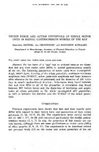
Figure Legends Figure 1. The magnitude and duration of action potential (AP)-induced DSI is enhanced by carbachol (CCh). A) Continuous voltage record of sIPSPs in the presence of 5 µM CCh alone and after a 20 Hz /1 s train of APs (denoted by arrowhead, APs blanked for clarity). Scale bar: 5 mV, 4 s. Expanded traces for baseline (prior to the AP train), DSI, and recovery are shown above (scale bar: 3 mV, 500 ms). B) Normalized sIPSP area illustrating the magnitude and time course of DSI in the absence (n = 10) or presence of CCh (n = 11). Gray symbols denote a significant difference from baseline (ANOVA, p < 0.05). In B and C, the AP train occurred at time zero and each data point represents the total synaptic area in a 2 s bin. C) Group time course of absolute sIPSP area for cells shown in B.
Figure 2. DSI is dependent on the number of postsynaptic APs and is modulated by reuptake inhibition. A) Group time course demonstrating the effect of varying the number of APs on normalized sIPSP area. For each condition, a 20 Hz train of 3, 5, 10, or 20 APs was delivered at time zero (n = 7 - 11 cells/condition). Gray symbols denote a significant difference from baseline (ANOVA, p < 0.05). Baseline data points were omitted for clarity. B) The average level of suppression measured 8 s after the AP train for each condition. C) Group time course based on a within cell comparison demonstrating the effect of 25 µM AM404, an endocannabinoid uptake inhibitor, on DSI evoked by a 20 Hz train of 5 APs (n = 5).
Figure 3. Frequency dependence of AP-induced DSI A) Group time courses showing the effect of different AP frequencies on sIPSP area. For each condition a train of 5 APs was delivered at 5, 10, 20, 40, or 80 Hz at time zero (n = 7 - 10 cells/condition). Gray bars denote a significant difference from baseline (ANOVA, p < 0.05). B) Modulation of sIPSP activity in a single cell following a 5 AP train at 5, 20, and 80 Hz.
Figure 4. Cannabinoids increase PN excitability. A) Left, sample traces from an individual cell demonstrating the threshold stimulus intensity required to generate a single AP during baseline and after 10 min of WIN55,212-2 exposure (WIN; 2 µM). Scale bar: 20 mV, 10 ms. Stimulus intensity was adjusted in 2 µA steps. The stimulus artifact was removed for clarity. Right, WIN significantly reduced the minimum stimulus intensity necessary to
18
evoke an AP (n = 5, p