ABSTRACT. In our previous work we proposed a dynamic image reconstruction procedure for gated cardiac imaging, of which the goal is to obtain a single ...
CARDIAC PERFUSION DEFECT DETECTION USING GATED DYNAMIC SPECT IMAGING Xiaofeng Niu, Yongyi Yang, and Miles N. Wernick Department of Electrical and Computer Engineering Illinois Institute of Technology, Chicago, IL 60616, USA ABSTRACT� In our previous work we proposed a dynamic image reconstruction procedure for gated cardiac imaging, of which the goal is to obtain a single image sequence that shows simultaneously both cardiac motion and tracer distribution change over time. In this work, we further develop and demonstrate the feasibility of this procedure for perfusion defect detection in gated cardiac imaging. We conduct Fisher’s linear discriminant analysis on the reconstructed dynamic images, and derive quantitative measures to differentiate defects from normal myocardial perfusion. Results are presented to demonstrate the proposed development using simulated gated cardiac imaging with the NURBS-based cardiac-torso (NCAT) phantom. Index Terms---Gated SPECT, dynamic SPECT, 5D reconstruction, spatio-temporal processing. 1. INTRODUCTION In nuclear cardiology myocardial perfusion imaging has become an important means for assessing regional coronary blood flow. Gated single-photon emission computed tomography (SPECT) offers an important additional benefit by permitting the evaluation of left ventricular function through quantitative assessment of wall motion [1]. Traditionally, in a gated study the distribution of the tracer is assumed constant over the entire study, and a static image is reconstructed for each gated frame. A logical and clinically important extension of gated imaging is the problem of reconstructing nuclear cardiac imaging studies using agents for which the tracer distribution changes during the course of imaging. In the early work in [2][3], a procedure was first developed in which a dynamic image sequence was reconstructed independently for each gate frame, followed by Wiener filtering across the gate frames for noise reduction. Subsequently, we proposed in [4][5] a joint reconstruction approach for gated dynamic imaging, where the dynamic images of the different gates are treated collectively as a single signal through motion compensation, and determined from the acquired data by using maximum a posterior �
(MAP) estimation. Such an approach allows us to exploit the statistical correlation among the different gate intervals during reconstruction, yielding an image sequence that shows both wall motion and time-varying trace distribution in the myocardium. The primary focus in [4][5] has been on development of reconstruction algorithms for unifying gated and dynamic imaging. Built upon this prior success, in this work we investigate the problem of perfusion defect detection with gated dynamic imaging. Our purpose is twofold: first, given the challenges associated with severely underdetermined data acquisition in SPECT, we want to investigate whether the reconstructed images can yield useful dynamic information which enables differentiation between normal and perfusion defects. Second, we want to explore efficient ways for characterization and visualization of information pertinent to perfusion defect in a gated dynamic image sequence; the purpose here is to determine an effective approach that allows for easy assessment of the presence of perfusion defect and wall motion from the dynamic images in a way that is similar to a traditional gated study. This is an important step toward our ultimate goal of making gated dynamic imaging a useful tool for clinical applications. Specifically, we will first apply statistical clustering analyses on the time activities curves (TACs) of the dynamic images, and demonstrate that the resulting TACs from gated dynamic reconstruction can indeed differentiate between normal and perfusion defects. We will next explore quantitative measures derived from compartmental modeling of the TACs of the dynamic images to differentiate defects from normal myocardial perfusion. In our experiments, we simulated gated cardiac imaging acquisition without the use of multiple fast camera rotations. This setting poses significant challenges to the reconstruction problem because, aside from the low data counts, the projection data are available for only one angular position of the rotating camera at any particular time instance, and therefore, are far from being sufficient (underdetermined by a factor over 20:1 in our experiments) for reconstructing the dynamic images. Moreover, with slow camera rotation which is more suitable for clinical applications [3], the fast-changing tracer dynamics can lead to significant inconsistencies in the projection data among the different angular positions. Despite such difficulties, our
This work was supported by NIH/NHLBI Grant HL65425.
978-1-4244-5654-3/09/$26.00 ©2009 IEEE
825
ICIP 2009
results demonstrate that the gated dynamic reconstruction procedure can still lead to adequate reconstruction results.
The term U m ( F) in (3) is used to enforce temporal smoothness along the motion trajectory. It is defined as
2. RECONSTRUCTION METHODS We assume that list-mode data are binned into K cardiac gating intervals and T time points (reflecting actual time during the imaging session). The dynamic imaging data are described by the following model E[ gt ,k ] H t f t ,k , t 1,!, T , k 1,..., K (1)
T
U m ( F)
F
T
U s ( F)
K
N
¦¦¦¦ ª¬ f t 1 k 1 j 1 iK j
t ,k
( j ) � f t ,k (i ) º¼
2
(4)
where f t ,k ( j ) is the jth voxel in frame f t ,k , K j is the unitdistance neighborhood around voxel j, and N is the number of voxels in f t ,k .
¦¦
J
1ª 2 º «¬1 � K min( k � l , K � k � l »¼ ft ,l o k C 1,l z k K
f t ,k �
t 1 k 1
where gt ,k , f t ,k represent the projection data and the image, respectively, at time interval t for gate frame k, E[·] is the expectation operator, H t is the system matrix which is timevarying in SPECT because of the rotation of the camera. In this study, both the distance-dependent spread function (PSF) and the attenuation effect are modeled in H t . Our goal is to reconstruct the images f t ,k from the data gt ,k , for all gates k 1,..., K and time points t 1,..., T . Collectively, the set of images f t ,k is a five-dimensional (5D) entity (3D space plus gate plus time). Hence this problem is also known as 5D reconstruction. As explained in the introduction, a direct inversion of the imaging equation in (1) can be challenging. Instead, we introduce temporal constraints to regulate the time activities at each voxel location, and apply motion-compensation to exploit the similarity among the different gate frames in the reconstruction algorithm. For convenience, let F { ^ f t ,k , t 1,..., T , k 1,..., K ` , i.e. a vector consisting of all the gate frames over time t; similarly, let G { ^gt ,k , t 1,..., T , k 1,..., K ` represent all the projection data. We propose to simultaneously reconstruct F based on the following constrained maximum a posterior (MAP) estimation Fˆ arg max ^ln p(G | F) � ln p( F)` subject to AF t 0 (2) where p(G | F) is the likelihood function, p(F) is a prior distribution on F, and the constraint AF t 0 is based on the condition that the dynamic activity at each voxel is either constant, increasing, decreasing, or first increasing and then decreasing over time t. Such a constraint was first introduced in [6], and is adapted here for the gated dynamic frames. We use a separable Gibbs prior for p( F) as following p( F) v exp( � E sU s ( F) � E mU m ( F)) (3) where U s ( F) and U m ( F) are two energy functions defined over space and gate intervals, respectively, E s and E m are the corresponding scalar weighting factors. The term U s ( F) in (3) is to enforce local spatial smoothness within each image frame. It is defined as
K
l
¦
2
(5)
where f t ,l ok denotes the motion compensated frame k from frame l, C is a normalization constant for unit DC gain, and J ! 0 is a parameter used to control the degree of smoothing. In U m ( F) temporally neighboring frames will have more contribution to the current frame than frames further away. Finally, Poisson distribution is assumed for the likelihood function p(G | F) . Owing to space limitation, we refer to [7] for details of the 5D reconstruction algorithm for (2). 3. TEMPORAL KINETIC MEASURES In this section we first apply Fisher’s linear discriminant analysis on the time activities curves (TACs) of the dynamic images. The purpose is to demonstrate whether the reconstructed images can differentiate defects from normal perfusion. We then derive quantitative measures to characterize the temporal kinetic information in the reconstructed dynamic images. 3.1. Fisher discriminant analysis Let’s denote the reconstructed TAC at each voxel location j by a vector fk ( j ) ª¬ f1,k ( j ),!, fT ,k ( j ) º¼c . Then the Fisher discriminant is given in the form h( j ) w cfk ( j ) , for which the discriminant vector w is computed as w S�w1 (ȝ n � ȝ d ) (6) where ȝ n and ȝ d denote the mean TAC vectors of normal and defect, respectively, and Sw is the within-class scatter matrix. In our experiment, Sw was estimated from the reconstructed TACs as S � f ( j ) � ȝ � f ( j ) � ȝ c w
¦
n
n
jnormal
�
¦
� f ( j ) � ȝ d � f ( j ) � ȝ d c
(7)
jdefect
in which the first summation is over the voxels in a normal region of interest (ROI) in the myocardium while the second summation is over the voxels in a defect ROI. The ROIs used in this study are shown in Fig 2. All voxels in defect and normal ROIs from K gating intervals are included. 3.2. Temporal kinetic measures Compartmental modeling has been widely used in kinetic analysis of TACs in dynamic imaging. We use a twocompartmental model which has been used in cardiac SPECT imaging with Tc-99m labeled Teboroxime [8][9]. It can be described as dQ(t ) (8) �k12Q(t ) � k21b(t ) dt where k21 and k12 denote, respectively, the uptake and washout rate constants, b(t) denotes the blood pool input function, and Q(t) denotes the time activity.
826
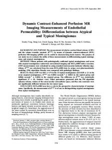
In this study, the kinetic model in Eq. (8) is fit to the reconstructed time activity f t ,k ( j ) at each voxel j in each gate k, based on which the uptake and washout parameters T k21 ( j ) and k12 ( j ) are obtained. Let k ( j ) � k12 ( j ), k21 ( j ) . We define a composite kinetic image as follows: C ( j ) � ĮT k ( j ) D1k12 ( j ) � D 2 k21 ( j ) (9) The definition in Eq. (9) is out of the consideration that the two parameters k21 and k12 are found to be highly correlated in previous studies [9]. Moreover, by setting the vector Į properly, the composite image will show different emphasis on washin and washout. For example, when Į (1,0)T , k ( j ) reflects solely the washout; likewise, when Į (0,1)T , k ( j ) reflects solely the uptake. In our experiments, we will obtain a composite image C ( j ) by using the Fisher discriminant, for which the vector Į in Eq. (9) is derived from the Fisher classifier for separating the perfusion defect from normal voxels based on their kinetic parameters. For convenience, this image will be referred to as the composite kinetic image. 4. EVALUATION STUDY 4.1. Image data To demonstrate the proposed 5D reconstruction method for perfusion defect detection, we used the NURBS-based cardiac-torso (NCAT) v2.0 phantom [10] with a tracerkinetic model simulating imaging with Tc99m labeled Teboroxime, as shown in Fig. 1, where different time activities were introduced among the different organs [3]. A perfusion defect was introduced in the anterior wall of the left ventricle (LV), as shown in Fig. 2 in a short-axis slice of the LV for two different gates (ED and ES). Note that the defect has slower uptake and washout rates than the normal myocardium, and there is a high concentration in the liver. In our simulation, a triple-head camera system was used, and 64 rotation stops covering a total of 360° by each head with 64×64 projection bins at each stop were used during 12 minute data acquisition. A total of eight gates were used. The field of view was 40.5 cm. The system had a distance dependent blur of approximately 13 mm full width at halfmaximum (FWHM) at the center. Attenuation effect was included. Poisson noise was introduced corresponding to a count level of 8 million total counts for the entire acquisition (i.e., for all projections and over all time). The image motion used for the temporal prior was determined from the noisy projection data. First, the reconstruction algorithm [6] was applied to reconstruct the dynamic sequence separately for each gate interval, i.e., by setting E m =0 in Eq. (3). The obtained images were then summed along the dynamic axis to get eight gate frames, which were subsequently processed by a lowpass filter (order-3 Butterworth filter with 0.2 cycles/voxel cutoff frequency). Afterward, the 3D extension of the optical flow method was applied to estimate the motion between the different gate frames.
827
4.2. Results 1) Reconstructed Dynamic Images: The proposed 5D algorithm was tested over 30 different noise realizations. For each noise run, the 5D algorithm yielded a sequence of 3D volumetric images for eight gate frames and 64 time points during the 12 minute period. We show in Fig. 3 some examples of reconstructed images. Due to space limitation, only one short-axis slice (Fig. 2) of the reconstructed LV volume is shown for four selected gates (#1, #3, #5, #7) at three time points (1.78, 6.09, and 11.91 min, representing early, middle, and late stages of the imaging period). For visualization purposes, these images were normalized at each time point such that the myocardium has the same maximum (as in clinical display). Interestingly, the following can be observed: the perfusion defect region (Fig. 2) was initially dimmer than the rest of the LV wall in the early stage, reflecting slower uptake; it then reversed to become brighter in the late stage, reflecting slower washout. These results were consistent with the dynamic TACs in Fig. 1. Such a temporal reversal behavior in tracer redistribution serves as an important indicator of the perfusion defect. 2) Fisher Discriminant Analysis of TACs: The Fisher discriminant analysis was applied to the reconstructed dynamic images from the 30 noise realizations when the defect was present. The TACs were extracted for the voxels in the defect and normal ROIs shown in Fig. 2 for each of the eight gates and 30 noise realizations. In Fig. 4, we show a distribution of the resulting Fisher discriminant values from all the extracted TACs. As can be seen, the Fisher discriminant values are clearly different for defect and normal voxels. Indeed, a t-test shows that the two groups are significantly different in mean values (p-valueĬ0). 3) Kinetic Parametric Images: In Fig. 5, we show the Fisher composite kinetic images for four selected gates of the same slice in Fig. 2 obtained from a typical noise realization. As can be seen, the intensity in these composite kinetic images is much lower in the defect region than in the normal region, reflecting slower tracer uptake and washout. In Fig. 6, we also show the Fisher linear discriminant values obtained directly from the reconstructed TACs for the same slice as above. The intensity of the defect region is also notably lower than that of the normal region in these images. Note that the different parametric images in the above were shown with the same colormap as in conventional SPECT, in which perfusion defects appear as cool spots. These results show that in spite of the low data counts in dynamic gated SPECT the derived parametric images could nonetheless provide good discrimination between normal and perfusion defects. 5. CONCLUSIONS In this work we demonstrated the feasibility of dynamic reconstruction for perfusion defect detection in gated cardiac SPECT imaging. We first conducted statistical clustering analyses on the reconstructed dynamic images. We then
derived parametric images for characterizing the temporal kinetic information in the reconstructed images for differentiating perfusion defects from normal perfusion. Our results demonstrated that the reconstructed images could yield useful dynamic information for detection of perfusion defects. Encouraged by these promising results, in the future we plan to further evaluate this procedure using more clinically relevant tasks.
Defect
Normal
6. REFERENCES
Liver
Fig. 2. Two gated frames (ED and ES) of a short-axis slice of the NCAT phantom with a simulated perfusion defect (indicated by x’s) in the anterior wall of the left ventricle.
1.78 min.
6.09 min.
11.91 min.
gate #1
Normalized radiotracer concentration
0.6
Defect Normal
3 2.5 2 1.5 1 0.5 0 -15
0.7
gate #7
3.5
0.9 0.8
gate #5
4
1 Defect myocardium Normal myocardium Left ventricular blood pool Liver Muscles Lungs
gate #3
Fig. 3. Reconstructed dynamic images at different time points.
Percentage (%)
[1] Garcia EG. Imaging guidelines for nuclear cardiology procedures Part 1. J. Nucl. Card. 1996; 3: G1-G46. [2] Farncombe TH, King MA, Celler AM, Blinder S. A fully 4D expectation maximization algorithm using gaussian diffusion based detector response for slow camera rotation dynamic SPECT. Proc. Of 6th meeting on Fully 3D Image Recon. in Rad. Nucl. Medi. 2001. [3] Feng B, Pretorius PH, Farncombe TH, Dahlberg ST, Narayanan MV, Wernick MN, et al. Simultaneous assessment of cardiac perfusion and function using 5-dimensional imaging with Tc-99m teboroxime. J. Nuclear Cardiology. 2006; 13(3): 354-361. [4] M. Jin, Y. Yang and M. Wernick, “Reconstruction of cardiacgated dynamic SPECT images,” Proc. IEEE Inter. Conf. on Image Processing, vol. 3, pp. III 752-755, 2005. [5] M. Jin, Y. Yang and M. Wernick, “Dynamic image reconstruction using temporally adaptive regularization for emission tomography,” Proc. IEEE Inter. Conf. on Image Processing, vol. 4, pp. IV 141-144, 2007. [6] Farncombe TH. Functional dynamic SPECT imaging using a single slow camera rotation. Ph.D. dissertation, Univ. of British Columbia, Vancouver, BC Canada, 2000. [7] Jin M, Yang Y, Wernick MN, King MA. Fully 5D reconstruction of gated dynamic cardiac SPECT images. IEEE Nucl. Sci. Sym. 2006. p. 3445-3448. [8] Smith AM, Gullberg GT, Christian PE. Experimental verification of technetium 99m-labeled Teboroxime kinetic parameters in the myocardium... J. Nucl. Card.. 1996; 3:130-142. [9] Chiao PC, Ficaro EP, et al. Compartmental analysis of Technetium-99m-Teboroxime kinetics employing fast dynamic SPECT at rest and stress. J. Nucl. Med. 1994; 35:1265-1273. [10] Segars W. Development of a new dynamic NURBS-based cardiac-torso (NCAT) phantom. Ph.D. dissertation, The University of North Carolina, 2001.
-10
-5
0 Fisher scores
5
10
15
Fig. 4. Normalized histogram of Fisher discriminant values for normal and defect voxels. 0.8 0.6
0.5
0.4
0.4
0.2
0.3
gate #1
gate #3
gate #5
gate #7
Fig. 5. Composite kinetic map derived from dynamic images.
0.2 0.1 0
15
0
2
4
6 8 Acquisition time (min.)
10
10
12
5
Fig. 1. Simulated time activity curves of different organs.
0 gate #1
gate #3
gate #5
gate #7
Fig.6. Fisher linear discriminant images derived from TACs.
828