PET Lung Ventilation/Perfusion Imaging Using 68Ga Aerosol (Galligas) and 68Ga-Labeled Macroaggregated Albumin S. J. Ament, S. Maus, H. Reber, H. G. Buchholz, N. Bausbacher, C. Brochhausen, F. Graf, M. Miederer and M. Schreckenberger
Abstract
Pulmonary imaging using ventilation/perfusion (V/P) single-photon emission tomography (V/P scan) with Tc-99m-labeled radiotracers is a well-established diagnostic tool for clinically suspected pulmonary embolism (PE). Ga-68 aerosol (Galligas) and Ga-68-labeled macroaggregated albumin (MAA) are potential tracers for positron emission tomography (PET) lung V/P imaging and could display an advantage over conventional V/P scans in terms of sensitivity and specificity. After radiochemical and animal studies, the clinical applicability of Ga-68 aerosol (Galligas) and Ga-68-labeled MAA was investigated in an exploratory study in patients with clinical suspicion of PE. PET scans were acquired using a 16-slice Gemini TF positron emission tomography/computed tomography (PET/CT) scanner. The acquisition protocol included low-dose computed tomography (CT) for attenuation correction (AC). Dosimetry calculations and continuative phantom measurements were performed. Structural analyses showed no modification of the particles due to the labeling process. In addition, in vitro experiments showed stability of Ga-68 MAA in various media. As expected, Ga-68-labeled human serum albumin microspheres (HSAM) were completely retained in the lung of the animals. In clinical use,
S. J. Ament (&) ! S. Maus ! H. Reber ! H. G. Buchholz ! N. Bausbacher ! F. Graf ! M. Miederer ! M. Schreckenberger Department of Nuclear Medicine, University Medical Centre, Johannes Gutenberg-University, Langenbeckstraße 1, 55131 Mainz, Germany e-mail:
[email protected];
[email protected] C. Brochhausen Institute of Pathology, University Medical Centre, Johannes Gutenberg-University, Mainz, Germany R. P. Baum and F. Rösch (eds.), Theranostics, Gallium-68, and Other Radionuclides, Recent Results in Cancer Research 194, DOI: 10.1007/978-3-642-27994-2_22, ! Springer-Verlag Berlin Heidelberg 2012
395
396
S. J. Ament et al.
PET lung ventilation and perfusion imaging using Ga-68 aerosol (Galligas) and Ga-68-labeled MAA was successful in all cases. In one case a clinically suspected PE could be detected and verified. The administered activity of Ga-68 aerosol (Galligas) and Ga-68-labeled MAA may be reduced by more than 50%, resulting in comparable radiation exposure to conventional V/P scans. In conclusion, Ga-68 aerosol (Galligas) and Ga-68-labeled MAA are efficient substitutes for clinical use and could be an interesting alternative with high accuracy for lung V/P imaging with Tc-99m-labeled radiotracers, especially in times of Mo-99 shortages and increasing use and spread of PET/CT scanners and Ga-68 generators, respectively. Abbreviations
AC CT CTA cGMP DLP DOTA DVT EANM HSA HSAM HU ICRP ITLC MAA NEC OSEM PE PET PET/CT RNP ROI rpm SEM SPECT SUV VOI V/P
Attenuation correction Computed tomography Computed tomography angiography Current good manufacturing practice Dose–length product 1,4,7,10-Tetraazacyclododecane-1,4,7,10-tetraacetic acid (chelate) Deep vein thrombosis European Association of Nuclear Medicine Human serum albumin Human serum albumin microspheres Hounsfield unit International Commission on Radiological Protection Instant thin-layer chromatography Macroaggregated albumin Noise-equivalent count Ordered-subsets expectation maximization (reconstruction) Pulmonary embolism Positron emission tomography Positron emission tomography/computed tomography Radionuclide purity Region of interest Revolutions per minute Scanning electron microscopy Single-photon emission computed tomography Standardized uptake value Volumes of interest Ventilation/perfusion
PET Lung Ventilation/Perfusion Imaging
397
Contents 1 Introduction.............................................................................................................................. 1.1 Background ..................................................................................................................... 1.2 Objective......................................................................................................................... 2 Materials and Methods............................................................................................................ 2.1 Materials ......................................................................................................................... 2.2 Methods........................................................................................................................... 2.3 Exploratory Studies ........................................................................................................ 3 Results...................................................................................................................................... 3.1 68Ga-Labeled Human Serum Albumin Microspheres ................................................... 3.2 Exploratory Studies ........................................................................................................ 3.3 Dosimetry........................................................................................................................ 4 Discussion ................................................................................................................................ 4.1 68Ga-Labeled Radiotracer for Lung Ventilation and Perfusion Imaging..................... 4.2 Dosimetry........................................................................................................................ 4.3 Perspectives .................................................................................................................... References......................................................................................................................................
1
Introduction
1.1
Background
397 397 398 399 398 399 401 402 402 405 411 415 415 419 421 421
Since being introduced to clinical medicine in the mid 1960s, pulmonary ventilation and perfusion imaging with single-photon emission tomography (V/P scan) has become a widely used method and a well-established diagnostic tool in the diagnosis of clinically suspected pulmonary embolism (PE) (Stein et al. 2009). Knipping et al. (1955) used xenon-133 to measure lung ventilation. Lung perfusion imaging was first performed by Wagner et al. (1964) using iodine-131-labeled macroaggregated albumin (MAA). Meanwhile, the standard technique for assessing lung ventilation and perfusion distribution is the use of an ultrafine aerosol of Tc-99mlabeled carbon particles (Technegas") and Tc-99m-labeled MAA. Used clinically since 1960, Tc-99m is the most widely available isotope in diagnostic nuclear medicine, firstly because of its attractive physical characteristics (half-life: 6 h, energy: 140 keV), and secondly because of the convenience of onsite supply via a molybdenum (Mo-99) generator. The daughter nuclide technetium (Tc-99m) is used in approximately 600,000 medical imaging procedures worldwide every week, corresponding to a demand of 450 TBq of Mo-99 (Ballinger 2010). The majority of the Mo-99 produced for Tc-99m medical use is covered by U-235 fission, mostly in highly enriched uranium nuclear reactors. Only five of these reactors are available around the world, and all of them are more than 40 years old, approaching the end of their lifetime. The now-frequent shutdowns are evidence of this. Consequently, any interruption in the routine operation of any of these reactors leads to supply shortages of Mo-99/Tc-99m in days, because of the short half-life of Mo-99 of 66 h (Ballinger 2010). To be prepared for the future, it is necessary to seek suitable alternatives to be more independent from any lack of Mo-99/Tc-99m.
398
S. J. Ament et al.
Use of an alternative c-emitting radionuclide (e.g., thallium-201, iodine-123, or indium-111) is associated with higher radiation exposure. In terms of sensitivity and specificity, PET scans, which provide quantitative images with high spatial resolution in three dimensions, could display an advantage over conventional V/P scans using single-photon emission computed tomography (SPECT). Cyclotron-produced positron-emitting fluorine-18 (F-18, half-life: 110 min) is the most used radionuclide in PET. FDA approval of sodium fluoride F-18 (F-18-NaF) as a substitute for Tc-99m-labeled radiotracer for bone scans is one answer to the Mo-99 supply problems. Otherwise, the dependence upon a cyclotron is a logistical and economical disadvantage of F-18-labeled radiotracers. Against this background, use of a generator-produced positron emitter instead of a cyclotron product could reduce cost effectively. Commercially available Ge-68/Ga-68 (271 days, 68 min) generators provide a decent lifetime of approximately 6 months and could be a potential alternative source (Ballinger 2010; Fani et al. 2008). In the past decade, one example using this source has been imaging of neuroendocrine tumors with Ga-68-labeled somatostatin receptor binding peptides as efficient substitutes for octreotide scintigraphy using SPECT (Baum et al. 2008). Consequently, in times of Mo-99/Tc-99m shortage, the renaissance of gallium68 aerosol and gallium-68-labeled MAA for lung ventilation/perfusion (V/P) imaging was a question of time.
1.2
Objective
The feasibility of producing an ultrafine dispersion of Ga-68-labeled carbon particles using a Technegas" generator was already published by Nozaki et al. (1995). Meanwhile, clinical use of PET aerosol lung scintigraphy was described and proposed for lung ventilation imaging by Kotzerke et al. (2010). These human studies could prove a constant activity over the lung (half-time corrected) and thus the stability of the deposition of Ga-68-labeled carbon particles (Galligas) over 3.5 h. Several methods for labeling of human serum albumin (HSA) microspheres with Ga-68 were described. An early approach was hydrolysis and precipitation of Ga(III)-68 ion in the presence of albumin particles (Hnatowich in 1976). PET scans were performed in dogs following intravenous administration of the microspheres (Chesler et al. 1975). Images obtained 6 h after injection showed that the Ga-68 activity was retained in the lung during this period, with negligible activity appearing in the liver. Wagner and Welch (1979) modified these method and used HSA microspheres as a model compound to attach Ga-68 by covalent conjugation via bifunctional high-affinity gallium chelation. The same group could detect deposition of Ga-68-DTPA-HSA in the lung of dogs over the entire PET study period. Even and Green (1989) performed the first direct labeling of MAA with Ga-68. They obtained radiochemical purities of 98.4 ± 0.3% and labeling yield of 97.0 ± 0.5%. Consequently, Ga-68 aerosol (Galligas) and Ga-68-labeled MAA are potential tracers for positron emission tomography (PET) lung V/P imaging and could display an advantage over conventional V/P scans in terms of sensitivity and
PET Lung Ventilation/Perfusion Imaging
399
specificity. After own radiochemical and animal studies, the clinical applicability of Ga-68 aerosol (Galligas) and Ga-68-labeled MAA was investigated in an exploratory study in patients with clinical suspicion of PE.
2
Materials and Methods
2.1
Materials
2.1.1 Chemicals MAA was purchased from GE Healthcare Buchler (Braunschweig, Germany) and HSA from ROTOP Pharmaka AG (Radeberg, Germany). HCl (0.6 N) for generator elution was prepared from TraceSELECT" Ultra HCl (30%) and sterile pyrogenfree water, both purchased from Sigma-Aldrich Chemie GmbH (Taufkirchen, Germany). Aseptic ethanol (99.5%) was obtained from the central pharmacy of the University Medical Center of the Johannes Gutenberg University (Mainz, Germany). TLC was carried out on ITLC-SG glass-fiber sheets from PALL Life Sciences (Port Washington, NY, USA), recorded with Gina Star TLC, and analyzed using miniGita software from Raytest (Straubenhardt, Germany). 2.1.2 Ge-68/Ga-68 Generator The short-half-life positron-emitting radionuclide Ga-68 (half-life: 68 min) was obtained as [Ga-68]Ga-chloride from a commercially available 30-mCi (1,110MBq) Ge-68/Ga-68 generator produced by iThemba LABS (Somerset West, South Africa) under current good manufacturing practice (cGMP) and distributed by IDB Holland (Baarle-Nassau, The Netherlands). The generator is a ready-to-use, closed system. The system contains a tin dioxide (SnO2) column in a polyethylene column with polyethylene tubing and no metal parts for the parent Ge-68 (half-life: 271 days) (Operating Instructions, 68Ga Generator iThemba LABS1).
2.2
Methods
2.2.1 Elution Characteristics The generator was eluted six times a week, one time per day. The equilibration time between elution was 4 h to allow regeneration of the Ge-68/Ga-68 generator. Ga-68 was obtained as [Ga-68]Ga-chloride. In analogy to Breeman et al. (2005) and the manufacturer instructions, fractionated elution of 10 fractions, each fraction composed of 0.5 mL 0.6 N HCl, was performed to concentrate and purify the eluate of metal impurities. The first eluate was discarded and served only to rinse the generator (Maus et al. 2011). Only fractions 3–5 containing the main activity ([80%) and a Ge68 breakthrough less than 0.001% were used for radiopharmaceutical production. The same technique was performed to evaluate Ge-68 breakthrough after 24 and 48 h. 1
Operating Instructions GMP Produced 68Ga Generator iThemba LABS (Somerset West, South Africa), distributed by IDB Holland (Baarle-Nassau, The Netherlands).
400
S. J. Ament et al.
2.2.2 Preparation of MAA and HSA Human Serum Albumin Kits Each kit was resuspended in 5 mL 0.9% sodium chloride and vigorously mixed for 0.5 h to wash the particles free of compounds such as stannous chloride, polysorbate, and hydrochloric acid (Maus et al. 2011). These steps were repeated three times before the microspheres were used for labeling with Ga-68. After centrifugation, the supernatants were discarded and the retained particles were reconstituted in 0.5 mL sterile water (B.Braun Melsungen AG, Melsungen, Germany). 2.2.3 Labeling With Ga-68 The reconstituted spheres were mixed in 0.5 mL sterile water with 1.5 mL generator eluate corresponding to the middle fraction and containing 600–1,200 MBq Ga-68. The mixture was adjusted to pH 4 with 265 mg HEPES buffer (GERBU Biochemicals GmbH, Gaiberg, Germany). The mixture was allowed to react for 20 min at 75#C under gentle agitation. This is the optimum reaction time indicated by the time– activity curve obtained (Maus et al. 2011). The volume of the reaction mixture was increased to 10 mL with sterile water and then purified for the application by a threefold centrifugation at 1,500 rpm for 10 min. 2.2.4 Determination of Labeling Yields Two different methods could be used to determine the labeling yield. First, instant thin-layer chromatography (ITLC) was performed using silica gel impregnated glass-fiber sheets and 0.9% NaCl solution as mobile phase. The Ga-68-labeled MAA and HSA microspheres remain at the start position, while free Ga-68 moves with the solvent front. The percentages of each fraction were determined relative to the total activity of the chromatogram (Maus et al. 2011). The second method was based on separation by centrifugation. Sterile water (10 mL) was added to the reaction mixture and centrifuged for 10 min at 1,500 rpm. Here, the activity of supernatant was defined as the free Ga-68 activity and the Ga-68-labeled microspheres as the labeled product (Maus et al. 2011). 2.2.5 Stability Experiments of Ga-68-MAA in Various Media To prove the stability of the Ga-68-MAA, tests in sterile water, 0.9% NaCl, and human plasma were performed. The plasma was obtained by taking blood samples from a human individual with immediate centrifugation at 2,700 rpm for 5 min over EDTA K monovettes from Sarstedt, Nümbrecht. Aliquots of the labeled particles containing activities between 100 and 120 MBq were added to 1 mL sterile water, 0.9% NaCl, and plasma obtained as described. After incubation for 1 h, each mixture was centrifuged for 5 min at 2,700 rpm, and the supernatants were removed and measured in a dose calibrator. The whole procedure was repeated three times. 2.2.6
Structural Analyses of Microspheres by Scanning Electron Microscopy (SEM) Particles were placed on a SEM specimen holder with a self-adhesive graphite pad for scanning electron microscopy. The microspheres were sputter-coated (Edward Sputter Coater S150B) with gold according to laboratory standard. The microspheres
PET Lung Ventilation/Perfusion Imaging
401
were imaged using a Zeiss DSM 962 scanning electron microscope with a voltage of 15 KV in case of macroaggregated albumin and 5 KV in case of HSA spheres.
2.3
Exploratory Studies
2.3.1 Animal Studies The animal study was approved by the animal study authority. For each formulation, two Sprague–Dawley rats (400–500 g) were anesthetized with 2% isoflurane/100% pure oxygen gas mixture. The biodistribution of the Ga-68labeled MAA and HSA microspheres was visualized and quantified using a dedicated microPET scanner Focus 120 (Siemens, Knoxville, TN). The scanner has axial field of view of 7.6 cm. The rats (n = 2 ? 2) were positioned on the scanner bed in supine position covering the lungs and the proximal part of the abdomen. The scans started simultaneously with i.v. application of about 10.3 ± 1.5 MBq via the tail vein. List-mode acquisition lasted 60 min, and in addition, a transmission scan using a Co-57 point source was performed for attenuation correction (AC). Data were histogrammed into 19 frames (3 9 20, 3 9 60, 3 9 120, and 10 9 600 s) and reconstructed iteratively with the ordered-subsets expectation-maximization (OSEM 2D) algorithm. Volumes-of-interest (VOI) analyses were performed using the PMOD 2.95 image analysis software package. VOIs for lung, liver, and unspecific binding were defined, and time–activity curves were derived from the VOI statistics. Taking into account the different amounts of injected activity and the different weights of the rats, standardized uptake values (SUV) were calculated (Maus et al. 2011). Additionally, lung ventilation and perfusion imaging with Ga-68 aerosol (Galligas) and Ga-68-labeled MAA were performed (n = 1) using a Gemini TF positron emission tomography/computed tomography (PET/CT) scanner (Philips, Best, The Netherlands) before clinical use. An ultrafine dispersion of Ga-68-labeled carbon particles was generated using a commercially available Technegas" generator (Tetley Manufacturing Ltd., Sydney, Australia) for lung ventilation imaging. A concentrated Ga-68 generator eluate solution (20 MBq/0.1 mL) was loaded into a graphite crucible (PulmotecTM) prefilled with ethanol using a 1-mL syringe. In analogy to the production of Technegas", the combination of graphite and an argon atmosphere was used for vaporization and subsequent generation of Ga-68 aerosol (Galligas). 2.3.2 Clinical Studies Due to the absence of alternatives in the Mo-99 crisis, the clinical applicability of Ga-68 aerosol (Galligas) and Ga-68-labeled MAA was investigated in an exploratory study in patients with clinical suspicion of PE. After detailed explanation and written consent, five patients (two male, three female; median age 75 years, range 66–83 years) with suspicion of PE each underwent PET/CT scan after inhalation of Ga-68-labeled carbon particles (3.4–12.6 MBq) and subsequent application of 42–105 MBq Ga-68-labeled MAA (injected activity calculated based on assessment of inhaled activity).
402
S. J. Ament et al.
According to European Association of Nuclear Medicine (EANM) guidelines for V/P scintigraphy and its key recommendations for investigation of regional pulmonary perfusion in PE diagnosis, Tc-99m-labeled MAA is the agent recommended for perfusion scintigraphy with evidence level IIa, grade B (Bajc et al. 2009b). Consequently, perfusion imaging was performed with Ga-68-labeled MAA microspheres (200,000–400,000 particles). In analogy to the administration of Tc-99mlabeled MAA, the vial was shaken gently before use and withdrawal of blood into the syringe during application was avoided (Bajc et al. 2009a). The suspension was given as intravenous bolus injection over 30 s while the patient breathed at normal tidal volume to ensure that the particles were infused over several respiratory cycles, facilitating uniform distribution within the pulmonary circulation (Bajc et al. 2009a). Analogously to the standard method for production of Technegas", an aerosol of Ga-68-labeled carbon particles was generated by vaporization of Ga-68 generator eluate solution (80–100 MBq/0.1 mL). Ga-68 aerosol (Galligas) was used within 10 min of generation to avoid aggregation of the particles. Administration was performed according to the manufacturer’s instructions for Tc-99m-labeled carbon particles (Technegas" generator user manual 2005). PET scans were acquired using a 16-slice Gemini TF PET/CT scanner (Philips, Best, The Netherlands) with axial field of view of 18 cm and transverse and axial resolution of 4.8 mm. Further specifications were published by Surti et al. (2007). The scatter fraction and noise-equivalent count (NEC) were measured by NEMA cylinder (20 cm diameter). The absolute sensitivity is 6.6 cps/kBq, whereas the scatter fraction is 27% and the peak NEC rate is 125 kcps at activity concentration of 17.4 kBq/mL. The system coincidence timing resolution is denoted by 585 ps (Surti et al. 2007). Lung ventilation and perfusion imaging was performed in 3D acquisition mode in two bed positions (from neck to abdomen, in one case three bed positions) with acquisition time of 5 min per bed position. Image reconstructions were performed using the standard Philips time-of-flight ordered-subsets expectation-maximization reconstruction (OSEM: 33 subsets, 3 iterations), and corrections for random events, scatter and attenuation were applied (Surti et al. 2007). The acquisition protocol included low-dose computed tomography (CT) [120 kV, 0.5 s/rotation, 5 mm slices, max. 60 mAs, dose–length product (DLP) 98–142 mGy!cm, field of view 600 mm] for AC with dose modulation and anatomic orientation. Additionally, dosimetry calculations using OLINDA (version 1.1; Vanderbilt University, 2007) and continuative phantom measurements were performed.
3
Results
3.1
68
Ga-Labeled Human Serum Albumin Microspheres
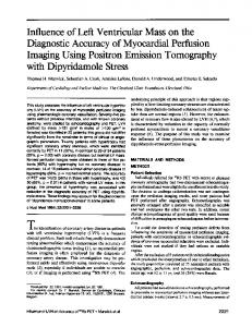
The experimental results for the labeling yields of both compounds with Ga-68 showed a dependence on the heating time as demonstrated exemplarily in Fig. 1. The determined labeling efficiency of MAA was 85 ± 2%, and of HSA was
PET Lung Ventilation/Perfusion Imaging
403
Fig. 1 a SEM images after the labeling procedure: left MAA, right HSA spheres, ultrastructural analysis (Maus et al. 2011). b Stability tests in sterile water, 0.9% sodium chloride (NaCl), and human plasma were performed (Sect. 2.2.5). No significant release of Ga-68 could be detected during the measurement period of 240 min
80 ± 5%. After washing with sterile water, the radiochemical purity for both was higher than 97% (Maus et al. 2011). MAA used for pulmonary perfusion scintigraphy contained approximately 2 9 106 microspheres with particle sizes between 3 and 150 lm. However, approximately 95% particles were specified within the range of 3–40 lm. HSA spheres, also used for perfusion diagnostics, contained 3–5 9 105 microspheres in the narrower size range of 10–30 lm (Maus et al. 2011). Structural analyses of MAA and HSA by SEM showed no modification of the particles due to the labeling process (Fig. 1a). In addition, in vitro experiments showed stability of Ga-68-labeled MAA in sterile water, 0.9% NaCl, and human plasma. No significant release of Ga-68 could be detected during the measurement period (Fig. 1b).
Ventilation
80-year-old female with acute dyspnea, tachycardia, known thrombosis, renal insufficiency, Ddimer at 2.06 lg/l
Inhomogeneous with central Homogeneous and peripheral radionuclide depositions and defect in the left lower lobe
Homogeneous
83-year-old male with acute dyspnea, Homogeneous two-level DVT, D-dimer at 4.4 lg/l, creatinine at 1.75 mg/dL, known prostate carcinoma
Inhomogeneous with defects left lateral and in projection to horizontal fissure of right lung (Fig. 3b)
Inhomogeneous, hypoperfusion in the right lower lobe dorsolateral (Fig. 3d)
Inhomogeneous with central depositions and defects left lateral and in projection to horizontal fissure of right lung (Fig. 3a)
Homogeneous
Perfusion
70-year-old female with chronic Inhomogeneous with central asthma with acute dyspnea, D-dimer radionuclide depositions at 8.0 lg/l, creatinine at 2.02 mg/dL (Fig. 3c)
66-year-old female with heart failure NYHA class III, chronic obstructive pulmonary disease, extreme obesity, type II diabetes, previous artificial pacemaker implantation, previous acute kidney failure, previous PE and DVT, increasing dyspnea since 1 week, D-dimer at 1.24 lg/l
77-year-old male with dizziness, Homogeneous decreased oxygen pressure, diabetic kidney disease, D-dimer at 5.66 lg/l
Clinical symptoms
Table 1 Clinical data and results of lung V/P imaging using PET/CT
Excluded, reverse mismatch in terms of pleural effusion with adjacent ventilation failure and suspicion of pneumonia
Excluded
Mismatch in terms of a PE
Excluded, combined V/P defects left lateral with suspicion of pneumonia and in projection to horizontal fissure of right lung
Excluded
Pulmonary embolism (PE)
Severe pleural effusion with adjacent ventilation failure, dystelectasis and signs of lung infiltrate in the left lower lobe, severe aortic sclerosis and carotid artery stenosis, degenerative changes in the spine, calcified, right dorsal flexed uterus (Fig. 3i)
Small pleural nodules, coronary atherosclerosis aortic sclerosis, multiple gallstones, suspect metastatic lesion in L1 vertebral body (Fig. 3f)
Pleural effusion, axillary lymph nodes increased but not enlarged, degenerative changes in the spine, calcification on the left lobe of the thyroid, enlarged left adrenal gland
Pleural effusion, ground-glass opacities particularly in left lower lobe, coronary atherosclerosis and severe aortic sclerosis, left ventricular dilatation, artificial pacemaker in the left upper chest
Coronary atherosclerosis, calcification of the aortic valve, axial hiatal hernia, solitary gallstone, degenerative changes in the spine
Additional information from low-dose CT
404 S. J. Ament et al.
PET Lung Ventilation/Perfusion Imaging
405
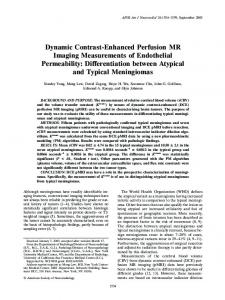
Fig. 2a, b a PET images (coronal slices) of the lung 30–60 min after i.v. injection of Ga-68labeled MAA using a dedicated microPET scanner Focus 120 (Sect. 2.3.1, Maus et al. 2011). b SUV curves after i.v. injection of Ga-68-labeled MAA and Ga-68-labeled HSA (Maus et al. 2011)
3.2
Exploratory Studies
3.2.1 Animal Study MAA and HSA particles were completely retained in the lung of the animals as demonstrated by the animal PET analyses (Fig. 2a). No significant differences in retention in the lung could be found. The time–activity curve indicated high in vivo stability, since no decrease of activity or migration of particles was observed within the
406
S. J. Ament et al.
Fig. 2c, d Lung (c) ventilation (top coronal slices, middle transaxial slices, bottom sagittal slices) and (d) perfusion (top coronal slices, middle transaxial slices, bottom sagittal slices) scan with an aerosol of Ga-68-labeled carbon particles (Galligas) and Ga-68-labeled MAA performed (n = 1) using a Gemini TF PET/CT scanner (Philips, Best, The Netherlands) showing complete incorporation in the lung of a Sprague–Dawley rat without any extrapulmonary activity being visualized
PET Lung Ventilation/Perfusion Imaging
407
Fig. 2c, d (continued)
first hour (Fig. 2b). No significant retention of Ga-68 MAA or Ga-68 HSA particles in the liver was detected (Maus et al. 2011). Additionally, we could demonstrate complete incorporation of Ga-68-labeled carbon particles (Galligas) in the lung of a Sprague–Dawley rat without any extrapulmonary activity being visualized (Fig. 2c, d).
3.2.2 Clinical Study In clinical use, PET lung ventilation and perfusion imaging using Ga-68 aerosol (Galligas) and Ga-68-labeled MAA was successful in all cases.
408
S. J. Ament et al.
Fig. 3a, b PET lung (a) ventilation and (b) perfusion imaging using Ga-68 aerosol (Galligas) and Ga-68-labeled MAA showing combined ventilation (top coronal slices, middle transaxial slices, bottom sagittal slices)/perfusion (top coronal slices, middle transaxial slices, bottom sagittal slices) defects left lateral and in projection to the horizontal fissure of right lung PET lung
Ventilation imaging was homogeneous in two cases. In three cases, inhomogeneous accumulation with central deposition of radionuclide could be observed (Table 1). The perfusion imaging was homogeneous in three cases. In one case, perfusion defects with corresponding ventilation defects were found (Fig. 3a, b). Case 1. 70-year-old female with acute dyspnea, elevated D-dimer to 8.0 lg/l, and creatinine to 2.02 mg/dL showed an inhomogeneous nuclide accumulation with a hypoperfusion in the right lower lobe dorsolateral (especially segment 6) and preserved ventilation in the same region in terms of PE (mismatch defect) (Fig. 3c, d). A follow-up study was performed with Tc-99m aerosol (Technegas") and Tc-99m-labeled MAA 3 days later (Fig. 3e).
PET Lung Ventilation/Perfusion Imaging
409
Fig. 3a, b (continued)
In all other cases PE as reason for the clinical symptoms could be excluded. Due to the low-dose CT performed for AC there was additional diagnostic information, for example, pleural effusion, infiltrates, dystelectasis, and more (Table 1). Case 2. 83-year-old male with acute dyspnea, known two-level deep vein thrombosis (DVT), elevated D-dimer to 4.4 lg/l, and subsequent suspicion of PE. CT angiography (CTA) was not applicable in regard of creatinine elevated to 1.75 mg/dL. The V/P imaging showed homogeneous accumulation in both scans without any signs for PE. Nevertheless, lowdose CT showed a significant metastatic suspect lesion in L1 vertebral body (Fig. 3f) especially in the knowledge of an operated prostate
410
S. J. Ament et al.
Fig. 3c, d c Ventilation (top coronal slices, middle transaxial slices, bottom sagittal slices) and (d) perfusion (top coronal slices, middle transaxial slices, bottom sagittal slices) imaging using Ga-68 aerosol (Galligas) and Ga-68-labeled MAA showing a hypoperfusion in the right lower lobe dorsolateral (especially segment 6) and preserved ventilation in the same region in terms of PE (mismatch defect)
carcinoma 2 years ago. Additional bone scintigraphy revealed multifocal metastatic bone disease (Fig. 3g). Case 3. 80-year-old female with acute dyspnea, known thrombosis, renal insufficiency, and elevated D-dimer to 2.06 lg/l. PE as reason for the clinical symptoms could be excluded. The V/P imagings showed a reversed mismatch according to morphological signs of pneumonia. In this patient a third bed position was performed with the lower abdomen. No activity in the urinary bladder could be observed (Fig. 3h). Concomitant low-dose CT revealed a calcified, right dorsal flexed uterus with suspicion of uterine myomatosis (Fig. 3i).
PET Lung Ventilation/Perfusion Imaging
411
Fig. 3c, d (continued)
3.3
Dosimetry
Dosimetry calculations were performed using OLINDA (version 1.1, Vanderbilt University, 2007). Residence times were calculated assuming purely physical disintegration of activity in the lung. Administered activity was recovered almost fully in the ‘‘whole-body’’ region of interest (ROI) of the PET images (in two or three bed positions) except for in one patient, in whom activity in the liver was detectable but not completely in the visual field. On average, 7.5 MBq Ga-68 aerosol (Galligas) was inhaled (3.4–12.6 MBq), determined from the ‘‘whole-body’’ ROI (two or three bed positions). Previously, validation with a Ge-68/Ga-68 homogeneous cylinder phantom (27 MBq) was obtained. The mean application activity for perfusion imaging was 70.4 MBq (42–105 MBq) as determined by decay-corrected measurement of the syringe
412
S. J. Ament et al.
Fig. 3e Follow-up 3 days later (case 1). Left Ventilation imaging with Tc-99m aerosol (Technegas"); Right Perfusion imaging with Tc-99m-labeled MAA
before and after injection. The remaining activities were adjusted to the time difference between ventilation and perfusion imaging (19.6–23.7 min) and were included in the dosimetry calculations for perfusion imaging. This activity correction was 6 MBq on average. Ventilation studies with Ga-68 aerosol (Galligas) showed a radiation exposure for the lung of 0.85 mSv/MBq and an effective dose of 0.11 mSv/MBq. According to the mean administered activity of 7.5 MBq, a lung dose of 6.4 mSv and an effective dose of 0.8 mSv could be calculated, respectively. In regard to image quality, 3.4 MBq inhaled Ga-68 aerosol (Galligas) was sufficient for ventilation imaging (Fig. 4a). The lung perfusion scans provided a lung dose of 0.71 mSv/MBq (0.09 mSv/ MBq effective dose). With the mean administered activity of 70.4 MBq, a lung dose of 49.4 mSv and an effective dose of 6.6 mSv were calculated. ‘‘Extrapulmonary activity’’ could be visualized and verified in dosimetry calculations in the rib skeleton (Fig. 4) and in one case in the liver but not completely
PET Lung Ventilation/Perfusion Imaging
413
Fig. 3f–i f Low-dose CT with significant metastatic suspect lesions in L1 vertebral body (case 2). g Bone scintigraphy with multifocal metastatic bone disease (case 2). h PET scan of lower abdomen without visible activity in the urinary bladder (case 3). i Low-dose CT showing a calcified, right dorsal flexed uterus (case 3)
414
S. J. Ament et al.
PET Lung Ventilation/Perfusion Imaging
415
b Fig. 4 a Ventilation imaging (top coronal slices, middle transaxial slices, bottom sagittal slices) performed with 3.4 MBq inhaled Ga-68 aerosol (Galligas). b Left ‘‘Extrapulmonary activity’’ could be visualized (ventilation imaging, transaxial slices; see a for coronal and sagittal slices) and verified in dosimetry calculations in the rib skeleton. Right Phantom measurements with lung mass density equivalent material (-850 HU, healthy lung: -910 to -700 HU) in a closed hollow cylinder showing inhomogeneous Ga-68 tracer accumulation. The hollow cylinder was in a torso phantom filled with (Ga-68-free) water. The quantification showed that 15% of the total applied activity dose was measurable (but ‘‘not existing’’) in the water
in the visual field for further quantification. Further phantom measurements showed that quantification of Ga-68 could be difficult. Of the administered activity in a closed hollow cylinder with lung mass density equivalent material (Fig. 4) 15% was recovered outside in activity-free water (inside of the torso phantom). Low-dose CT (120 kV, 0.5 s/rotation, 5 m slices, max. 60 mAs, DLP 73.9–132.9 mGy!cm, field of view 600 mm) were obtained for AC with additional radiation exposure of 1.5 mSv on average.
4
Discussion
4.1
68
Ga-Labeled Radiotracer for Lung Ventilation and Perfusion Imaging
4.1.1 Ga-68 Aerosol (Galligas) The feasibility of producing radioactive-labeled carbon aerosol with other radionuclides (F-18, K-42, Cu-64, Ga-68) using a Technegas" generator was already investigated by Nozaki et al. in 1995. The latest preparation and assessment of F-18 fluorogas for lung scintigraphy was presented by Henriksen et al. (2011). Three healthy male volunteers underwent PET/CT scan after inhalation of [37 MBq fluorogas (185 MBq loaded into the crucible). Homogeneous distribution of activity over the entire lung parenchyma was described. These findings correlate with the data of Nozaki et al. (1995), who demonstrated a good distribution of F-18-labeled carbon particles in the lung periphery, but also rapid elimination from the lungs with half-life of 10 min. F-18 had labeling yield of 20–25%. In contrast, Ga-68 and Tc-99m showed similar labeling yields of 30–45% and a similar washout fraction in cold water of 5–15% and \15%, respectively. The combination of graphite and an argon atmosphere was used to vaporize generator-eluted [Ga-68]Ga-chloride for generating Ga-68-labeled particles. The assumption was that charged Ga-68 binds by adsorption to the carrier particles (carbon particles) in the aerosol (Nozaki et al. 1995). The physical and chemical nature of Technegas" were described in detail by Senden et al. (1997). The generated particles consist of hexagonal platelets of metallic Tc-99m, which are tightly enclosed by a thin layer of carbon. The average size of the particles is about 30–60 nm with approximately 80% of particles smaller than 100 nm. The ratio
416
S. J. Ament et al.
of thickness to diameter is approximately 1:10. Similar findings are expected for Ga-68-labeled carbon particles (Galligas). Animal studies using ‘‘Gallgas’’ (Ga-68-labeled pseudogas) showed greater differences between poorly and well-ventilated regions, suggesting higher resolution using PET/CT (Borges et al. 2011). In comparative studies on healthy piglets and piglets with diffuse airway obstructions induced by methacholine infusion, ventilation scans were performed using Technegas" (SPECT) and Ga-68 aerosol (PET/CT). Good visualization was obtained with both techniques. However, the distribution of Ga-68-labeled pseudogas in the ventilated lung tissue was more detailed in the PET image. No additional stability analyses were performed. In analogy, we could demonstrate complete incorporation of Ga-68 aerosol (Galligas) in the lung of a Sprague–Dawley rat using a PET/CT scanner (Fig. 2c). No activity in the abdomen, urinary bladder, or blood pool could be detected. Human studies with 20 healthy subjects using Ga-68 aerosol Galligas (named by Kotzerke et al. 2010a) could prove constant activity over the lung (half-time corrected) and thus the stability of the deposition of Ga-68-labeled carbon particles over 3.5 h without elimination via blood, urine, or feces being observed. Nevertheless, our results only allow information over the measurement period in clinical routine; later images are not available. In consequence, first clinical studies were performed by Kotzerke et al. (2010b). Fifteen patients with clinical suspicion of PE were investigated after inhalation of Galligas for ventilation imaging and subsequent administration of Ga-68-labeled HSAM for perfusion imaging (Sect. 4.1.2). In correlation to these clinical studies and the previously described animal studies (Borges et al. 2011), ventilation imaging with Ga-68 aerosol (Galligas) revealed inhomogeneous distribution with central and peripheral depositions of the radionuclide observed in cases with severe obstructive ventilation disorders or pneumonic infiltrate (Table 1, Fig. 3a, c). However, the automated Technegas" generator does not allow researchers to change the operating conditions according to the experimental setup. Consequently a series of questions remain unanswered. Further studies on the physical and chemical properties of Ga-68 aerosol (Galligas) are awaited.
4.1.2 Ga-68-Labeled HSAM Due to the high binding affinity of Ga(III)-68 to the blood serum protein transferrin (given by log K1 = 20.3), the main requirement is thermodynamic stability towards hydrolysis and formation of Ga(OH)3 (Harris and Pecoraro 1983). A prerequisite for a radiopharmaceutical, especially when used as a perfusion tracer, is avoidance of ligand exchange with human transferrin (Fani et al. 2008). For this reason, an early approach was to attach bifunctional metal chelating agents to macromolecules such as human serum albumin. Wagner and Welch (1979) used HSA as a model compound to attach In-111 and Ga-68 to albumin by covalent conjugation via a bifunctional high-affinity Ga-68 chelating ligand because of the high stability
PET Lung Ventilation/Perfusion Imaging
417
constant of Ga-68 to transferrin. Deposition of Ga-68-DTPA-HSA in the lungs of dogs could be demonstrated over the entire PET study period. Recently, Wunderlich et al. (2010) compared the stability of Y-90-, Lu-177-, and Ga-68-labeled HSAM using DOTA as a high-affinity Ga-68 chelating ligand. Animal studies with Ga-68-DOTA-HSA microspheres showed that PET could detect and quantify regional pulmonary blood flow in animals as small as the rat (Richter et al. 2010). First human studies were performed by Kotzerke et al. (2010b). Fifteen patients with clinical suspicion of PE were investigated after inhalation of 5–20 MBq Galligas and subsequent administration of Ga-68-labeled HSAM [injected activity four to eight times higher than the activity deposition (5–20 MBq) from the previous ventilation study with Galligas]. The deposition of Galligas was homogeneous in 11 cases and slightly inhomogeneous in 2 cases. In two other cases, inhomogeneous accumulation could be observed. For the production of Galligas, 100 MBq [Ga-68]Ga-chloride was loaded into the graphite crucible of a commercially available Technegas" generator. Subsequently, the program for Technegas" generation was initiated to produce an ultrafine dispersion of Ga-68-labeled carbon particles. In three patients the lung perfusion scan showed perfusion defects with preserved ventilation in the same region in terms of PE. Another five patients had inhomogeneous deposition of the Ga-68-labeled HSA microspheres and a homogeneous ventilation scan. Perfusion imaging was obtained in this study with Ga-68-labeled HSA microspheres (Rotop Pharmaka GmbH, Radeberg, Germany). In contrast to our animal studies using same HSA microspheres (Sect. 3.2.1), labeling of HSA with Ga-68 was performed using a high-affinity Ga-68 chelating ligand for stabilization of the product. Nevertheless, using a chelator results in additional purification steps to remove unbounded chelator from the matrix. Another labeling procedure was described by Hnatowich in 1976. Labeling of tin-soaked albumin microspheres with Ga-68 was performed by hydrolysis and precipitation of Ga(III)-68 ions in the presence of albumin particles. The labeling procedure itself is simple, since gallium forms complexes without use of reducing agents. Even and Green (1989) and Maziere et al. (1986) first described direct labeling of commercial MAA kits with Ga-68. They used a generator with a tin dioxide column as carrier for the mother nuclide Ge-68. The generator was eluted with 2–2.5 mL 1 N HCl and provided [Ga-68]Ga-chloride. Due to the solvent volume, this method includes an evaporating step under nitrogen gas. Mathias and Green (2008) described a method using a TiO2-based generator which could be eluted with 0.1 N HCl, which made an evaporating step unnecessary. The final Ga-68-labeled MAA product was obtained with 81.6 ± 5.3% decay-corrected radiochemical yield and radiochemical purity of 99.8 ± 0.1%. We could verify the results of Even and Green (1989) and Mathias and Green (2008), especially the prewashing step to free the particles of compounds such as stannous chloride, polysorbate, and hydrochloric acid. Conscientious preparation could increase the labeling yield up to 10%. MAA was labeled with efficiency of 85 ± 2%, and HSA with 80 ± 5%. The minor differences are caused by leakages
418
S. J. Ament et al.
during the process of purification as a consequence of the differences in particle size. After washing with sterile water, the radiochemical purity for both kits was higher than 97%. The whole procedure from elution up to the final product takes a total of less than 30 min (Maus et al. 2011). In contrast, we used a SnO2-based generator eluted with 0.6 N HCl. In the past, SnO2 showed the best parameters in terms of Ge-68 breakthrough and Ga-68 elution (Rösch and Knapp 2003). The major requirement for clinical use of Ga-68 is a specified Ge-68 breakthrough of \0.001%. We could show that our final Ga-68 solution had radionuclide purity (RNP), expressed as the ratio of activities of Ge-68 over Ga-68, of 0.0005%. It was already demonstrated that the eluate of the iThemba Labs SnO2-based generator has low metal impurities (de Blois et al. 2011). Consequently, the eluate could be used directly for labeling with DOTA peptides and showed full incorporation in radiopeptides. As expected, both MAA and HSA particles were retained in the lungs of animals as demonstrated by the animal PET analyses. No significant differences in retention in the lung could be found. Time–activity curves suggested high in vivo stability, since no decrease of activity or migration of particles was observed within the first hour (Fig. 2b). No significant retention and no visual uptake of Ga-68 MAA or Ga-68 HSA particles in the liver were detectable (Fig. 2b, c). In conclusion, we could demonstrate that HSAM could be successful and stably labeled with Ga-68 by a simple and fast preparation method. Adapted to an automatic synthesis unit and produced in compliance with GMP guidelines, Ga-68-labeled MAA or HSA spheres are an effective substitute as PET perfusion tracer for human use. According to EANM guidelines for V/P scintigraphy and its key recommendations for investigation of regional pulmonary perfusion in PE diagnosis, 99 mTc-MAA is the agent recommended for perfusion scintigraphy with evidence level IIa, grade B (Bajc et al. 2009b). Consequently, perfusion imaging was performed with Ga-68-labeled MAA. Our findings showed correlated and complementary results to previously described studies. Although for a very small group of patients, our results show that Ga-68 aerosol (Galligas) and Ga-68-labeled MAA are efficient for clinical use and could be an interesting alternative with high accuracy to lung V/P imaging with Tc-99m-labeled radiotracers. The image quality of PET lung ventilation and perfusion scans using Ga-68 aerosol (Galligas) and Ga-68-labeled MAA was excellent, succeeding in safe diagnosis of PE (Fig. 3c, d). A follow-up 3 days later with Tc-99m aerosol (Technegas") and Tc-99m MAA (Fig. 3e) enables an implicit comparison of the two methods, showing the advantage of the PET/CT scan in providing images with high spatial resolution. In one case a third bed position was performed with the lower abdomen, but no activity in the urinary bladder could be detected (Fig. 3h). In addition, in vitro experiments showed high stability of Ga-68-labeled MAA in sterile water, 0.9% NaCl, and human plasma. No significant release of Ga-68 could be detected during the measurement period of 4 h (Fig. 1). The complexes seemed to be more stable than the Ga(III)-68-transferrin complex, or kinetically inert. In result there is
PET Lung Ventilation/Perfusion Imaging
419
no exchange with this protein, which in turn would also lead to an accumulation in the blood pool that could not be visualized in our studies. Nevertheless, in contrast to our animal studies, activity in the liver was visually detectable in one patient, but not completely in the visual field for further quantification. After exclusion of other sources, an arteriovenous shunt was most likely the reason for this. However, the detailed chemical nature of the Ga-68 binding to the MAA particles is still unknown. Mathias and Green (2008) hypothesized that Ga-68 adsorbs to the surface of the MAA particles after hydrolysis to insoluble gallium hydroxide. Another example of efficient clinical use of Ga-68-labeled MAA is provided by its intraarterial application into the hepatic artery as an integral part of patient evaluation before selective intraarterial radioembolization (SIRT) of hepatic neoplasm to quantify the hepatopulmonary shunt, possible dystopic extrahepatic flow, and regional intrahepatic distribution of particles (Gartenschlaeger et al. 2011).
4.2
Dosimetry
4.2.1 V/P Ratio Dosimetry calculations showed higher radiation exposure in comparison with SPECT using Tc-99m-labeled radiotracers for lung V/P imaging. Due to the heterogeneity in the V/P ratio (coincidence rate 37,040–118,810 for ventilation scans; coincidence rate 409,230–811,680 for perfusion scans, V/P ratio from 1:4 up to 1:16) and taking into account the influence of the true coincidence, an additional exemplary dosimetry calculation was performed in the case with a reliable V/P ratio. In this case (patient with PE) the radiation exposure for 6 MBq inhaled Ga-68 aerosol (Galligas) (lung dose 5.7 mSv, effective dose 0.7 mSv, coincidence rate 71,000) was comparable to that for 40 MBq inhaled Technegas" for lung ventilation imaging (lung dose 4.4 mSv, effective dose 0.6 mSv, International Commission on Radiological Protection 1998) and 50% higher for the application of 42 MBq Ga-68-labeled MAA (lung dose 28.7 mSv, effective dose 3.7 mSv, coincidence rate 415,000) compared with 200 MBq Tc-99m-labeled MAA for lung perfusion imaging (lung dose 13.2 mSv, effective dose 2.2 mSv, International Commission on Radiological Protection 1998). Formally, the administrated activity could still have been reduced up to 50% without any influence on image resolution. In the study of Kotzereke et al. (2010b), the administered activity of Ga-68labeled HSAM for perfusion imaging was four to eight times higher than the activity deposition (5–20 MBq) from the previous ventilation study with Galligas. The image quality was so good that use of 3 MBq administered activity for ventilation imaging was proposed. In comparison, we could exemplarily show very good results in terms of image quality in ventilation imaging performed with
420
S. J. Ament et al.
3.4 MBq inhaled Ga-68 aerosol (Galligas) (Fig. 4a). In this case, the radiation exposure (0.31 mSv/3.4 MBq, coincidence rate 37,040) was even 50% lower than with Technegas" (0.6 mSv/40 MBq, ICRP 80) for lung ventilation imaging (Fig. 4a). Further phantom measurements with Ga-68 revealed a possible reduction of the acquisition time to 90 s for each scan without any influence on image quality and subsequent reduction of the additional radiation exposure of the obtained low-dose CT.
4.2.2 Ga-68 Properties The visualized ‘‘extrapulmonary activity’’ in the rib skeleton (Fig. 4b, left) as a result of free Ga-68 is predicted to be implausibly as it is well established that Ga(III)-68 distributes through the circulatory system bound to the serum irontransport protein transferrin (Harris and Pecoraro 1983). The high kinetic energy and consecutive high positron range of Ga-68 (max 1.9 MeV, median 0.84 MeV) could have an unwanted effect on PET imaging, especially in regard to quantification of Ga-68. Kemerink et al. (2011) could show in phantom measurements that Ga-68 sources are visualized sharply in air but tail with a large fraction of annihilations extending over several centimeters. Nevertheless, the central peak of Ga-68 was even slightly narrower in lung-like material than in soft tissue. They proposed that only annihilations in the source could be detected, because once a positron leaves the source in air it is likely to be lost from detection, its range being about 800 times larger than in water (i.e., several metres). Otherwise, in medium with increasing density (e.g., bone), positrons that escape in air are increasingly scattered, stopped, and annihilated in a region outside the source, making the volume with annihilations larger than in air and visualizable by the scanner (Kemerink et al. 2011). This phenomenon was also described with another high-energy positron emitter I-124 with similar profile to Ga-68 (Kemerink et al. 2011). In human studies with patients with differentiated thyroid cancer and lesions around the air-filled trachea, shine-through artifacts were described due to annihilations of I-124 in the opposite wall of the trachea, incorrectly suggesting activity at that location (Adul-Fatah et al. 2009). In terms of sensitivity and specificity of the performed lung ventilation and perfusion imaging, this aspect is mostly neglected. Nevertheless, for quantification of Ga-68, large VOI are needed for complete activity recovery (Kemerink et al. 2011). With VOI around the lung excluding the rib skeleton, extrapulmonary activity could be detected. Analogously, in our phantom measurements, 15% of activity administered in a closed hollow cylinder with lung mass density equivalent material (Fig. 4b, right) was recovered outside in activity-free water (inside of a torso phantom). In our clinical study, iterative reconstruction using ordered-subset expectation maximization (OSEM) with 33 subsets and 3 iterations was performed. However, other reconstruction algorithms did not show any effects on the visualized ‘‘extrapulmonary activity.’’
PET Lung Ventilation/Perfusion Imaging
4.3
421
Perspectives
Currently, CT angiography (CTA) and multidetector CT angiography have replaced ventilation/perfusion (V/P) scintigraphy as the primary imaging modality for PE in many centers (Pipavath and Godwin 2008; Strashun 2007). However, in comparison, CTA is more expensive and the radiation exposure is much higher. Radiation exposure should always follow the ‘‘as low as reasonably achievable’’ (ALARA) principle, and CT pulmonary angiography delivers a nonnegligible radiation dose of at least, e.g., 20 mGy to the breasts of an average-sized woman (Parker et al. 2005). Moreover, it is not applicable in patients who have contraindications to iodinated contrast material (Freeman et al. 2007; Sostmann et al. 2008). Consequently, V/P scans will continue to retain a high priority in diagnosis of PE (Gutte et al. 2009; Zöpfel et al. 2009). To be equipped for the future, especially in times of Mo-99 shortages and increasing use and spread of PET/CT scanners and Ga-68 generators, it is worth performing further studies on Ga-68-labeled radiotracers for ventilation and perfusion imaging. Further information on the binding affinities and complete biodistribution of Ga-68-labeled HSAM needs to be obtained. In terms of reconstruction artifacts, further investigation on the broad range of positrons due to the high kinetic energy of Ga-68 and the possible direct effects of both on the quantification of PET lung V/P imaging should be performed. Nevertheless, the higher resolution of PET imaging and additional information from the low-dose CT performed for AC could display an advantage over conventional V/P scans in terms of sensitivity and specificity. Although the radiation exposure of a low-dose CT for a lung scan is 1.5 mSv on average, the morphological imaging can provide a different specific diagnosis such as, e.g., pneumonia, pleural effusion, and dystelectasis (Table 1). Gutte et al. (2009) also found better estimation in a subset of 81 patients using V/P SPECT and low-dose CT (sensitivity 97%, specificity 100%) than in a subset of 77 patients using V/P SPECT alone (sensitivity 97%, specificity 88%). In comparison, multidetector CT angiography alone (81 patients) had sensitivity of 68% and specificity of 100% (Gutte et al. 2009). However, a comparative study of Ga-68-labeled radiotracers using PET/CT and Tc-99m-labeled radiotracers using SPECT/CT for lung ventilation and perfusion imaging with a larger group of patients is necessary to ensure clinical utility.
References Abdul-Fatah SB, Zamburlini M, Halders SG et al (2009) Identification of a shine through artifact in the trachea with I-124 PET/CT. J Nucl Med 50:909–911 Bajc M, Neilly JB, Miniati M et al (2009a) EANM guidelines for ventilation/perfusion scintigraphy, part 1-pulmonary imaging with ventilation/perfusion single photon emission tomography. Eur J Nucl Med Mol Imaging 36:1356–1370
422
S. J. Ament et al.
Bajc M, Neilly JB, Miniati M et al (2009b) EANM guidelines for ventilation/perfusion scintigraphy, part 2-algorithms and clinical considerations for diagnosis of pulmonary emboli with V/PSPECT and MDCT. Eur J Nucl Med Mol Imaging 36:1528–1538 Ballinger JR (2010) Mo-99 shortage in nuclear medicine: crisis or challenge? J Labelled Compd Radiopharm 53:167–168 Baum RP, Prasad V, Hommann M et al (2008) Receptor PET/CT imaging of neuroendocrine tumors. Recent Results Cancer Res 170:225–242 Borges JB, Velikyan I, Langström B et al (2011) Ventilation distribution studies comparing technegas and ‘‘gallgas’’ using 68GaCl3 as the label. J Nucl Med 52:206–209 Breeman WA, de Jong M, de Blois E et al (2005) Radiolabelling DOTA-peptides with 68Ga. Eur J Nucl Med Mol Imaging 32:478–485 Chesler DA, Hales C, Hnatowich DJ et al (1975) Three-dimensional reconstruction of lung perfusion image. J Nucl Med 16:80–82 de Blois E, Sze Chan H, Naidoo C et al (2011) Characteristics of SnO2 based Ge-68/Ga-68 generator and aspects of radiolabelling DOTA-peptides. Appl Radiat Isot 69:308–315 Even GA, Green MA (1989) Gallium-68-labeled macro aggregated human serum albumin, 68 Ga-MAA. Nucl Med Biol 16:319–321 Fani M, Andreé JP, Maecke HR et al (2008) 68Ga-PET: a powerful generator-based alternative to cyclotron-based PET radiopharmaceuticals. Contrast Media Mol Imaging 3:67–77 Freeman LM (2007) Don’t bury the V/P Scan: it’s as good as multidetector CT angiograms with a lot less radiation exposure. J Nucl Med 49:5–8 Gartenschlaeger M, Maus S, Buchholz H et al (2011) Investigation for extrahepatic shunt before SIRT by PET/CT with 68Ga-. Nuklearmedizin 50(4):N37–N38 (in German) Gutte H, Mortensen J, Jensen CV et al (2009) Detection of pulmonary embolism with combined ventilation-perfusion SPECT and low-dose-CT: Head-to-head comparison with multidetector CT angiography. J Nucl Med 50:1987–1992 Harris WR, Pecoraro VL (1983) Thermodynamic binding constants for gallium transferrin. Biochemistry 22(2):292–299 Henriksen G, Scheidhauer K, Schwaiger M et al. (2011) Preparation and assessment of F-18 fluorogas for lung scintigraphy. J Nucl Med 52(1):315P(1493) Hnatowich DJ (1976) Labeling of tin-soaked albumin microspheres with 68Ga. J Nucl Med 17:57–60 International Commission on Radiological Protection (1998) Radiation dose to patients from radiopharmaceuticals (addendum to ICRP publication 53). Ann ICRP 28(3):1–126 (ICRP Publication 80) Kemerink GJ, Visser MG, Franssen R et al (2011) Effect of the positron range of 18F, 68Ga, 124I on PET/CT in lung–equivalent materials. Eur J Nucl Med Mol Imaging 38:940–948 Knipping HW, Bolt W, Venrath H et al (1955) A new method of heart and lung function testing, the regional functional analysis in the lung and heart clinic by the radioactive noble gas xenon 133 (isotope thoracography). Dtsch Med Wochenschr 80(31–32):1146–1147 (in German) Kotzerke J, Andreeff M, Wunderlich G (2010a) PET aerosol lung scintigraphy using galligas. Eur J Nucl Mol Imaging 37:175–177 Kotzerke J, Andreeff M, Wunderlich G et al (2010b) Ventilation/perfusion lung scintigraphy using PET and 68Ga-labeled radiopharmaceuticals. Nuklearmedizin 49:203–208 (in German) Mathias CJ, Green MA (2008) A convenient rote to [68Ga]Ga-MAA for use as a particulate PET perfusion tracer. Appl Radiat Isot 66:1910–1912 Maus S, Buchholz HG, Ament S et al (2011) Labelling of commercially available human serum albumin kits with 68Ga as surrogates for 99mTc-MAA microspheres. Appl Radiat Isot 69(1):171–175 Maziere B, Loc’h C, Steinling M et al (1986) Stable labeling of serum albumin microspheres with gallium-68. Appl Radiat Isot 37:360–361 Nozaki T, Muraoka H, Hara T et al (1995) Production of fine aerosols labelled with various radionuclides by sublimation from a graphite boat, and their properties an tracer use. Appl Radiat Isot 46(3):157–165
PET Lung Ventilation/Perfusion Imaging
423
Parker MS, Hui FK, Camacho MA, Chung JK, Broga DW, Sethi N (2005) Female breast radiation exposure during CT pulmonary angiography. Am J Roentgenol 185(5):1228–1233 Pipavath SNJ, Godwin JD (2008) Acute pulmonary thromboembolism: a historical perspective. Am J Roentgenol 191(3):639–641 Richter T, Bergmann R, Pietzsch J et al (2010) Effects of posture on regional pulmonary blood flow in rats as measured by PET. J Appl Physiol 108(2):422–429 Rösch F, Knapp FF (2003) Radionuclide generators. In: Vértes A, Nagy S, Klencsár Z, Rösch F (eds) Handbook of nuclear chemistry, vol 4. Kluwer Academic, The Netherlands Senden TJ, Moock KH, Gerald JF et al (1997) The physical and chemical nature of technegas. J Nucl Med 38:1327–1333 Sostmann DH, Miniati M, Gottschalk A et al (2008) Sensitivity and specificity of perfusion scintigraphy combined with chest radiography for acute pulmonary embolism in PIOPED II. J Nucl Med 49:1741–1748 Stein PD, Freeman LM, Sostman HD et al (2009) SPECT in acute pulmonary embolism. J Nucl Med 50(12):1999–2007 Strashun AM (2007) A reduced role of V/Q scintigraphy in the diagnosis of acute pulmonary embolism. J Nucl Med 48:1405–1407 Surti S, Kuhn A, Werner ME et al (2007) Performance of philips gemini TF PET/CT scanner with special consideration for its time of flight imaging capabilities. J Nucl Med 48:471–480 Technegas" generator user manual (2005) European english version/revision A http://www.cyclopharm.com/system/asset/location/95/TGP_Manual_Euro_-8_06.qxp.pdf. Last accessed 12 Aug 2011 Wagner HN Jr, Sabiston DC Jr, Ilo M et al (1964) Regional pulmonary blood flow in man by radioisotope scanning. J Am Med Assoc 187:601–603 Wagner SJ, Welch MJ (1979) Gallium-68 labeling of albumin and albumin microspheres. J Nucl Med 20:428–433 Wunderlich G, Schiller E, Bergmann R et al (2010) Comparison of the stability of Y-90-, Lu-177and Ga-68-labeled human serum albumin microspheres (DOTA-HSAM). Nucl Med Biol 37(8):861–867 Zöpfel K, Bacher-Stier C, Pinkert J et al (2009) Ventilation/perfusion lung scintigraphy: what is still needed? A review considering technetium-99 m-labeled macro-aggregates of albumin. Ann Nucl Med 23(1):1–16