237-243
11/12/07
14:25
Page 237
ONCOLOGY REPORTS 19: 237-243, 2008
237
CD1c+ and CD303+ dendritic cells in peripheral blood, lymph nodes and tumor tissue of patients with non-small cell lung cancer JACEK TABARKIEWICZ1, PAWEL RYBOJAD2, ANDRZEJ JABLONKA2 and JACEK ROLINSKI1 Departments of 1Clinical Immunology and 2Thoracic Surgery, Skubiszewski Medical University, Jaczewskiego 8, 20-954 Lublin, Poland Received August 27, 2007; Accepted October 15, 2007
Abstract. Dendritic cells (DCs) are the most potent antigen presenting cells, which can stimulate a cellular immune response against malignant tumor cells. Many authors have described the phenomenon of tumor infiltration by dendritic cells and emphasized an immunosuppressive tumor influence on DC function. In the present study, we examined the presence of myeloid CD1c+ (BDCA-1+) dendritic cells and lymphoid/plasmacytoid CD303+ (BDCA-2+) dendritic cells in peripheral blood, lymph nodes and cancer tissue of patients with non-small cell lung cancer (NSCLC). Fifty male patients treated surgically for NSCLC stages I-IIIa without neoadjuvant chemotherapy were included. Employing a multiparameter flow cytometry for CD1c, CD19, CD123 and CD303, we observed an accumulation of immature DCs in the tissues involved in the neoplasmatic process with the predominance of lymphoid/plasmacytoid over myeloid DCs. Moreover, in peripheral blood NSCLC patients had a significantly lower percentage of CD1c+ DCs than healthy donors. Our results suggest that NSCLC cells might hamper the maturation of DCs, thus escaping an efficient immune response. Introduction Dendritic cells (DCs) are the most potent antigen presenting cells with an ability to prime both a primary and secondary immune response to tumor cells. Many authors have described the phenomenon of tumor infiltration by dendritic cells. A positive correlation between the grade of tumor tissue
_________________________________________ Correspondence to: Dr Jacek Tabarkiewicz, Department of Clinical Immunology, Skubiszewski Medical University, Jaczewskiego 8, 20-095 Lublin, Poland E-mail:
[email protected] Key words: dendritic cells, non-small cell lung cancer, blood dendritic cells antigen, BDCA
infiltration by DCs and a favorable prognosis has been observed (1-5). DCs in tumors might play a stimulating and protective role for effector T lymphocytes. Neoplasmatic (e.g. non-small cell lung carcinoma) cells are able to induce lymphocyte apoptosis. DCs that infiltrate tumor tissue could prevent (by costimulating molecules and secreted cytokines) tumor-specific lymphocytes from tumor-induced cell death (6). A direct inhibition of tumor proliferation and an induction of tumor cell apoptosis through DCs have also been observed in vitro and in animal models (7-9). Most authors emphasize an immunosuppressive tumor influence on DCs function. The tumor infiltrating DCs are mainly immature and remain in this stage. This leads to a lack of effective antigen presentation (10-12). Thus, tumor tissue constitutes a ‘black hole’ which attracts and absorbs DCs. Tumor-infiltrating DCs do not migrate to lymphatic nodes after engulfing antigens, but persist in tumor tissue and become apoptotic (13,14). Neoplastic cells secrete many chemokines which attract DCs, and on the other hand, other cytokines, e.g. IL-10, block their physiological migration (15,16). The immunosuppressive tumor influence on DCs is not limited to tumor tissue. Numerous aberrations of circulating dendritic cells, decrease in the blood DCs percentage, and a lack of their maturation have been described in cancer patients (17-19). Proliferating tumor negatively influences DCs through many noxious factors: TGF-ß, IL-10, VEGF, IL-4, PGE2, H2O2, NO, soluble IL-12 receptors, complement inhibitors, proteases, gangliosides, hexosamines, α-fetoprotein, fibronectin and phosphatidyloserine (16,20-25). Lung cancer is one of the most frequent neoplasms in the world (26,27). High mortality and an increasing morbidity among men and women makes this cancer a serious economical and social problem, especially in highly developed countries (26). Since conventional therapies, such as chemotherapy, irradiation and surgery are limited, there is a fervent need for new therapeutical approaches. Immunotherapy, with the use of DCs is becoming of interest for different solid tumors, including NSCLC (28-30). DCs might not only become a useful tool in immunotherapy, but also in the understanding of tumor influence on DCs which could help to explain a tumor's escape from an immune response.
237-243
11/12/07
238
14:25
Page 238
TABARKIEWICZ et al: CD1C+ AND CD303+ DENDRITIC CELLS IN TISSUES OF PATIENTS WITH NSCLC
The aim of our study was to investigate the presence and distribution of CD1c+ and CD303+ dendritic cells in NSCLC patients. Materials and methods Patients. Fifty male patients treated surgically for NSCLC, without preoperative chemotherapy, were included in the study. All of them were hospitalized in the Department of Thoracic Surgery at the Medical University of Lublin. The age of the patients ranged from 43 to 73 years (61.27±7.32, median 61). Peripheral blood WBC of the studied group ranged from 4.8 to 19.5 G/L (9.9±3.6, median 9.5). Peripheral blood from 17 healthy men, at the mean age of 58.42±15.04 (median 67), was used as a control. In the healthy donors, peripheral blood WBC was within the normal range between 4 and 10 G/L. The diagnosis of NSCLC was established by histopathology of tumor samples. Patients were at different stages of the disease: 21% were at stage I, 21% at stage II, and 58% at stage IIIa. Thirty-two percent of tumors were diagnosed as planoepithelial carcinoma, 29% as carcinoma macrocellulare, 21% as mixed cell carcinoma and 18% adenocarcinoma; 82% were at stage G2, others at G3. Patients were surgically treated according to their disease status. Lobectomy was performed in 53% of the cases, pulmonectomy in 23%, videothoracoscopy in 8%, both bilobectomy and explorative surgery in 3%. None of the patients had signs of infection at the time of investigation and for a month before surgery none had been taking drugs of known influence on the immune system. None of the patients had undergone blood transfusion. Patients with allergic diseases in anamnesis were excluded from the study. The research protocol was approved by the Ethics Committee of the Medical University of Lublin and all patients gave written informed consent. Samples. Peripheral blood (20 ml) was obtained from all patients. Tumor fragments (without necrotic areas and healthy lung tissue) and draining lymph nodes were taken during surgery and immersed in 0.9% NaCl solution (Polfa, Lublin, Poland). Immediately after sampling they were exposed to further processing. Isolation of mononuclear cells. Peripheral blood (20 ml) was collected into sterile heparinized tubes. Peripheral blood mononuclear cells were isolated by density gradient centrifugation (Gradisol-L, Aqua Medica, Lodz, Poland) and washed twice in phosphate-buffered saline (PBS, Biochrome AG, Berlin, Germany) without Ca2+ and Mg2+, containing 0.5% bovine serum albumin (BSA, Sigma-Aldrich, Germany) and 2 mM EDTA (Sigma-Aldrich). Draining lymph nodes and cancer tissue, without necrotic areas and healthy lung tissue, were taken during surgical treatment. Solid tissue samples were homogenized using a MediMachine (Dako, Glostrup, Denmark). Mononuclear cells were separated by gradient centrifugation and washed twice in PBS without Ca2+ and Mg2+, containing 0.5% BSA and 2 mM EDTA. Cell immunophenotyping. In each case, cell surface antigens were determined on fresh cells at the time of sample
submission. The following directly conjugated monoclonal antibodies were used: mouse anti-human BDCA-1(CD1c)FITC (Miltenyi-Biotec, Bergisch Gladbach, Germany), BDCA-2(CD303)-FITC (Miltenyi-Biotec), CD123-PE (Becton Dickinson, San Jose, CA, USA) and CD19CyChrome (BD Pharmingen, San Diego, CA, USA). Immunofluorescent staining was prepared according to the manufacturers' protocols. A class-matched isotype control was used to establish unspecific binding. Cells were collected using a double-color FACSCalibur flow cytometer equipped with a 488-nm argon laser (Becton Dickinson), and analyzed by the CellQuest Software (Becton Dickinson). A total of 300,000 events were collected. Cell debris and dead cells were excluded from the analysis based on scatter signals and PI staining. BDCA-1 -CD1c marker is also expressed on a subpopulation of CD19 + small resting B lymphocytes. Mononuclear cell analysis region was analysed for CD1c and CD19 staining. CD1c+ B cells were excluded from CD1c + blood DCs by counter-staining for CD19. CD1c+/CD19- cells were counted as immature myeloid DCs (31). Then, the mononuclear cell analysis region was analyzed for BDCA-2 -CD303 and CD123 antigens. CD303+/CD123+ cells were considered to be immature lymphoid/plasmacytoid DCs (31). Results are expressed as the percentage of cells in the mononuclear cell gate. Cytometric analysis is shown in Fig. 1. The myeloid/plasmacytoid DCs ratio was calculated by a division of the percentage of CD1c+/CD19- cells by the percentage of CD303+/CD123+ cells, and this parameter was used for checking any disturbances in a balance between these two subpopulations. Statistical analysis. The fit of the data to the normal distribution was tested with the Kolmogorov-Smirnov's test, since the distribution of the data was significantly different from normal Wilcoxon and U Mann-Whitney non-parametric tests. The Statistica 6.0 PL software (Statsoft, Krakow, Poland) was used for statistical analysis and results are shown as median and minimum-maximum values. A survival curve comparison was made with the Mantel-Cox test and is presented as mean values ± standard deviation. Results Assessment of the percentage of immature dendritic cells (DCs) in tissues of NSCLC patients. We evaluated the percentage of immature myeloid and lymphoid/plasmacytoid DCs, the number of both DC subpopulations (total DCs) and the ratio of myeloid and plasmacytoid cells in peripheral blood, draining lymph nodes and tumor tissue. The percentage of myeloid CD1c+/CD19- dendritic cells was the lowest in peripheral blood (0.19%; 0.04-0.61%), but the differences among examined tissues were not statistically significant. The highest percentage of lymphoid/plasmacytoid DCs CD303+/CD123+ was detected in lymph nodes (0.69%, 0.03-18.8%). It was significantly higher (p=0.001) than the DCs content in the peripheral blood (0.23%; 0.05-0.61%), and not significantly higher (p=0.14) than in the tumor tissue (0.4%; 0.06-2.84%). The difference in the DCs content between peripheral blood and tumor tissue was statistically significant (p=0.02). The total number of both DC
237-243
11/12/07
14:25
Page 239
ONCOLOGY REPORTS 19: 237-243, 2008
239
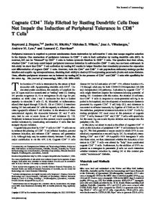
Figure 1. Cytometric analysis of dendritic cells (DCs) in peripheral blood, lymph nodes and tumors of NSCLC patients. The R1 region was gated on live mononuclear leukocytes and the R2 region was set to assess myeloid or lymphoid/plasmacytoid DCs, respectively. (A) Analysis of CD1c+/CD19- myeloid DCs. (B) Analysis of CD303+/CD123+ lymphoid/plasmacytoid DCs.
subpopulations was the highest in lymph nodes (0.96%; 0.3919.61%). It was significantly higher (p=0.003) than in peripheral blood (0.47%; 0.25-1.00%), but not significantly (p=0.47) higher in tumor tissue (0.81%; 0.19-4.34%). The difference between peripheral blood and tumor was statistically significant (p=0.04). We observed that the
myeloid DCs/plasmacytoid DCs ratio was significantly lower in malignant tissue (tumor and lymph nodes). The ratio of CD1c+/CD19- cells to CD303+/CD123+ cells was the lowest in lymph nodes: 0.26 (0.04-17.66), and was significantly (p=0.018) different from the peripheral blood ratio: 1.18 (0.47-3.88) and tumor tissue: 0.62 (0.17-6.33) (p=0.03). The
237-243
11/12/07
240
14:25
Page 240
TABARKIEWICZ et al: CD1C+ AND CD303+ DENDRITIC CELLS IN TISSUES OF PATIENTS WITH NSCLC
Figure 2. Comparison of percentages of CD1c + and CD303 + dendritic cells, total percentage of both subpopulation of DCs, and myeloid to lymphoid/plasmacytoid DCs ratio in peripheral blood (PB), lymph nodes (LN) and tumor (T) of NSCLC patients. Dot, median; box, 25-75 percentiles; whiskers, minimum and maximum. (A) Tissue distribution of CD1c+/CD19- myeloid DCs. (B) Tissue distribution of CD123+/CD303+ plasmacytoid DCs. (C) Tissue distribution of total DCs. (D) Comparison of myeloid to lymphoid/plasmacytoid DCs ratio in different tissue.
Analysis of patient survival depending on the percentage of CD1c + and CD303 + dendritic cells in tissues of NSCLC patients. Patients were divided into two groups based on the values below and above appropriate median percentages of dendritic cells. We concluded that patients with a low percentage of CD1c+ dendritic cells in the tumor survive significantly longer than patients with a high percentage, respectively 42.9±15.9 and 19.2±17.2 months (Fig. 3). Other differences in survival depend on the percentage of CD1c+ and CD303+ dendritic cells in tissues of NSCLC patients. Figure 3. Differences in survival between patients with a low and high percentage of tumor-infiltrating CD1c+ dendritic cells.
difference between peripheral blood and tumor tissue was not statistically significant (p=0.33). The results are summarized in Fig. 2. Comparison of CD1c+ or CD303+ dendritic cells in tissues of patients with different histological types of NSCLC. Between examined tissues there were no significant differences in the percentages of CD1c+/CD19- and CD303+/CD123+ and in the total number of CD1c+/CD19- and CD303+/CD123+ among each histological type of NSCLC, such as planoepithelial carcinoma, carcinoma macrocellulare, adenocarcinoma and mixed cell carcinoma. There was no significant difference in the CD1c+/CD19- and CD303+/CD123+ ratio among different histological types of lung cancer.
Comparison of immature peripheral blood DCs between surgically treated NSCLC patients and healthy donors. There was a statistically significantly lower (p=0.009) percentage of CD1c+/CD19- and not significantly lower (p=0.41) percentage of CD303+/CD123+ in the study group in comparison with the age-matched control group of healthy men. The total number of myeloid DCs and lymphoid/plasmacytoid DCs was significantly lower in the study group (p=0.04). The myeloid DCs/plasmacytoid DCs ratio was also significantly lower in the study group (p=0.04) in comparison with the healthy donors. The results are summarized in Fig. 4. Discussion We investigated the presence of both myeloid and plasmacytoid dendritic cells (DCs) in peripheral blood, lymph nodes and tumor tissue of NSCLC patients. DC infiltration of solid
237-243
11/12/07
14:25
Page 241
ONCOLOGY REPORTS 19: 237-243, 2008
241
Figure 4. The presence of myeloid and plasmacytoid DCs in peripheral blood of NSCLC patients and healthy donors (HD). Dot, median; box, 25-75 percentiles; whiskers, minimum and maximum. (A) Frequency of CD1c+/CD19- myeloid DCs. (B) Frequency of CD123+/CD303+ plasmacytoid DCs. (C) Frequency of all DCs. (D) Comparison of myeloid to lymphoid/plasmacytoid DCs ratio.
tumors has been described for several solid tumors (1-3). In pancreatic carcinoma, Dallal and associates observed a decreased level of dendritic cells or even their complete absence (32). The results of our study confirm the findings of these reports, demonstrating the presence of immature DCs in tumor tissue without molecules needed for the induction of an efficient immune response (24-33). We observed the highest frequency of immature lymphoid/plasmacytoid and myeloid dendritic cells in draining lymph nodes, which is in conflict with the physiological status, where mature DCs in lymphoid tissue are able to stimulate the immune system. The presence of so many immature DCs can be explained by cancer development. Many authors have demonstrated a blockade of DCs maturation by the proliferating tumor, e.g. of lung cancer (34-36). Lung cancer cells secreting bombesine-like peptides (BLP) blocked the maturation and function of DCs. BLP also shortened the viability of DCs (35). Other factors considered as inhibitors of DC maturation, such as VEGF, IL-10 and gangliosides are produced by tumor cells (37-39). It was also shown that DCs that are in direct contact with live cancer cells were not able to stimulate lymphocytes towards the lysis of tumor cells (11,40). Inhibition of DCs maturation and their functional status might constitute one of the tumor escape mechanisms. Tsujitani et al proved that tumor tissue
infiltrating DCs do not prevent the spread of tumor invasion, but do prevent nodal involvement (4,41). This confirms clinical observation of tumor growth and metastasis despite the DCs presence in the tumor tissue. The results of our study suggest that NSCLC also has a disadvantageous influence on DCs. A higher percentage of DCs in tumor tissue seems to prove that cancer tissue acts as a kind of DCs trap. Although DCs migrate to tissues involved in a neoplasmatic process, their maturation and function are disturbed and DCs are eventually eliminated. These findings seem to be confirmed by the analysis of the survival of patients with a low and high percentage of tumor-infiltrating myeloid dendritic cells. Patients who accumulated CD1c+ DCs in the tumor show shorter survival time than patients with a low percentage of CD1c+ in the tumor. Myeloid dendritic cells seem to be entrapped in cancer tissue and do not migrate to lymph nodes, where they could promote an anti-cancer immune response. Our results are contrary to some authors (1-5), probably due to the fact that we focused on immature dendritic cells. We noted a prevalence of CD303+/CD123+ lymphoid/ plasmacytoid DCs in tumor tissue and lymph nodes. Comparing those tissues to peripheral blood, we found that the ratio of myeloid DCs and lymphoid/plasmacytoid DCs was significantly lower in the majority of our patients and was
237-243
11/12/07
14:25
242
Page 242
TABARKIEWICZ et al: CD1C+ AND CD303+ DENDRITIC CELLS IN TISSUES OF PATIENTS WITH NSCLC
even inverse. This fact proves a prevalence of lymphoid/ plasmacytoid line DCs in tissues involved in a neoplasmatic process. As lymphoid/plasmacytoid DCs physiologically stimulate humoral immune responses and immunological tolerance, this might be considered as another tumor-escape mechanism. It seems that in order to guarantee a favorable immunological response, cancer cells may secrete adequate chemotactic substances which can attract a suitable subpopulation of DCs. The presence of examined DC subpopulations was confirmed also in lung tissue non-directly affected by cancer. Demedts et al found that in mononuclear cells isolated from lung 1.18%±0.19 were CD3 -/CD19 -/ CD1c +/HLA-DR + and 0.57%±0.1 were CD303 +/CD123 + (42,43). The percentage of plasmacytoid DCs described in normal lung tissue was comparable to our findings in the tumor, but the percentage of myeloid DCs was higher than in tumor tissue and lymph nodes. We did not find any significant differences in examined parameters between patients at different stages of NSCLC in contrast to the observations for other cancer types, e.g. breast cancer (3,11). We also compared the percentage of immature dendritic cells in the peripheral blood of NSCLC patients and healthy donors. We found a statistically lower percentage of DCs among NSCLC patients when compared with the healthy control group. This suggests a systemic influence of cancer on dendritic cells. A similar effect was observed in patients with cancers of the gastrointerstitial tract and in patients with breast, lung and head and neck cancer (18,37,44). A lower dendritic cell count in peripheral blood may be explained by a negative influence of cancer cells on the generation of DCs. Many authors described inhibitory effects on the generation of DCs by substances secreted by cancer cells, e.g. VEGF, IL-10, gangliosides, or PSA (25,34,45). Another explanation of the smaller amount of circulating DCs in the peripheral blood of NSCLC patients might be the theory of cancer tissue acting as a specific ‘black hole’ for DCs (14). DCs have been described to be attracted by cancer cells through specific chemotactic substances, where they are inactivated or eliminated (13,14). We studied not only the absolute amount of DCs in peripheral blood, but also their lymphoid/plasmacytoid and myeloid subpopulations. A decrease in the entire amount of DCs was mainly due to a significant decrease in myeloid DCs, whereas the lymphoid/plasmacytoid subpopulation was significantly higher in the NSCLC patients. Similar results were found by Sakakura et al (46) and Hoffmann et al (47) in squamous cell carcinoma of the head and neck. In summary, our data suggest that the development of NSCLC promotes the generation and survival of lymphoid/plasmacytoid DCs. Thus it may constitute a tumor escape mechanism, since lymphoid/plasmacytoid DCs promote immunological tolerance. Acknowledgements The authors thank K. Wojas MSc and I. Baran MSc for their technical contribution to this study and B. Rybojad MD and S. Chocholska PhD for their help in the preparation of the manuscript. They acknowledge Professor M. Schmitt
(Department of Internal Medicine III, University of Ulm, Germany) for his scientific discussion on our topic. References 1. Coppola D, Fu L, Nicosia SV, Kounelis S and Jones M: Prognostic significance of p53, bcl-2, vimentin, and S100 protein-positive Langerhans cells in endometrial carcinoma. Hum Pathol 29: 455-462, 1998. 2. Goldman SA, Baker E, Weyant RJ, Clarke MR, Myers JN and Lotze MT: Peritumoral CD1a-positive dendritic cells are associated with improved survival in patients with tongue carcinoma. Arch Otolaryngol Head Neck Surg 124: 641-646, 1998. 3. Tsuge T, Yamakawa M and Tsukamoto M: Infiltrating dendritic/Langerhans cells in primary breast cancer. Breast Cancer Res Treat 59: 141-152, 2000. 4. Tsujitani S, Kakeji Y, Maehara Y, Sugimachi K and Kaibara N: Dendritic cells prevent lymph node metastasis in patients with gastric cancer. In Vivo 7: 233-237, 1993. 5. Zeid NA and Muller HK: S100 positive dendritic cells in human lung tumors associated with cell differentiation and enhanced survival. Pathology 25: 338-343, 1993. 6. Mailliard RB, Dallal RM, Son YI and Lotze MT: Dendritic cells promote T-cell survival or death depending upon their maturation state and presentation of antigen. Immunol Invest 29: 177-185, 2000. 7. Chapoval AI, Tamada K and Chen L: In vitro growth inhibition of a broad spectrum of tumor cell lines by activated human dendritic cells. Blood 95: 2346-2351, 2000. 8. Shimamura H, Cumberland R, Hiroishi K, Watkins SC, Lotze MT and Baar J: Murine dendritic cell-induced tumor apoptosis is partially mediated by nitric oxide. J Immunother 25: 226-234, 2002. 9. Vanderheyde N, Aksoy E, Amraoui Z, Vandenabeele P, Goldman M and Willems F: Tumoricidal activity of monocytederived dendritic cells: evidence for a caspase-8-dependent, Fas-associated death domain-independent mechanism. J Immunol 167: 3565-3569, 2001. 10. Chaux P, Favre N, Bonnotte B, Moutet M, Martin M and Martin F: Tumor-infiltrating dendritic cells are defective in their antigen-presenting function and inducible B7 expression. A role in the immune tolerance to antigenic tumors. Adv Exp Med Biol 417: 525-528, 1997. 11. Gabrilovich DI, Corak J, Ciernik IF, Kavanaugh D and Carbone DP: Decreased antigen presentation by dendritic cells in patients with breast cancer. Clin Cancer Res 3: 483-490, 1997. 12. Nestle FO, Burg G, Fah J, Wrone-Smith T and Nickoloff BJ: Human sunlight-induced basal-cell-carcinoma-associated dendritic cells are deficient in T cell co-stimulatory molecules and are impaired as antigen-presenting cells. Am J Pathol 150: 641-651, 1997. 13. Pirtskhalaishvili G, Shurin GV, Esche C, et al: Cytokinemediated protection of human dendritic cells from prostate cancer-induced apoptosis is regulated by the Bcl-2 family of proteins. Br J Cancer 83: 506-513, 2000. 14. Pirtskhalaishvili G, Shurin GV, Gambotto A, et al: Transduction of dendritic cells with Bcl-xL increases their resistance to prostate cancer-induced apoptosis and antitumor effect in mice. J Immunol 165: 1956-1964, 2000. 15. Qin Z, Noffz G, Mohaupt M and Blankenstein T: Interleukin-10 prevents dendritic cell accumulation and vaccination with granulocyte-macrophage colony-stimulating factor gene-modified tumor cells. J Immunol 159: 770-776, 1997. 16. Vicari AP, Caux C and Trinchieri G: Tumour escape from immune surveillance through dendritic cell inactivation. Semin Cancer Biol 12: 33-42, 2002. 17. Almand B, Clark JI, Nikitina E, et al: Increased production of immature myeloid cells in cancer patients: a mechanism of immunosuppression in cancer. J Immunol 166: 678-689, 2001. 18. Lissoni P, Brivio F, Ferrante R, et al: Circulating immature and mature dendritic cells in relation to lymphocyte subsets in patients with gastrointestinal tract cancer. Int J Biol Markers 15: 22-25, 2000. 19. Wojas K, Tabarkiewicz J and Rolinski J: Dendritic cells in cancer patients. Folia Histochem Cytobiol 42: 45-48, 2004.
237-243
11/12/07
14:25
Page 243
ONCOLOGY REPORTS 19: 237-243, 2008
20. Enk AH, Jonuleit H, Saloga J and Knop J: Dendritic cells as mediators of tumor-induced tolerance in metastatic melanoma. Int J Cancer 73: 309-316, 1997. 21. Esche C, Lokshin A, Shurin GV, et al: Tumor's other immune targets: dendritic cells. J Leukoc Biol 66: 336-344, 1999. 22. Fanger NA, Maliszewski CR, Schooley K and Griffith TS: Human dendritic cells mediate cellular apoptosis via tumor necrosis factor-related apoptosis-inducing ligand (TRAIL). J Exp Med 190: 1155-1164, 1999. 23. Ohm JE, Shurin MR, Esche C, Lotze MT, Carbone DP and Gabrilovich DI: Effect of vascular endothelial growth factor and FLT3 ligand on dendritic cell generation in vivo. J Immunol 163: 3260-3268, 1999. 24. Shurin M and Gabrilovich D: Regulation of dendritic cell system by tumor. Cancer Res Ther Control 11: 65-78, 2001. 25. Shurin GV, Shurin MR, Bykovskaia S, Shogan J, Lotze MT and Barksdale EM Jr: Neuroblastoma-derived gangliosides inhibit dendritic cell generation and function. Cancer Res 61: 363-369, 2001. 26. Hill G, Millar W and Connelly J: ‘The great debate’ 1: Smoking, lung cancer, and cancer epidemiology. Can Bull Med Hist 20: 367-386, 2003. 27. Radzikowska E, Glaz P and Roszkowski K: Lung cancer in women: age, smoking, histology, performance status, stage, initial treatment and survival. Population-based study of 20,561 cases. Ann Oncol 13: 1087-1093, 2002. 28. Hirschowitz EA, Foody T, Kryscio R, Dickson L, Sturgill J and Yannelli J: Autologous dendritic cell vaccines for non-small-cell lung cancer. J Clin Oncol 22: 2808-2815, 2004. 29. Ishikawa A, Motohashi S, Ishikawa E, et al: A phase I study of alpha-galactosylceramide (KRN7000)-pulsed dendritic cells in patients with advanced and recurrent non-small cell lung cancer. Clin Cancer Res 11: 1910-1917, 2005. 30. Iwashita Y, Tahara K, Goto S, et al: A phase I study of autologous dendritic cell-based immunotherapy for patients with unresectable primary liver cancer. Cancer Immunol Immunother 52: 155-161, 2003. 31. Dzionek A, Fuchs A, Schmidt P, et al: BDCA-2, BDCA-3, and BDCA-4: three markers for distinct subsets of dendritic cells in human peripheral blood. J Immunol 165: 6037-6046, 2000. 32. Dallal RM, Christakos P, Lee K, Egawa S, Son YI and Lotze MT: Paucity of dendritic cells in pancreatic cancer. Surgery 131: 135-138, 2002. 33. Bell D, Chomarat P, Broyles D, Netto G, et al: In breast carcinoma tissue, immature dendritic cells reside within the tumor, whereas mature dendritic cells are located in peritumoral areas. J Exp Med 190: 1417-1426, 1999. 34. Aalamian M, Pirtskhalaishvili G, Nunez A, et al: Human prostate cancer regulates generation and maturation of monocyte-derived dendritic cells. Prostate 46: 68-75, 2001.
243
35. Makarenkova VP, Shurin GV, Tourkova IL, et al: Lung cancer-derived bombesin-like peptides down-regulate the generation and function of human dendritic cells. J Neuroimmunol 145: 55-67, 2003. 36. Redlinger Jr RE, Mailliard RB and Barksdale Jr EM: Neuroblastoma and dendritic cell function. Semin Pediatr Surg 13: 61-71, 2004. 37. Bonfanti A, Lissoni P, Bucovec R, Rovelli F, Brivio F and Fumagalli L: Changes in circulating dendritic cells and IL-12 in relation to the angiogenic factor VEGF during IL-2 immunotherapy of metastatic renal cell cancer. Int J Biol Markers 15: 161-164, 2000. 38. Lissoni P, Malugani F, Bonfanti A, et al: Abnormally enhanced blood concentrations of vascular endothelial growth factor (VEGF) in metastatic cancer patients and their relation to circulating dendritic cells, IL-12 and endothelin-1. J Biol Regul Homeost Agents 15: 140-144, 2001. 39. Shurin MR, Yurkovetsky ZR, Tourkova IL, Balkir L and Shurin GV: Inhibition of CD40 expression and CD40-mediated dendritic cell function by tumor-derived IL-10. Int J Cancer 101: 61-68, 2002. 40. Gabrilovich DI, Ciernik IF and Carbone DP: Dendritic cells in antitumor immune responses. I. Defective antigen presentation in tumor-bearing hosts. Cell Immunol 170: 101-110, 1996. 41. Tsujitani S, Kakeji Y, Watanabe A, Kohnoe S, Maehara Y and Sugimachi K: Infiltration of dendritic cells in relation to tumor invasion and lymph node metastasis in human gastric cancer. Cancer 66: 2012-2016 1990. 42. Demedts IK, Brusselle GG, Vermaelen KY and Pauwels RA: Identification and characterization of human pulmonary dendritic cells. Am J Respir Cell Mol Biol 32: 177-184, 2005. 43. Demedts IK, Bracke KR, Maes T, Joos GF and Brusselle GG: Different roles for human lung dendritic cell subsets in pulmonary immune defense mechanisms. Am J Respir Cell Mol Biol 35: 387-393, 2006. 44. Almand B, Resser JR, Lindman B, et al: Clinical significance of defective dendritic cell differentiation in cancer. Clin Cancer Res 6: 1755-1766, 2000. 45. Aalamian M, Tourkova IL, Chatta GS, et al: Inhibition of dendropoiesis by tumor derived and purified prostate specific antigen. J Urol 170: 2026-2030, 2003. 46. Sakakura K, Chikamatsu K, Takahashi K, Whiteside TL and Furuya N: Maturation of circulating dendritic cells and imbalance of T-cell subsets in patients with squamous cell carcinoma of the head and neck. Cancer Immunol Immunother 55: 151-159, 2006. 47. Hoffmann TK, Muller-Berghaus J, Ferris RL, Johnson JT, Storkus WJ and Whiteside TL: Alterations in the frequency of dendritic cell subsets in the peripheral circulation of patients with squamous cell carcinomas of the head and neck. Clin Cancer Res 8: 1787-1793, 2002.