Acta neurol. belg., 2006, 106, 87-89
Case report
Central nervous system metastases of pulmonary adenocarcinoma mimicking neurofibromatosis type 2 B. M. P. WILLEKENS1, S. ROOKER2, G. DUA2, B. APPEL3, J. J. MARTIN4, R. CROLS1 and P. P. DE DEYN1,5 Department of Neurology, Middelheim Hospital, ZNA, Antwerp, Belgium ; 2Department of Neurosurgery, Middelheim Hospital, ZNA, Antwerp, Belgium ; 3Department of Neuroradiology, Middelheim Hospital, ZNA, Antwerp, Belgium ; 4Department of Neuropathology, Institute Born-Bunge , University of Antwerp, Belgium ; 5Department of Neurochemistry and Behavior, Institute Born-Bunge, University of Antwerp, Belgium 1
————
Abstract We report a case of a 51-year-old man presenting with rapidly progressive unilateral tinnitus, hearing loss and imbalance. Neuroimaging revealed bilateral VIIIth cranial nerve masses and multiple cerebral and spinal cord lesions that were interpreted as being acoustic schwannomas and multiple meningeomas. An initial tentative diagnosis of neurofibromatosis type 2 (NF2) was made. Both clinical and radiological evolution were atypical for NF2 and the initial diagnosis of NF2 was questioned. Additional technical investigations demonstrated a pulmonary adenocarcinoma. Postmortem examination confirmed that this patient had multiple central nervous system metastases of a primary pulmonary adenocarcinoma, presenting clinically and neuroradiologically as a probable neurofibromatosis type 2. Clinicians should be aware of the rare possibility of central nervous system metastases mimicking NF2. Key words : Acoustic schwannomas ; neurofibromatosis ; metastasis ; inner auditory canal ; pulmonary adenocarcinoma ; cerebellopontine angle. Introduction We report the case of a patient with bilateral masses in the internal auditory canal and multiple cerebral lesions, that were initially attributed to neurofibromatosis type 2 but later correctly diagnosed as metastases of a pulmonary adenocarcinoma. Previous similar cases mimicking acoustic schwannomas, had a known history of pulmonary adenocarcinoma, melanoma, systemic lymphoma and breast adenocarcinoma (Nakada et al. 1983 ; Yamakami et al. 1999 ; Currie and Tomma 2001 ; Marques et al. 2002). Other cases describe isolated predominantly unilateral metastases to the inner auditory canal of pulmonary adenocarcinoma, pancreatic adenocarcinoma, melanoma or an unknown primary tumor as the first manifestation of malignancy (Moloy et al. 1989 ; Minami et al. 1996 ;
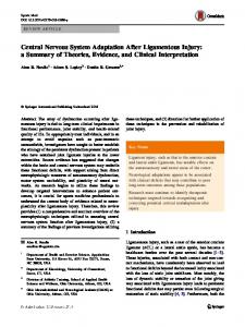
Kingdom et al. 1993 ; Hariharan et al. 2005 ; Lee and Weber 2005). Our case is the first that presents with symptomatic bilateral cerebellopontine angle masses and multiple other asymptomatic central nervous system lesions, suggestive of NF2, that later demonstrated to be metastases of pulmonary adenocarcinoma. Case report A 51-year old male patient presented to the ENT-specialist on referral of his general physician, with progressive tinnitus and a hearing deficit of the right ear since a few days. On walking he noticed a slight deviation to the left. He had flu-like symptoms 3 weeks before onset of these symptoms. His medical history was unremarkable. He was not taking any medication. He smoked 15 cigarettes per day for 20 years. In the family history there were no remarkable problems. Audiogram showed right sided deafness (250 Hz at 110 dB) and left sided perceptive hearing loss. Brainstem auditory evoked potentials showed a flat trace on stimulation on the right. MRI of the fossa posterior showed bilateral lesions in the cerebellopontine region, suspect for acoustic schwannomas (Fig. 1A). There were several cerebral lesions, suspect for meningeomas (Fig. 1B). Subsequently, the patient was referred to the neurology department. On neurological examination there was a perceptive hearing deficit on the right and Romberg’s sign was positive. Further cranial nerve examination was normal, as were motor functions. There was a slight dysmetria on the finger-nose and heel-shin test, more pronounced on the right than on the left side. Reflexes were brisk, but symmetrical and there was a bilateral Babinski’s sign. There was significant oedema surrounding only one brain lesion. For this reason, treatment with corticoids was initiated. Ophtalmological examination showed bilateral cataract, as result of nuclear sclerosis as well as subcapsular posterior cataract.
88
B. M. P. WILLEKENS ET AL.
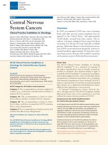
A FIG. 1. — A. Axial T1-weighted thin slice through the petrosal bones after injection of Gadolinium shows bilateral masses in the internal auditory canals, first considered as possible vestibularis schwannoma, indicated with arrows ; B. Sagittal T1weighted slice through the cervical region after injecC tion of Gadolinium with multiple enhancing lesions. The extra-axial were considered possible meningeo- B ma or schwannomas and the intramedullar lesion a possible ependymoma ; C. — One axial reconstruction of a 3 D series after injection of Gadolinium. Three peripheral lesions considered as possible meningioma, one of them with broad oedema.
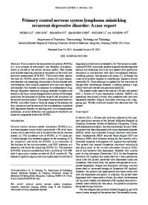
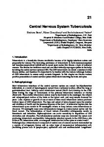
MRI of the spinal cord demonstrated several contrast-enhancing lesions on C2, C4-C5 and D6-7, that were suspect for multiple schwannomas, with one intramedullary lesion, interpreted as being a possible ependymoma (Fig. 1C). Based on this imaging, a preliminary diagnosis of neurofibromatosis type 2 was made. Blood sampling for genetics was performed, but was later on found to be negative. One month later this patient was referred to our neurological outpatient clinic with complaints of sensory loss in the right buttocks. In the last month he had lost approximately 4 kg of body weight. On neurological examination we found sensory loss in the S1 dermatome, and an absent ankle reflex on the right side. Subsequent MRI of the lumbosacral region demonstrated several lesions, considered as schwannomas, on the cauda equina (Fig. 2A). One month after the initial evaluation, follow-up MRI of the brain and cervical spine, demonstrated dramatic growth of almost all lesions, not consistent with classical evolution of NF2. The possibility of cerebral metastases with metastases to the whole neuraxis was further investigated. Plain radiography of the thorax showed a lesion, suggestive of an infiltrate, in the perihilar region on the left side. CT thorax showed a mass in the lingula, suspect for a neoplastic formation. In both lungs, multiple small nodules were seen. In both adrenals there were masses visualized, suggestive for metastatic lesions. Bronchoscopy was done, and biopsies taken. Histological examination revealed a non-small cell lung carcinoma, type adenocarcinoma. Palliative cranial and cervical radiotherapy was started, but the patient deteriorated progressively and deceased 2,5 months after initial symptoms. Postmortem examination of brain and spinal cord (see figure 2B) demonstrated that all the lesions were microscopically compatible with metastases of the pulmonary adenocarcinoma,
A
B
FIG. 2. — A. Sagittal T1-weighted slice through the lumbar region after injection of Gadolinium. Multiple enhancing lesions located on the cauda equina described as schwannoma or meningeoma ; B. Autopsy specimen of lumbar spinal cord and cauda equine with multiple nodules on the radices.
including the inner auditory canal masses. No meningeomata or schwannomata were seen. Discussion Metastases to the internal auditory canal are very rare. In the literature there are some cases of unilateral metastasis to the cerebellopontine angle (Nakada et al. 1983 ; Moloy at al 1989 ; Minami et al 1996 ; Kingdom et al. 1993 ; Currie and Tomma 2001 ; Marques et al. 2002 ; Ferri et al. 1998), mimicking unilateral acoustic schwannomas, and only a few case reports on metastases mimicking bilateral acoustic schwannomas (Yamakami et al. 1999 ; Hariharan et al. 2005 ; Lee and Weber 2005). This is the third case in anglosaxan literature
CENTRAL NEURVOUS SYSTEM METASTASES
that describes lesions in both inner auditory canals as initial manifestation of a malignancy. This report however, is the first case description where additional asymptomatic central nervous system lesions mimicked the neuroradiological image of neurofibromatosis type 2 with bilateral acoustic schwannomas, meningeomas and spinal cord schwannomas. Imaging studies alone are insufficient to diagnose neurofibromatosis type 2. In this case, clinical and neuroimaging follow-up studies, prompted us to reconsider the initial diagnosis. A diagnosis of pulmonary adenocarcinoma with multiple central nervous system metastases was made. Key points to reconsider the diagnosis in this case were the weight loss, new radicular symptoms and fast evolutive disease on neuro-imaging. Clinicians should always consider the rare possibility of bilateral masses in the cerebellopontine angle with other neuraxis lesions to be of metastatic origin, even if the history of the patient is unremarkable. REFERENCES CURRIE L., TOMMA A. Malignant melanoma presenting as sudden onset of complete hearing loss. Ann. Plast. Surg., 2001, 47 : 336-7. FERRI G. G. et al. Metastasis in the inner auditory canal. Acta Otorhinolaryngol. Ital., 1998, 18 : 269-75. HARIHARAN S., ZHU J., NADKARNI M. A., DONAHUE J. E. Metastatic lung cancer in the cerebellopontine angles mimicking bilateral acoustic neuroma. J. Clin. Neurosci., 2005, 12 : 184-6.
89
KINGDOM T. T., LALWANI A. K., PITTS L. H. Isolated metastatic melanoma of the cerebellopontine angle : case report. Neurosurgery, 1993, 33 : 1424. LEE W. T., WEBER P. C. Melanoma metastasis masquerading as bilateral acoustic neuromas. Otolaryngol. Head Neck Surg., 2005, 132 : 505-6. MARQUES E., BRANDIS A., SAMII M., TATAGIBA M. Late metastasis of breast adenocarcinoma into internal auditory canal and cerebellopontine angle : case report. Arq. Neuropsiquiatr., 2002, 60 : 639-42. MINAMI M. et al. Solitary metastasis of lung cancer to the cerebellopontine angle-case report. Neurol. Med. Chir., 1996, 36 : 172-4. MOLOY P. J., DEL JUNCO R., PORTER R. W., BRACKMANN D. E. Metastasis from an unknown primary presenting as a tumor in the internal auditory meatus. Am. J. Otol., 1989, 10 : 297-300. NAKADA T., ST JOHN J. N., KNIGHT R. T. Solitary metastasis of systemic malignant lymphoma to the cerebellopontine angle. Neuroradiology, 1983, 24 : 225-28. YAMAKAMI I., OISHI H., IWADATE Y., YAMAURA A. Isolated metastases of adenocarcinoma in the bilateral internal auditory meatuses mimicking neurofibromatosis type 2-case report. Neurol. Med. Chir., 1999, 39 : 756-61.
Prof. Dr. P. P. DE DEYN, Department of Neurology, Middelheim Hospital, ZNA, Antwerp (Belgium). E-mail :
[email protected].