H&0 CLINICAL CASE STUDIES
Primary Central Nervous System Lymphomatoid Granulomatosis in a Patient Receiving Azathioprine Therapy L. Katherine Martin, MD 1 Pierluigi Porcu, MD 2 Robert A. Baiocchi, MD, PhD 2 Jack W. Erter, MD 2 Abhik Ray Chaudhury, MBBS 3
Department of Internal Medicine, 2Department of Hematology and Oncology, 3Department of Pathology at The Ohio State University Medical Center, Columbus Ohio 1
Introduction Lymphomatoid granulomatosis (LYG) is a rare lymphoproliferative disorder seen in both immunocompetent and immunosuppressed patients that exhibits characteristics of both lymphomatous and granulomatous disease. Histologically, it is a necrotizing angiocentric and angiodestructive large B-cell lymphoma that is composed of small numbers of Epstein-Barr virus (EBV)-positive neoplastic B-cells amid a background of reactive, predominantly CD4+ T-cells.1 Fever and cough are the most common presenting symptoms, followed by malaise, weight loss, shortness of breath, neurologic symptoms, and chest pain.2 Patients typically present with extranodal disease. The lungs are affected in 90% of cases, and the skin, kidney, and liver, and central nervous system (CNS) are less frequently involved. Neurologic involvement is typically secondary to metastatic spread of primary pulmonary lesions3 and confers a worse prognosis.2 Very few cases of isolated or initial CNS involvement have been reported.3-6 We present a case of LYG confined to the CNS in an immunosuppressed patient. Case Presentation A 69-year-old Caucasian woman with autoimmune hepatitis treated with azathioprine 50 mg/day for the past 10 months presented with progressively worsening confusion, dysarthria, aphasia, and mild right-sided weakness. Other symptoms included a 30-pound weight loss (23% of body weight) over 4 months, drenching night sweats, Address correspondence to: L. Katherine Martin, MD, The Ohio State University Medical Center, Department of Internal Medicine, 1654 Upham Drive, 202 Means Hall, Columbus, OH 43210; Phone: 614-292-6445; Fax: 614-293-7037; Email:
[email protected].
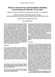
fatigue, low-grade fevers, occasional cough, and mild shortness of breath. At the time of initial assessment, the patient’s only measurable neurologic deficits were wordfinding difficulties and confusion; there was no lymphadenopathy, hepatosplenomegaly, or skin rash. Blood counts, chemistry, and liver function were normal. Noncontrast computed tomography (CT) scan of the head showed left temporal, parietal, and frontal masses with surrounding edema and midline shift. Magnetic resonance imaging (MRI) of the brain with gadolinium revealed multiple enhancing lesions in the cerebellar hemispheres with significant surrounding edema and inferior cerebellar tonsillar herniation. Multiple supratentorial areas of abnormal enhancement were also noted with the largest lesion located in the region of the left sylvian fissure and the adjacent frontotemporal region, measuring 2.3 1.6 cm, with significant edema and rightward midline shift (Figure 1). Infectious workup, including tuberculin skin test, was negative. Azathioprine was discontinued, and treatment with intravenous dexamethasone was instituted for cerebral edema. On hospital day 4, the patient underwent stereotactic biopsy for diagnosis, after which her right-sided weakness became more noticeable and she developed complete expressive aphasia. Biopsy demonstrated large atypical lymphoid elements positive for CD20, CD30, EBV-encoded RNA (EBER-1), and EBER-2 in an infiltrative as well as a prominent angiocentric pattern with focal areas of necrosis, and many background CD4+ T-cells, consistent with type-C LYG (Figure 2). CT scans of the chest, abdomen, and pelvis showed 2 sub-centimeter nodules in the right lower lobe, a cirrhotic liver, a 1.2 cm left adrenal nodule, and an enhancing mass projecting into the endometrial canal. Positron emission tomography (PET)CT showed hypermetabolic foci corresponding to CNS disease seen on previous scans. None of the abnormalities
Clinical Advances in Hematology & Oncology Volume 7, Issue 1 January 2009
65
MARTIN ET AL
Figure 1. T1 weighted magnetic resonance imaging scans with gadolinium demonstrating several enhancing lesions in the left frontotemporal and cerebellar areas.
B
A
C
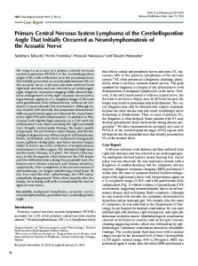
Figure 2. a) Hematoxylin and Eosin (H&E) stain at 400 and b) 600 magnification demonstrating a polymorphous lesion with large atypical lymphoid elements in an angiocentric and angiodestructive pattern as well as large numbers of plasma cells, histiocytes, and lymphocytes. At 600, the large lymphoid elements are clearly visible; c) EBER immunostain at 400 magnification showing positivity in the large atypical cells. Eber=Epstein-Barr virus encoded RNA
seen on chest and abdominal imaging was hypermetabolic. Lumbar puncture was done with cerebrospinal fluid revealing an abnormal population of CD19+ B-cells with aberrant loss of immunoglobulin light chain expression. Bone marrow and peripheral blood analyses showed no evidence of involvement by LYG. Whole blood EBV viral load by polymerase chain reaction (PCR) was modestly elevated (1,000 copies/mL), and enzyme-linked immunosorbent assay (ELISA) for human immunodeficiency virus (HIV) was nonreactive. On hospital day 22, treatment was begun with systemic high-dose methotrexate 3.5 mg/m2 (day 1), followed by intravenous rituximab (Rituxan, Genentech/ Biogen Idec) 375 mg/m2 (day 3), and antiviral therapy with intravenous zidovudine (AZT) 1.5 g twice daily and ganciclovir 1 g twice daily beginning on day 3.
66
When chemotherapy was commenced, the patient’s aphasia was slightly improved. On treatment day 3, she became more lethargic and somnolent, and developed an oxygen requirement of 2 L/min by nasal canula. Chest radiography showed alveolar infiltrates consistent with mild pulmonary edema. The patient did not respond clinically or radiographically to diuresis. Over the next 3 days her ability to communicate diminished. Serum sodium levels declined to a nadir of 119, and a diagnosis of syndrome of inappropriate antidiuretic hormone was entertained but not confirmed. Pancytopenia developed on treatment day 4, as well as liver function abnormalities that peaked on treatment day 6 (ALT, 1230; AST, 727; total bilirubin, 3.5; direct bilirubin, 2.3; alkaline phosphatase normal) with right upper quadrant ultrasonography revealing only cirrhosis.
Clinical Advances in Hematology & Oncology Volume 7, Issue 1 January 2009
P R I M A R Y C N S LY M P H O M A T O I D G R A N U L O M A T O S I S W I T H A Z A T H I O P R I N E T H E R A P Y
Immunologic workup was negative, as were cytomegalovirus and herpes simplex virus DNA by PCR. Serial measurements by PCR revealed no significant increase in EBV viral load. On treatment day 7 (hospital day 29), the patient developed acute hypoxic respiratory failure with hypotension and became unresponsive. She required intubation and vasopressors, and was transferred to the medical intensive care unit for cardiopulmonary support. Chest radiography showed markedly worsened diffuse alveolar infiltrates. Head CT showed worsening edema in the left temporal and parietal lobes and new dilation of the left temporal horn of the left lateral ventricle. Antiviral treatment was discontinued, and empiric broad-spectrum antibiotic therapy, antifungal prophylaxis, supportive blood product transfusions, and filgrastim were given. Blood cultures grew E. coli and M. catarrhalis on hospital day 30. Serum sodium normalized with normal saline, but the patient was unable to be weaned from vasopressor or ventilatory support. On hospital day 34, life support devices were withdrawn and the patient expired. Discussion LYG was first described by Leibow and colleagues in 1972 and was initially thought to be a T-cell lymphoproliferative disorder due to the prominence of CD4+ T-cells seen on biopsy specimens. It was later shown that LYG is an EBV-associated B-cell lymphoproliferative disorder (B-LPD) in which the numerous infiltrating T-cells are reactive.7 Unlike post-transplant lymphoproliferative disorders (PTLD), the EBV latency pattern in LYG has not been extensively studied. However, association with type III latency has been documented.8 As with PTLD and other EBV positive B-LPDs, a considerable percentage of patients with LYG are immunosuppressed. At least 3 cases of LYG following drug-induced immune suppression have been reported. In 2 of these cases, the authors reported clinical responses following discontinuation of immune suppression.9-11 CNS involvement was not reported in any of these cases. The natural history of LYG can be variable, and spontaneous remission can occur, although generally, prognosis is poor with a 5-year mortality rate of 60–90% and a median survival of 14 months. A standard of therapy has yet to be established due to the rarity of the disease. Treatments have included observation, cyclophosphamide, and prednisone alone or as part of multi-agent chemotherapy, with few long-term remissions.2 Rituximab has shown success in small numbers of patients alone12,13 or as part of multi-agent chemotherapy.14,15 Interferon alfa with or without autologous stem cell transplantation7,16 has also been shown to induce durable remissions.
Radiation therapy has not been as well studied, but successful CNS cases have been reported.5 To date, no prospective controlled clinical trials have been conducted to further test these treatment modalities. Our patient developed LYG clinically confined to the CNS, as determined by imaging studies, while receiving therapy with the immunosuppressive drug azathioprine. The resultant decreased CD8+ T-cell surveillance may have resulted in EBV-induced malignant B-cell transformation, similar to that which occurs in PTLD, where EBV is thought to increase the growth and survival of infected B-cells in the setting of impaired immune surveillance.1 We stopped azathioprine, but it was difficult to assess the effect of this intervention in our patient because she underwent an invasive neurosurgical procedure and developed superimposed complications that altered her clinical course. Prior to institution of chemotherapy, some improvement in neurologic status was noted without radiologic change. The treatment we selected for this patient was based on our prior experience with combined antiviral therapy plus or minus chemotherapy or radiation in a preclinical model of, as well as in, patients with EBVpositive primary CNS lymphomas and HIV-related lymphomas.17 In our previous studies, the addition of radiotherapy was necessary for response to antiviral drugs in cases exhibiting latency I or II, whereas tumors exhibiting latency III appeared to respond to antiviral therapy alone. Chemotherapy has also been shown to induce latency III and lytic phases of EBV replication and enhance response to antiviral therapy.18,19 Because we were not able to immediately establish the type of latency in our patient, we did not feel justified in using antiviral therapy alone. Moreover, because of the patient presentation with prominent constitutional symptoms, we concluded that it was necessary to provide therapy for occult systemic disease while at the same time targeting the CNS compartment. Thus, we elected to start high-dose methotrexate in addition to antiviral therapy and rituximab. The death of our patient likely occurred due to multiorgan failure associated with sepsis. Unfortunately, we were not able to obtain an autopsy. In conclusion, our patient is an example of an atypical presentation of a rare disease. LYG is most often a pulmonary process but is occasionally confined to other organ systems. Although in the past CNS involvement was often documented only post-mortem, a recent study shows that CNS involvement can be demonstrated with MRI imaging in more than 50% of cases.20 Therefore, LYG should be included in the differential diagnosis of immunosuppressed patients presenting with neurologic symptoms even if there is no evidence of pulmonary involvement.
Clinical Advances in Hematology & Oncology Volume 7, Issue 1 January 2009
67
MARTIN ET AL
References 1. Rezk SA, Weiss LM. Epstein-Barr virus-associated lymphoproliferative disorders. Hum Pathol. 2007;38:1293-1304. 2. Katzenstein AL, Carrington CB, Leibow AA. Lymphomatoid granulomatosis: a clinicopathologic study of 152 cases. Cancer. 1979;43:360-373. 3. Bae WK, Lee KS, Kim PN, et al. Lymphomatoid granulomatosis with isolated involvement of the brain- case report. J Korean Med Sci. 1991;6:255-259. 4. Mizuno T, Takanashi Y, Onodera H, et al. A case of lymphomatoid granulomatosis/angiocentric immunoproliferative lesion with long clinical course and diffuse brain involvement. J Neurol Sci. 2003;213:67-76. 5. Petrella TM, Walker IR, Jones GW, Leber B. Radiotherapy to control CNS lymphomatoid granulomatosis: a case report and review of the literature. Am J Hematol. 1999;62:239-241. 6. Kokmen E, Billman JK Jr, Abell MR. Lymphomatoid granulomatosis clinically confined to the CNS. A case report. Arch Neurol. 1977;34:782-784. 7. Wilson WH, Kingma DW, Raffeld M, et al. Association of lymphomatoid granulomatosis with Epstein–Barr viral infection of B lymphocytes and response to interferon-alpha 2b. Blood. 1996;87:4531-4537. 8. Taniere P, Thivolet-Bejui F, Vitrey D, et al. Lymphomatoid granulomatosisa report on four cases: evidence for B phenotype of tumoral cells. Eur J Respir. 1998;12:102-106. 9. Yazdi, AS, Metzler G, Weyrauch S, et al. Lymphomatoid Granulomatosis Induced by Imatinib-Treatment. Arch Dermatol. 2007;143:1222-1223. 10. Kameda H, Okuyama A, Tamaru J, et al. Lymphomatoid granulomatosis and diffuse alveolar damage associated with methotrexate therapy in a patient with rheumatoid arthritis. Clin Rheumatol. 2007;26:1585-1589. 11. Shimada K, Matsui T, Kawakami M, et al. Methotrexate-related lymphoma-
Review
Lymphomatoid Granulomatosis: Challenges in Diagnosis and Treatment Elena Gitelson, MD, 1 Tahseen Al-Saleem, MD, 2 and Mitchell R. Smith, MD, PhD 1 Departments of Medical Oncology 1 and Pathology 2 Fox Chase Cancer Center, Philadelphia, Pennsylvania
Martin and colleagues1 report a case of primary central nervous system (CNS) lymphomatoid granulomatosis (LYG) in a patient treated with azathioprine immunosuppressive therapy for presumed autoimmune hepatitis. Magnetic resonance imaging (MRI) of the brain showed multiple supra- and infra-tentorial lesions; brain biopsy was consistent with LYG with a prominent angiocenAddress correspondence to: Mitchell R. Smith, MD, PhD, Department of Medical Oncology, Fox Chase Cancer Center, 333 Cottman Avenue, Philadelphia, PA 19111; Tel: 215-728-2674; Fax: 215-728-3639; E-mail:
[email protected].
68
toid granulomatosis: a case report of spontaneous regression of large tumors in multiple organs after cessation of methotrexate therapy in rheumatoid arthritis. Scand J Rheumatol. 2007;36:64-67. 12. Jordan K, Grothey A, Grothe W et al. Successful treatment of mediastinal lymphomatoid granulomatosis with rituximab monotherapy. Eur J Haematol. 2005;74:263-266. 13. Zaidi A, Kampalath B, Peltier WL, Vesole DH. Successful treatment of systemic and central nervous system lymphomatoid granulomatosis with rituximab. Leuk Lymphoma. 2004;45:777-780. 14. Hu Y, Liu C, Chiu CH, Hsaio LT. Successful treatment of elderly advanced lymphomatoid granulomatosis with rituximab-CVP combination therapy. Eur J Haematol. 2007;78:176-177. 15. Alinari L, Pant S, McNamara K, et al. Lymphomatoid Granulomatosis presenting with gingival involvement in an immune competent elderly male. J. Surg. Pathol. 2008. (First submission) (IF: 2.384). 16. Johnston A, Coyle L, Nevell D. Prolonged remission of refractory lymphomatoid granulomatosis after autologous hemopoietic stem cell transplantation with post-transplantation maintenance interferon. Leuk Lymphoma. 2006;47:323-328. 17. Roychowdhury S, Peng R, Baiocchi RA, et al. Experimental treatment of Epstein-Barr virus- associated central nervous system lymphoma. Cancer Res. 2003;63: 965-971. 18. Feng WH, Hong G, Delecluse HJ, et al. Lytic induction therapy for EpsteinBarr virus positive B-cell lymphomas. J Virol. 2004;78:1839-1902. 19. Feng WH, Israel B, Raab-Traub N, et al. Chemotherapy induces lytic EBV replication and confers ganciclovir susceptibility to EBV-positive epithelial cell tumors. Cancer Res. 2002;62:1920-1926. 20. Patsalides AD, Atac G, Hedge U, et al. Lymphomatoid granulomatosis: abnormalities of the brain at MR imaging. Radiology. 2005;237: 265-273.
tric pattern, areas of necrosis, and presence of EBER-1 (Epstein Barr Virus [EBV] Encoded RNA) and EBER-2. LYG is an EBV–related T-cell–rich monoclonal B-cell lymphoproliferative disorder with a broad pathologic spectrum and variable malignant potential. Angiocentric infiltration with vascular damage and necrosis are pathognomonic and result from the angiodestructive potential of the polymorphous lymphoid infiltrate, predominantly T lymphocytes admixed with plasma cells, immunoblasts, and histiocytes, in contrast to sparse clonal EBV–positive B-cells. Uncertain whether it represented an inflammatory process with granulomalike formation or lymphomatous proliferation, Leibow designated the term "lymphomatoid granulomatosis" for the lung lesions2 almost 40 years ago. Histologic grading of LYG depends on the number of atypical large B-cells present in the biopsy. Grade 1 lesions lack cytologic atypia, and EBV–positive transformed large B-cells are rare. The EBV–positive cells can be demonstrated by EBER in situ hybridization, and are less than 5 per high power field (hpf ) in grade 1. They are more frequent in grade 2 lesions (5–20 per hpf ), whereas grade 3 lesions still show an inflammatory background, but contain readily identifiable enlarged atypical B-cells sometimes in aggregate (>20 per hpf ). Areas of infarctlike necrosis are almost always present. If only sheets
Clinical Advances in Hematology & Oncology Volume 7, Issue 1 January 2009
P R I M A R Y C N S LY M P H O M A T O I D G R A N U L O M A T O S I S W I T H A Z A T H I O P R I N E T H E R A P Y
of large transformed B-cells are present without the polymorphous background, the lesion should be designated as diffuse large B-cell lymphoma and not lymphomatoid granulomatosis. Monoclonal immunoglobulin gene rearrangement can be demonstrated in most cases of grade 2 and 3 disease, but not always in grade 1.3 The variety of histologic presentations translates into a large spectrum of potential clinical outcomes, ranging from a self-limited disorder with spontaneous resolution in 15–20%—an indolent disease with waxing and waning course—to an extensive multisystem disease with active vasculitis and rapid progression to an aggressive EBV–positive large B-cell lymphoma with median overall survival of less than 2 years. Recurrent disease often demonstrates a higher histologic grade. The pathogenesis of the lymphomatoid granulomatosis can be explained by the presence of the EBV genome that encodes a series of products interacting with, or exhibiting homology to, a variety of anti-apoptotic molecules, cytokines, and signal transducers, hence promoting EBV infection, immortalization, and transformation.4 The tissue necrosis and vascular damage may be due, at least in part, to the production of monokines induced by interferon gamma. Chemokines, such as IP-10 and Mig,5 elaborated as a result of the EBV infection, may be responsible for the vascular damage by promoting T-cell adhesion to endothelial cells in at least partially immunocompetent hosts. Clinicopathologically, LYG frequently presents as a systemic disease with prominent pulmonary and skin lesions; other typical sites of involvement include CNS, liver, kidney, and bone marrow. Constitutional symptoms such as weight loss, fatigue, sweats, and chills are typical.6 Uncommonly, LYG presents as localized disease limited to an extra-pulmonary site such as CNS.7-9 CNS involvement is the third most common manifestation site of LYG after pulmonary and skin lesions and has been reported to occur in at least 30% of patients diagnosed with LYG.6 In addition, peripheral nerve damage has been reported in 7% of cases.10 Remarkably, brain involvement does not indicate transformation of LYG into aggressive lymphoma and may be found in symptomatic, as well as in asymptomatic, patients. Patsalides and colleagues11 conducted a comprehensive retrospective analysis of MRI results of 25 patients with LYG and found a wide spectrum of CNS lesions in 53% (13/25) of patients. Most frequently, patients presented with multifocal intraparenchymal brain lesions which exhibited punctate or linear enhancement (n=7), followed by leptomeningeal and cranial nerve involvement (n=6). Abnormal cerebrospinal fluid with circulating clonal B-cells was detected in 5 patients; 2 had involvement of the choroid plexus, and isolated dural lesions were encountered in one case.
Whereas most of the above abnormalities resolved upon successful treatment of LYG, some lesions evolved into lacunar infarctions. Importantly, only 10 of 13 patients developed neurologic symptoms, whereas 3 patients with MRI–confirmed CNS abnormalities remained asymptomatic. This finding points to more prevalent CNS involvement in LYG than prior estimates of 30%. It also suggests that patients diagnosed with LYG may benefit from a screening MRI of the brain. LYG may develop in immunocompromised as well as in immunocompetent individuals, although patients with underlying immunodeficiency are at increased risk of LYG.12-15 The hypothesis is that innate or acquired defects in cellular immunity result in incomplete eradication of EBV–infected B-cells, permitting propagation of a B-cell clone surrounded by activated T- and other reactive cells. Spontaneous regression of LYG after removing immunosuppressive therapy is consistent with this hypothesis.13 Agarwal and colleagues16 presented a patient with rheumatoid arthritis treated with oral methotrexate who developed systemic LYG with evidence of progression to large B-cell lymphoma. His LYG went into remission after discontinuation of methotrexate. Alternatively, LYG may manifest with symptoms of systemic autoimmune disease such as arthritis or nonspecific cutaneous vasculitis. This may mask the diagnosis of LYG before the development of more typical pulmonary nodules and other systemic symptoms and confirmation of LYG from diagnostic biopsy.16 A differential diagnosis of Wegener’s granulomatosis should always be considered when pulmonary nodules and constitutional symptoms develop. The patient reported here manifested with autoimmune hepatitis and was treated with azathioprine for 10 months prior to diagnosis of biopsy-proven CNS LYG. Systemic symptoms of weight loss and cough developed 4 months prior to diagnosis of LYG. It is possible that hepatitis may have been the presenting sign of LYG, with regression of liver lesions either spontaneously or as a result of azathioprine, and then eventual relapse at a novel site with symptomatic brain lesions. We have observed a patient with LYG who initially presented with skin, pulmonary, and peripheral nerve damage and dramatically responded to steroids in all affected areas. However, she soon relapsed with extensive multifocal lesions in a new site, skeletal muscle, and biopsy revealed grade 3 LYG, while all her initial sites remained significantly improved. Treatment of LYG remains a challenge and a standard treatment paradigm is not yet established. Whereas some patients have self-limited disease or respond well to steroids, many will eventually progress to large B-cell lymphoma with generally poor response to conventional chemotherapy and median overall survival of less than 2 years. Several reports of rituximab (Rituxan, Genen-
Clinical Advances in Hematology & Oncology Volume 7, Issue 1 January 2009
69
MARTIN ET AL
tech/Biogen Idec) given as a single agent have been published with variable success.17-19 There are at least 2 reports of successful treatment of brain lesions with intravenous weekly rituximab with sustained complete resolutions of brain abnormalities.18,20 There is a report of 14 months observation followed by pulse steroids, which resulted in sustained complete remission of LYG with multiple brain lesions.7 The above observations emphasize that the brain lesions do not necessarily correlate with LYG transformation into aggressive lymphoma and may respond to less aggressive therapies. More intense treatment regimens such as CHOP (cyclophosphamide, doxorubicin, vincristine, prednisone), R-CHOP (rituximab plus CHOP) or R-CVP (rituximab plus cyclophosphamide, vincristine, prednisone) interferon alpha–containing regimens, and cranial irradiation have been reported with various responses.21-23 R-CHOP or other similar regimens are usual recommendations for treatment of grade 3 LYG due to the high rate of rapid progression into EBV–positive large B-cell lymphoma. It is not clear, however, whether such an aggressive initial approach is of benefit in grade 1 or 2 LYG, and a trial of rituximab given as a single agent or in combination with steroids may present a reasonable first choice. However, many patients are, or will become, refractory to rituximab, R-CHOP, or to interferon-based chemotherapy and will die of LYG shortly after diagnosis. Multicenter clinical trials, which are currently lacking, are essential for better management of this relatively uncommon and poorly understood peculiar monoclonal B-cell proliferation with propensity to evolve to frank lymphoid malignancy. References 1. L. Martin K, Porcu P, Baiocchi RA, Erter JW, Chaudhury AR. Primary central nervous system lymphomatoid granulomatosis in a patient receiving azathioprine therapy. Clin Adv Hematol Oncol. 2009;7:65-68. 2. Liebow AA, Carrington CR, Friedman PJ. Lymphomatoid granulomatosis. Hum Pathol. 1972;3:457-558. 3. Pittaluga S, Wilson WH, Jaffe ES In: Swerdlow SH, Campo E, Harris NL, Jaffe ES, Pileri, SA, Stein H, Thiele J, Vardiman JW, eds. WHO Classification of Tumours of Haematopoietic and Lymphoid Tissues. 4th ed. Lyon: IARC; 2008:247-249. 4. Carbone A, Gloghini A, Dotti G. EBV-associated lymphoproliferative disorders: classification and treatment. Oncologist. 2008;13:577-585.
70
5. Teruya-Feldstein J, Jaffe ES, Burd PR, et al. The role of Mig, the monokine induced by interferon-gamma, and IP-10, the interferon-gamma-inducible protein-10, in tissue necrosis and vascular damage associated with Epstein-Barr viruspositive lymphoproliferative disease. Blood. 1997;90:4099-4105. 6. Lundell RB, Weenig RH, Gibson LE. Lymphomatoid granulomatosis. Cancer Treat Res. 2008;142:265-272. 7. Okuda T, Akai F, Kataoka K, Taneda M. A case of lymphomatoid granulomatosis followed for 14 months on the basis of clinical and histological findings. Brain Tumor Pathol. 2008;25:33-38. 8. Kokmen E, Billman JK, Jr Abell MR. Lymphomatoid granulomatosis clinically confined to the CNS. A case report. Arch Neurol. 1977;34:782-784. 9. Mizuno T, Takanashi Y, Onodera H, et al. A case of lymphomatoid granulomatosis/angiocentric immunoproliferative lesion with long clinical course and diffuse brain involvement. J Neurol Sci. 2003;213:67-76. 10. Katzenstein AL, Carrington CB, Liebow AA. Lymphomatoid granulomatosis: a clinicopathologic study of 152 cases. Cancer. 1979;43:360-373. 11. Patsalides AD, Atac G, Hedge U, et al. Lymphomatoid granulomatosis: abnormalities of the brain at MR imaging. Radiology. 2005;237:265-273. 12. Varoczy L, Gergely L, Szakall S, Illes A. Angiocentric lymphomatoid granulomatosis and severe hypogammaglobulinaemia. Haematologia (Budap). 2002;32:535-541. 13. Shimada K, Matsui T, Kawakami M, et al. Methotrexate-related lymphomatoid granulomatosis: a case report of spontaneous regression of large tumours in multiple organs after cessation of methotrexate therapy in rheumatoid arthritis. Scand J Rheumatol. 2007;36:64-67. 14. Issakhanian M, Chang L, Cornford M, et al. HIV-2 infection with cerebral toxoplasmosis and lymphomatoid granulomatosis. J Neuroimaging. 2001;11:212216. 15. Sebire NJ, Haselden S, Malone M, Davies EG, Ramsay AD. Isolated EBV lymphoproliferative disease in a child with Wiskott-Aldrich syndrome manifesting as cutaneous lymphomatoid granulomatosis and responsive to anti-CD20 immunotherapy. J Clin Pathol. 2003;56:555-557. 16. Agarwal V, Agarwal A, Pal L, Misra R. Arthritis in lymphomatoid granulomatosis: report of a case and review of literature. Indian J Med Sci. 2004;58:67-71. 17. Jaffre S, Jardin F, Dominique S, et al. Fatal haemoptysis in a case of lymphomatoid granulomatosis treated with rituximab. Eur Respir J. 2006;27:644-646. 18. Zaidi A, Kampalath B, Peltier WL, Vesole DH. Successful treatment of systemic and central nervous system lymphomatoid granulomatosis with rituximab. Leuk Lymphoma. 2004;45:777-780. 19. Jordan K, Grothey A, Grothe W, Kegel T, Wolf HH, Schmoll HJ. Successful treatment of mediastinal lymphomatoid granulomatosis with rituximab monotherapy. Eur J Haematol. 2005;74:263-266. 20. Ishiura H, Morikawa M, Hamada M, et al. Lymphomatoid granulomatosis involving central nervous system successfully treated with rituximab alone. Arch Neurol. 2008;65:662-665. 21. Rao R, Vugman G, Leslie WT, Loew J, Venugopal P. Lymphomatoid granulomatosis treated with rituximab and chemotherapy. Clin Adv Hematol Oncol 2003;1:658-660; discussion 660. 22. Petrella TM, Walker IR, Jones GW, Leber B. Radiotherapy to control CNS lymphomatoid granulomatosis: a case report and review of the literature. Am J Hematol. 1999;62:239-241. 23. Bushunow PW, Casas V, Duggan DB. Lymphomatoid granulomatosis causing central diabetes insipidus: case report and review of the literature. Cancer Invest. 1996;14:112-119.
Clinical Advances in Hematology & Oncology Volume 7, Issue 1 January 2009