Gillian VENN* and Roger M. MASON. Department of Biochemistry, Charing ..... Sandy, J. D.,Adams, M. E., Billingham, M. E., Plaas, A. &. Muir, H. (1984) Arthritis ...
475
Biochem. J. (1986) 234, 475-479 (Printed in Great Britain)
Changes in mouse intervertebral-disc proteoglycan synthesis with age Hereditary kyphoscoliosis is associated with elevated synthesis Gillian VENN* and Roger M. MASON Department of Biochemistry, Charing Cross and Westminster Medical School, University of London, Fulham Palace Road, London W6 8RF, U.K.
Mice with hereditary kyphoscoliosis (ky/ky) develop intervertebral-disc degeneration at the cervico-thoracic junction. Disc proteoglycans were investigated to determine whether changes in synthesis or structure were associated with this. Elevated 35S-proteoglycan synthesis was found in one or more cervico-thoracic discs in 80-day-old ky/ky mice. The hydrodynamic size and aggregation properties of ky/ky-mouse disc 35S-proteoglycans extracted with 4 M-guanidinium chloride were normal. Increased proportions of small 35S-proteoglycans were extracted with 0.5 M-guanidinium chloride from discs of normal and ky/ky mice with increasing age.
INTRODUCTION Kyphoscoliosis in the BDL mouse is due to an autosomal recessive gene defect (Mason & Palfrey, 1984). Histology revealed that in all adult homozygous recessive (ky/ky) mice studied one or more intervertebral discs at the cervico-thoracic junction show degenerative changes, notably loss ofcells, loss of distinct disc structure and disc wedging, sometimes leading to herniation (Mason & Palfrey, 1984). Histological changes are well developed by 60 days after birth. Pressure by the herniated disc on the spinal cord may affect motor function and hence development of kyphoscoliosis at lower spinal levels. We have compared the proteoglycans of intervertebral discs from young and old affected mice with those of normal CBA mice and clinically unaffected heterozygous BDL mice to investigate whether a defect in proteoglycan biochemistry is present. Earlier experiments on newly synthesized proteoglycans of 30-day-old-CBA-mouse disc showed that the major species is a large chondroitin sulphate-containing molecule, with a metabolic half-life of 10-12 days. This proteoglycan can form aggregates with hyaluronic acid both at the time of synthesis and 6 weeks later (Venn & Mason, 1983). A small proteoglycan with a slightly longer half-life, which is unable to form aggregates, is also synthesized. Neither species contains keratan sulphate (Venn & Mason, 1985). EXPERIMENTAL Materials All reagents were ofAnalaR grade or the highest purity available (BDH Chemicals Poole, Dorset, U.K.). In addition, the following were used: Sepharose CL2B, Sephadex G-50 and Sephadex G-25 (PD10 columns) (Pharmacia, Uppsala, Sweden), whale and shark cartilage *
chondroitin sulphate, and twice-recrystallized papain (Sigma Chemical Co., Poole, Dorset, U.K.). Animals Breeding pairs of homozygous recessive males (ky/ky) and heterozygous females (+/ky) were set up. Affected offspring were detected by failure to perform the 'placing reflex' (Dickinson & Meikle, 1973). Methods All methods have been described previously in detail (Venn & Mason, 1983).
Labelling procedure in vivo. Briefly, male mice of ages varying from 30 to 100 days were injected intraperitoneally with 20 mCi of Na235SO4/kg body wt., and 3 h later with a chase dose of 1 mg of Na2SO4. Animals were killed at times indicated in the text. Costal-cartilage slivers and individual intervertebral discs were removed. Discs were either extracted individually or pooled as indicated in the text.
Extraction of 35S-proteoglycans or 35S-glycosaminoglycans. 35S-glycosaminoglycans were extracted from individual discs by papain digestion (18 h at 65 °C) (Venn & Mason, 1983). 35S-proteoglycans were sequentially extracted from tissues labelled in vivo under associative conditions and then under dissociative conditions. 35S-glycosaminoglycans were extracted from the residues by papain digestion (Venn & Mason, 1983). Unincorporated radioactivity was removed from extracts and residues with added Dl or aAl carrier proteoglycans (Oegema et al., 1975; Faltz et al., 1979) by chromatography on PDIO columns (Venn & Mason, 1983).
To whom correspondence and requests for reprints should be addressed.
Vol. 234
Gel-permeation chromatography. 35S-proteoglycans
were chromatographed with carrier aA 1 or Dl chondro-
G. Venn and R. M. Mason
476 15
r (a)
(b)
E
-d
10op (U C U cn
0 C
Em
tn
0
cn
cn
0 x
51"
m
0 C C C C C7/ T T T T C C C C C7/ T T T T 3/4 4/5 5/6 6/7 T1 1/2 2/3 3/4 4/5 3/4 4/5 5/6 6/7 Ti 1/2 2/3 3/4 4/5 Intervertebral-disc level
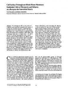
Fig. 1. Total 35S-glycosaminoglycan synthesis in individual intervertebral discs of young and mature mice ) mice by papain digestion. (a) CBA 35S-glycosaminoglycans were released from discs of 30-day-old (----) and 80-day-old ( mice; (b) ky/ky mice. Each point is the mean +S.E.M. for 7-11 animals. ISignificantly different to ky/ky, P 0.0 197; *significantly different to ky/ky, P 0.004. =
=
sarcoma proteoglycans on Sepharose CL2B columns eluted under dissociative or associative conditions (see Figure legends) (Venn & Mason, 1983).
RESULTS Synthesis of 35S-glycosaminoglycans in individual discs Individual discs from C3/4 in the cervical region to T4/5 in the thoracic region were excised from 30- and 80-day-old mice after a 3 h pulse with Na235SO4 and 24 h chase with unlabelled sulphate. Each disc was digested with papain and the 35S-glycosaminoglycans were determined quantitatively. The results for normal (CBA) and scoliotic (ky/ky) 30-day-old mice are shown in Figs. l(a) and l(b) (broken lines). Although the synthesis of 35S-glycosaminoglycan in any particular disc varies considerably between different animals and also between adjacent discs, the amount and pattern of synthesis of 35S-glycosaminoglycans in the 30-day-old normal and ky/ky mice are similar. This was confirmed by an unpaired t-test analysis of the data, which showed no significant differences in synthesis for 30-day-old normal, + /ky and ky/ky mice at any disc level. Below the cervico-thoracic junctional area the amount of 35S-glycosaminoglycan in individual discs increased progressively into the lumbar region (results not shown). This is probably related to the increasing size of the discs between the mid-thoracic and lower lumbar levels. Cervico-thoracic discs of 80-day-old CBA mice synthesized less 35S-glycosaminoglycan than did those of 30-day-old CBA mice (Fig. la). The difference is statistically significant at and below the C7/T1 level
(P 0.03-0.002, unpaired t test). The variation in 35S-glycosaminoglycan synthesis in any specific disc was generally less between different animals in the older mice =
than in younger mice. In contrast, 35S-proteoglycan synthesis in the cervicothoracic discs of 80-day-old ky/ky mice (Fig. lb, continuous line) was little or no different from that in 30-day-old ky/ky mice (Fig. lb, broken line). There was, however, a significant increase in synthesis of 35Sglycosaminoglycan in 80-day-old ky/ky mice compared with 80-day-old CBA mice in the T1/T2 disc (P = 0.0004) and C7/T1 disc (P = 0.02). These discs correspond to the region of the spine where disc degeneration is invariably detected histologically in older ky/ky mice.
Synthesis and metabolism of 30-day-old-kyphoscolioticmouse disc 35S-proteoglycans The metabolic half-life, solubility, hydrodynamic size, aggregation properties and glycosaminoglycan composition of 35S-proteoglycans synthesized by 30-day-oldkyphoscoliotic-mouse disc and costal cartilage were the same as those described previously for 30-day-old normal mice (Venn & Mason, 1983) (results not shown). Sequential extraction of 35S-proteoglycan from disc and costal cartilage of mice of different ages 35S-proteoglycans were extracted from discs and costal cartilage of 30-, 60-, 75- and 100-day-old mice after a 3 h pulse with Na235SO4 and 1 h chase with unlabelled Na2SO4. For 30-day-old mice the cervical, thoracic and lumbar discs were pooled separately. For older mice a separate pool was made of the discs in the area of 1986
Proteoglycan synthesis in kyphoscoliotic-mouse discs Associative Dissociative extract
extract
Residue
(a)
50-
13
25 it n 50 v
I
c
(b) 11
I
C.) m
25 50
0) 0. oL cn
-
iL (c)
X 50 0
ae 25 -
0 .
0
0
0.
4
I
2
50
co 25
i(d) 1
i
1~I
4 0
c0
477
Thus during aging the mouse disc synthesizes a larger proportion of proteoglycans that are less entrapped in the matrix. This contrasts with costal cartilage, where the proteoglycans become increasingly entrapped in the tissue during skeletal maturation. Changes in 35S-proteoglycan solubility were constant over 60, 75 and 100 days of age (60 and 75 days not shown), indicating that the changes in the tissue that they reflect are complete by the time of skeletal maturation. By 60 days and thereafter the total synthesis of 35S-proteoglycans by intervertebral discs (sum of every disc) in normal CBA mice was approx. 25% that of 30-day-old mice. Solubilities of ky/ky-mouse disc and cartilage proteoglycans (Fig. 2, 0 symbols) were similar to normal. Thus histological changes in the discs are not reflected by solubility changes and, therefore, organization of disc proteoglycans within the matrix. The amount of 35S-proteoglycan in ky/ky-mouse discs from every region was slightly higher than in CBA-mouse discs, but nevertheless declined with aging as described for normal mice. The amount of 35S-proteoglycan in the cervicothoracic pool of 60- and 100-day-old ky/ky mice was 2.2-2.5 times that in CBA mice of similar age, in agreement with the results in Fig. 1.
o
50 - (e)
t
25 -iF 01
30100
30100 Age (days)
30100
Fig. 2. Age-related changes in the solubility of newly synthesized mouse disc and cartilage 35Sproteoglycans The Figure shows the relative proportions of total 35S-proteoglycans extracted by 0.5 M-guanidinium chloride (associative extract), 4.0 M-guanidinium chloride (dissociative extract) and papain (residue). (a) Cervical discs; (b) cervico-thoracic discs; (c) thoracic discs; (d) lumbar discs;
(e) costal cartilage. 0, CBA mice; 0, ky/ky mice. Results are the means for three animals (six for 30-day-old CBA mice). Bars show the ranges of values.
degeneration, namely the last three cervical discs and the first thoracic disc. The remaining cervical, thoracic and lumbar discs were pooled separately. Similar proportions of35S-proteoglycans were extracted from the discs and costal cartilage of 30-day-old CBA mice (Fig. 2, * symbols) with about 22% solubilized by the associative solvent and 50% by the dissociative solvent. The costal-cartilage 35S-proteoglycans become much less soluble in 100-day-old mice, with only 50% of the total extractable. This is due to a decrease in the proportion of dissociatively extracted proteoglycan from 51% at 30 days to 28% at 100 days. The changes in solubility of intervertebral-disc proteoglycans with aging are different. In 100-day-old mice the proportion of the total disc proteoglycans extracted by associative solvent increases, and that extracted by dissociative solvent decreases. This change is particularly marked in the certical discs. The proportion of insoluble disc proteoglycans is about the same at all ages at each spinal level.
Vol. 234
Hydrodynamic size of 35S-proteoglycans extracted from cervical discs 35S-proteoglycans extracted from cervical (30-day-old) or cervico-thoracic-junction (60-100-day-old) intervertebral discs of ky/ky mice were chromatographed on Sepharose CL2B columns (Fig. 3). Those extracted from 30-day-olddiscsbyO.5 M-guanidiniumchloridechromatographed on associative columns as a broad peak with two partially resolved components (Ka, = 0.56 and 0.65; Fig. 3a) and are similar to those reported previously for normal mouse disc proteoglycans (Venn & Mason, 1983). Dissociative Sepharose CL2B chromatographs (results not shown) of these extracts were similar to the associative profiles. In 60- and 100-day-old discs this peak is narrower and more retarded on the column (Kay. = 0.8; Figs. 3d and 3g), showing that the proteoglycans are smaller and less heterogeneous than those from young mice. 35S-proteoglycans extracted sequentially from the discs of 30-day-old ky/ky mice with 4.0 M-guanidinium chloride chromatographed on dissociative Sepharose CL2B columns as a broad peak (Kav. = 0.36; Fig. 3b) and were of larger hydrodynamic size than those in the associative extract (Fig. 3a). Approx. 5000 of these proteoglycans were capable of forming aggregates when dialysed to associative conditions in the presence of carrier aAl proteoglycans and exogenous hyaluronate (Fig. 3c). There were no differences in hydrodynamic size or extent of aggregate formation by dissociatively extracted disc proteoglycans from either 60- or 100-day-old mice (Figs. 3e-3h). Disc proteoglycans from 30-100-day-old normal mice gave exactly the same Sepharose CL2B chromatographic profiles as those for ky/ky mice (results not shown). Therefore the hydrodynamic properties of newly synthesized disc proteoglycans from ky/ky mice up to 100 days old were no different from those of normal mice, even though the older kyphoscoliotic animals show marked
histological changes.
G. Venn and R. M. Mason
478 75
(a)
75 . (b)
(c) 53%
75
50 50-
25 25O
-d
(e)
A
c 50 U
2 25 I-
A
50 u
QooL
,
50 -
25
(f)
|
60%
rn
10 0
r
30 -(g)
30- L i.%
20~
20
-
lo0
10
-
I
t
0'
(i)
t 51%
40
0
120 Vo
40 1
vt
t
20-
120
0
401
vo vt Elution volume (ml)
0
1 20 vo
404 Vt
Fig. 3. Hydrodynamic size and aggregation properties of sequentially extracted 36S-proteoglycans from cervico-thoracic discs of ky/ky mice The Figure shows the Sepharose CL2B profiles of 35S-proteoglycans extracted from pooled cervical discs (a-c) or cervico-thoracic discs (d-i) of 30-day-old (a-c), 60-day-old (d-f) and 100-day-old (g-i) mice by 0.5 M-guanidinium chloride, pH 5.8 (a, d and g), and 4.0 M-guanidinium chloride, pH 5.8 (b, c, e, f, h and i). Samples were chromatographed alone (a, b, d, e, g and h) or after dialysis to associative conditions with exogenous hyaluronate (1O,ug) and aAl proteoglycan (1 mg) (c,f and i). Columns were eluted at 2.5 ml/h with 0.5 M-sodium acetate, pH 5.8 (a, c, d, f, g and i), or with 4.0 M-guanidinium chloride/0.05 M-sodium acetate, pH 5.8 (b, e and h). Percentages refer to the proportion of total radioactivity recovered from the column that was eluted in the void-volume fractions (V0).
DISCUSSION We have compared the proteoglycans synthesized by intervertebral discs of normal and kyphoscoliotic mice during the period of aging when development and progression of spinal curvature occurs in the latter. During aging the solubility of mouse disc proteoglycans increased and cartilage proteoglycan solubility decreased. Similar results were obtained for human discs (Adams & Muir, 1976) and rat, human and pig cartilage (Oohira & Nogami, 1980; Roughley & White, 1980; gimuinek & Muir, 1972). Overall synthesis ofmouse disc proteoglycans decreases with age and probably results in decreased amounts of proteoglycan in discs of older mice, as has been reported for dog and man (Ghosh et al., 1977; Beard & Stevens, 1980). Older discs contain proportionally more small, non-aggregated, 35S-proteoglycan, which could be extracted in associative conditions. In addition, the size of these proteoglycans decreased with aging, whether by synthesis of small molecules or by proteolytic cleavage of larger molecules is not known. Irrespective of their origin, they constitute a large proportion of the total proteoglycans in the cervico-thoracic region. The change in ratio of non-aggregated small proteoglycans to large
aggregatingmoleculesmaychangethemolecularorganization and mechanical properties of the discs in this region, making them more prone to degenerative changes if subject to abnormal forces. Intervertebral-disc proteoglycans in pig, dog and man decrease in size and ability to aggregate and increase their glucosamine/galactosamine ratio with aging (Stevens et al., 1979). It has been suggested that this is due to degradation of the chondroitin sulphate-rich region ofthe proteoglycan and loss of the hyaluronic acid-binding region, resulting in smaller keratan sulphate-rich proteoglycans (Stevens et al., 1979; McDevitt et al., 1981). However, other studies have shown that the size of the large aggregating proteoglycan of human and rabbit discs is constant with age (Pedrini-Mille et al., 1980, 1983; Lipson & Muir, 1981) even though there is an increase in the proportion of small non-aggregating proteoglycans in human discs with age (Adams & Muir, 1976). Mouse disc proteoglycans follow the same pattern of age-related change. The solubility, size and aggregation properties of the proteoglycans synthesized by discs in the cervico-thoracic area of kyphoscoliotic mice are the same as in normal mice. However, the amount of 35S-glycosaminoglycan 1986
Proteoglycan synthesis in kyphoscoliotic-mouse discs
synthesis is significantly higher in the C7/T1 and T1/T2 discs of 80-day-old ky/ky mice than in normal mice. These discs are in the region of the spine where histologically detectable disc degeneration always occurs, with accompanying loss in cellularity (Mason & Palfrey, 1984). It is likely, therefore, that proteoglycan synthesis is increased in the remaining cells, perhaps as a consequence of mechanical changes, which are known to affect chondrocytes (Fitton-Jackson, 1970). Similar increased synthesis has been seen in an experimental rabbit model (Lipson & Muir, 1981) and in human disc degeneration (Lyons et al., 1981). In the rabbit the proteoglycan size and aggregation properties were unchanged, as in our study, but in humans degenerate disc proteoglycans were larger and more aggregated than normal. It is noteworthy that elevated proteoglycan synthesis is one of the first biochemical changes detectable in experimental osteoarthrosis, another degenerative disease involving chondrocytes (Sandy et al., 1984; Oegema & Thompson, 1986). We cannot determine on the basis of the present results whether increased proteoglycan synthesis in the junctional discs is a primary or a secondary effect of the ky gene. Nevertheless the results suggest that changes occur in the level of transcription of proteoglycan genes in particular discs in mice with hereditary kyphoscoliosis. We thank Sarah Whitfield and Katharine Dyne for excellent technical assistance. The financial support of the Arthritis and Rheumatism Council of Great Britain is gratefully acknowledged.
REFERENCES Adams, P. & Muir, H. (1976) Ann. Rheum. Dis. 3, 289-296
Received 27 August 1985/13 November 1985; accepted 3 December 1985
Vol. 234
479 Beard, H. & Stevens, R. L. (1980) in Lumbar Spine and Back Pain (Jayson, M. I. V., ed.), pp. 407-436, Pitman Medical, Tunbridge Wells Dickinson, A. G. & Meikle, V. M. H. (1973) Lancet i, 1186 Faltz, L. L., Reddi, A. H., Hascall, G. K., Martin, D., Pita, J. C. & Hascall, V. C. (1979) J. Biol. Chem. 254, 1375-1380 Fitton-Jackson, J. S. (1970) Proc. R. Soc. London Ser. B 175, 405-455 Ghosh, P., Taylor, T. F. K. & Braund, K. G. (1977) Gerontology 23, 87-98 Lipson, S. J. & Muir, H. (1981) Spine 6, 194-210 Lyons, G., Eisenstein, S. M. & Sweet, M. B. E. (1981) Biochim. Biophys. Acta 673, 443-453 Mason, R. M. & Palfrey, A. J. (1984) J. Orthop. Res. 2, 333335 McDevitt, C. A., Billingham, M. E. J. & Muir, H. (1981) Semin. Arthritis Rheum. 11, Suppl. 1, 17-18 Oegema, T. R. & Thompson, R. C. (1986) in Articular Cartilage Biochemistry (Kuettner, K. E., Schleyerbach, R. & Hascall, V. C., eds.), Raven Press, New York, in the press Oegema, T. R., Hascall, V. C. & Dziewiatkowski, D. D. (1975) J. Biol. Chem. 250, 6151-6159 Oohira, A. & Nogami, H. (1980) J. Biol. Chem. 255, 13461350 Pedrini-Mille, A., Pedrini, U. A. & O'Connor, R. (1980) Trans. Orthop. Soc. 5, 104 Pedrini-Mille, A., Pedrini, V. A., Thelisco, C., Ponseti, I. V., Weinstein, S. L. & Maynard, J. A. (1983) J. Bone Jt. Surg. Am. Vol. 65, 815-823 Roughley, P. J. & White, R. J. (1980) J. Biol. Chem. 255, 217-224 Sandy, J. D., Adams, M. E., Billingham, M. E., Plaas, A. & Muir, H. (1984) Arthritis Rheum. 27, 388-397 gimCinek, Z. & Muir, H. (1972) Biochem. J. 126, 515-523 Stevens, R. L., Ewins, R. J. F., Revell, P. A. & Muir, H. (1979) Biochem. J. 179, 561-572 Venn, G. & Mason, R. M. (1983) Biochem. J. 215, 217-225 Venn, G. & Mason, R. M. (1985) Biochem. J. 228, 443-450