May 11, 2001 - chlamydial protein with properties of integration host factor (IHF) bound to the same site as the protein in the extracts. IHF-binding activity was ...
Molecular Microbiology (2001) 41(2), 451–462
Characterization of integration host factor (IHF) binding upstream of the cysteine-rich protein operon (omcAB ) promoter of Chlamydia trachomatis LGV serovar L2 Jianmin Zhong, Annemarie L. Douglas and Thomas P. Hatch* Department of Molecular Sciences, University of Tennessee, Memphis, TN 38163, USA. Summary Chlamydiae are bacterial parasites that carry out a distinct developmental cycle within host cells; however, the mechanisms by which these organisms regulate stage-specific gene expression are not known. We identified a DNA element located between nucleotide (nt) 2135 and 290 upstream from the transcription start point of the late stage-specific CRP operon (omcAB ) of Chlamydia trachomatis, to which a protein in extracts of chlamydiae harvested at 23 h after infection binds. A recombinant protein of C. trachomatis open reading frame (ORF) CT267, which is homologous to bacterial integration host factor (IHF) and the heat-unstable nucleoid protein (HU), bound to the same element and produced the same DNase I footprint as the protein in chlamydial extracts. Recombinant ORF CT267 protein bound with high affinity to the DNA element and induced a sharp bend in a DNA fragment containing the binding site, suggesting that ORF CT267 encodes a protein with IHF-like activity, and recombinant protein had a positive effect on in vitro transcription of the CRP operon. IHF-binding activity and IHF protein were detected in extracts of C. trachomatis during the early to intermediate phases of the late stage of the developmental cycle (between 17 and 30 h after infection), but were absent in the extreme late phase of the cycle and in the infectious form of chlamydiae. The presence of an IHF binding site upstream of the CRP operon and the presence of chlamydial IHF-like protein when late stage genes are transcribed suggests that the chlamydial IHF may play a role in stage-specific gene expression.
Accepted 11 May, 2001. *For correspondence. E-mail thatch@ utmem.edu; Tel. (11) 901 448 4664; Fax (11) 901 448 8462.
Q 2001 Blackwell Science Ltd
Introduction Members of the genus Chlamydia are important agents of disease in humans and animals. They are obligate intracellular bacteria that progress through a distinct developmental cycle that begins with the entry of an infectious but metabolically inactive elementary body (EB) into a host cell and is followed by the conversion of the EB to a metabolically active reticulate body (RB) by 4–6 h postinfection (pi), multiplication of RBs during the middle phase of the cycle and reorganization of RBs to EBs late in the cycle, starting at 18 –24 h pi. For most strains of chlamydiae, the cycle is completed between 30 and 48 h pi. Very little is known about how gene expression is controlled in chlamydiae, and essentially nothing is known about the mechanisms by which stage-specific genes are regulated. Chlamydial genomes encode three sigma factors: a major sigma factor s66 (rpoD ) and two alternative sigma factors, s54 (rpoN ) and s28 (rpsD ) (Stephens et al., 1998; Kalman et al., 1999; Read et al., 2000). The expression of several s66-dependent genes has been investigated by in vitro transcription assay systems (Mathews and Sriprakash, 1994; Douglas and Hatch, 1995; 1996; Fahr et al., 1995; Tan and Engel, 1996; Tan et al., 1998; Shaumburg and Tan, 2000; Shen et al., 2000). Interestingly, all known late stage-specific genes that have been investigated in vitro, the cysteine-rich protein (CRP) operon (omcAB ), the histone-like protein genes (hctA and hctB ) and two small late transcription unit genes (ltuA and ltuB ), appear to have major sigma factorlike promoters and are dependent on s66 (Fahr et al., 1995). No chlamydial genes have been demonstrated experimentally to be transcribed by alternative sigma factors. However, two Chlamydia trachomatis genes that are transcribed late in the cycle, open reading frame (ORF) CT652.1 and CT683, have promoters that resemble the consensus (TGGCAC-N5-TTGC) that is recognized by s54 of other bacteria (Mathews and Timms, 2000). Transcript levels of C. trachomatis s54 have been investigated by reverse transcription –polymerase chain reaction (RT– PCR). Shaw et al. (2000) detected rpoN transcripts early in the infection cycle, whereas Douglas and Hatch (2000) first detected transcripts in the middle stage of the cycle. In contrast, Mathews et al. (1999) detected transcripts only
452 J. Zhong, A. L. Douglas and T. P. Hatch late in the cycle, and Mathews and Timms (2000) proposed that s54 may initiate transcription of a subset of late genes, including ORFs CT652.1 and CT683. Three chlamydial proteins have been shown to bind to DNA experimentally. Two DNA-binding histone-like proteins, which are unique to chlamydiae, are major structural proteins that are made late in the developmental cycle and may be responsible for the condensation of DNA into the nucleoid seen in EBs and the general silencing of gene expression late in the developmental cycle (Wagar and Stephens, 1988; Barry et al., 1992; 1993; Pedersen et al., 1994; Remacha et al., 1996). Their binding specificity is not known, but it is likely to be broad to account for the large number of molecules bound per chromosome (Hackstadt et al., 1993). A third chlamydial DNA-binding protein, EUO, binds preferentially to AT-rich sequences (Zhang et al., 1998; 2000). EUO transcripts can be detected within minutes after infection, but EUO protein can be detected throughout the growth phase of the cycle (Zhang et al., 1998). EUO is a quantitatively minor protein of unknown function; it has been speculated to be either a histone-specific protease (Kaul et al., 1997) or a general activator of transcription (Zhang et al., 2000). The purpose of our study was to identify transcription factors that play a role in the expression of the late stagespecific CRP operon (reviewed by Hatch, 1996). We identified a DNA binding site for a protein in extracts of RBs of C. trachomatis between the CRP operon and a divergent late stage gene (ORF CT444.1) encoding a putative lipoprotein. We found that a recombinant chlamydial protein with properties of integration host factor (IHF) bound to the same site as the protein in the extracts. IHF-binding activity was present in chlamydial extracts up to 30 h pi, and recombinant IHF stimulated in vitro transcription of the CRP operon, suggesting that chlamydial IHF may play a role in late developmental stage gene expression.
C. trachomatis L2 CRP operon transcription is dependent on the chlamydial major sigma factor s66. A small ORF (CT444.1) of 74 codons is located upstream of [predicted translation initiation codon at nucleotide (nt) 2246, relative to the CRP operon transcription start point] and in the opposite orientation to the CRP operon. We used an RNase protection assay to demonstrate that ORF 444.1 RNA is expressed in C. trachomatis L2 late in the developmental cycle (Fig. 2). ORF 444.1 appears to be poorly expressed: we had difficulty detecting two small transcripts of , 500 bases by Northern blot and were unable to detect a clear transcription start point by primer extension analysis (data not shown). Several potential bacterial major sigma factor recognition 210 hexanucleotides, including one that matches the Escherichia coli consensus and is separated by 16 bp from a 235 hexanucleotide with only two mismatches to the consensus (Fig. 1), are present upstream of the translation start site. The predicted peptide of ORF CT444.1 lacks strong homology to known proteins, and its function remains a mystery. SIGNALP analysis (http://www.cbs.dtu.dk/services/SignalP/) predicted a signal sequence with cleavage between amino acids 12 and 13, and PSORT analysis (http://psort.nibb.ac. jp) predicted cleavage between amino acids 18 and 19. However, close examination of the predicted N-terminus of ORF CT444.1 reveals characteristics similar to the small CRP (OmcA), including two charged amino acids after the initiating methionine, a continuous stretch of hydrophobic amino acids and a cysteine residue at position 19, which is believed to be lipid modified in the small CRP of C. psittaci 6BC (Everett et al., 1994). It is possible, therefore, that ORF 444.1 encodes a lipoprotein, the mature form of which would have a peptide molecular weight of 6371 and a pI of 5.65.
Identification of a DNA binding site upstream of the CRP operon Results Analysis of DNA sequence upstream of the CRP operon The C. trachomatis L2 DNA sequence upstream of the CRP operon, which is identical to the corresponding sequence in C. trachomatis serovar D (Stephens et al., 1998), is shown in Fig. 1. The transcription start point (11) of the operon was determined by primer extension analysis by Fahr et al. (1995) and is preceded by a putative major sigma factor promoter sequence that is identical to that of C. trachomatis L1 (Lambden et al., 1990) and similar to those in Chlamydia psittaci and Chlamydia pneumoniae (Watson et al., 1995). Fahr et al. (1995) confirmed by in vitro transcription analysis that
We examined the upstream region of the CRP operon for potential regulatory elements by gel mobility shift assay with extracts prepared from RBs at 23 h pi and a DNA probe extending from nucleotide 2285 to 115 and found activity in RB extracts that was not present in uninfected cells (Fig. 3). Zhang et al. (1998) demonstrated previously that EUO protein is present in early mid-cycle RBs of C. psittaci 6BC (16 h pi) and binds to the CRP operon promoter region. However, recombinant EUO (rEUO) produced a different shift pattern (Fig. 3), and the addition of anti-EUO failed to alter the migration rate of the DNA– protein probe, although it did supershift the rEUO – DNA complex (data not shown). Treatment of the RB extract with proteinase K eliminated gel shift activity, indicating Q 2001 Blackwell Science Ltd, Molecular Microbiology, 41, 451–462
IHF binding in Chlamydia
453
Fig. 1. C. trachomatis serovar L2 DNA sequence upstream of the CRP operon. Sequence from nt 2320 to 1109, relative to the CRP operon transcription start point (11), is shown. The first three amino acids of the small CRP (OmcA) and the first 25 amino acids of ORF CT444.1 are indicated above and below the DNA sequence respectively. Putative 210 and 235 s66 recognition sites and the region protected from DNase I digestion by RB extracts (see Fig. 5) are underlined. Sequences similar to the IHF 30 binding site consensus (WATCAANNNNTTR; where W ¼ A or T; N ¼ any nucleotide; R ¼ A or G) are indicated below the bottom strand.
that one or more proteins were responsible for the DNA binding (data not shown). Gel shift competition assays were used to approximate the location of the DNA binding site in the intergenic region between the CRP operon and ORF 444.1 to between nt 2154 and 2114 (data not shown). The ability of RB extracts to shift probes of < 40–50 bp within the 2210 to 255 range was generated to confirm the competition assays. Probes from 2210 to 2155, 2154 to 2115 and 2114 to 255 were not shifted, whereas the 2139 to 279 probe was retarded, suggesting that the only site bound by proteins in RB extracts from 23 h pi RBs is located within nt 2139 and 279 and that binding probably requires sequence on both sides of the 2114/2115 junction (Fig. 4). A DNase I protection (footprint) assay was used to locate more precisely the site bound by protein in 23 h RB extracts. The protected region on the bottom strand of the 2285 to 115 probe extended from 294 to 2135, and the footprint on the upper strand extended from 290 to 2135 (Fig. 5). Weakly hypersensitive sites were noted above Q 2001 Blackwell Science Ltd, Molecular Microbiology, 41, 451–462
and below the protected region on the bottom strand. Careful inspection of the assay gels failed to detect other protected regions, suggesting that binding of only one protein in 23 h pi extracts can be detected by footprint assay.
Identification of a protein that binds DNA sequence upstream of the CRP operon Extracts from RBs harvested and purified at 23 h pi were fractionated by SDS–PAGE, and proteins were eluted from gel slices and tested for their ability to bind to upstream CRP operon DNA probes. In a preliminary experiment in which slices below the 103 kDa protein standard were examined, eluate from the 7.4 –21.9 kDa slice bound to upstream CRP operon DNA. Extracts were then fractionated on a second gel, and 3 mm slices between 21.9 and 7.4 kDa were examined for binding activity. Nearly all the binding activity to both the long 2285 to 115 probe (covering the entire ORF CT444.1– CRP operon intergenic region) and the short 2136 to 290
454 J. Zhong, A. L. Douglas and T. P. Hatch
Fig. 2. Expression of ORF CT444.1 during the developmental cycle. RNA was extracted from uninfected host cells (L cell, lane 1), infected cells at the times indicated after infection (lanes 2 – 9) and EBs isolated at 48 h pi (EB), and ORF CT444.1 transcripts and chlamydial 23S rRNA were detected by RNase protection assay. The amount of chlamydial RNA analysed in each lane was roughly balanced on the basis of the strength of the protection signal to the 23S rRNA probe. Lanes 11 and 12 show the protection by 30 h pi RNA of the ORF CT444.1 and rRNA probes separately. Lanes 13 (yeast 1 RNase) and 14 (yeast – RNase) are controls in which irrelevant yeast RNA was substituted for RNA extracted from chlamydiae. The ORF probe and the rRNA probe (see single small spot), incubated in the absence of RNase, are shown in lanes 15 and 16 respectively. A reference DNA sequencing ladder is shown in the far left four lanes. The weak signals to ORF 444.1 that were noted at 1 and 4 h pi may reflect residual transcripts present in infecting EBs. Strong signals to ORF 444.1 were first detected at 25 h pi and were present in EBs.
probe (covering the footprint sequence) was in the 13 – 16 kDa gel slice (Fig. 6). Matrix-assisted laser desorption/ionization time-of-flight (MALDI-TOF) mass spectrometric analysis was used to detect proteins within the 13 –16 kDa gel slice. Whereas many chlamydial proteins were detected in the slice, only one was likely to bind DNA on the basis of homology to other bacterial proteins. This protein is encoded by ORF CT267 (predicted molecular weight 11 451), which bears similarity to the a- (IhfA) and b (IhfB)-subunits of integration host factor (IHF) and the heat-unstable nucleoid protein (HU). IHF has many functions in bacteria and bacteriophages, including DNA replication and recombination and regulation of transcription. The primary function of IHF appears to be architectural, introducing a sharp bend in DNA that facilitates the interaction of other nucleoproteins (reviewed by Goosen and van de Putte, 1995). Alpha and beta dimers of IHF bind to DNA with high affinity to specific sites of about 30–35 bp consisting of a conserved 3 0 element of WATCAANNNNTTR (where W ¼ A or T; N ¼ any nucleotide; and R ¼ G or A) and a degenerate, AT-rich 50 domain (Craig and Nash, 1984). HU, a homodimer in most bacteria but a heterodimer in E. coli, plays a role in chromosome organization (reviewed by Schmid, 1990). HU appears to
bind to intrinsically bent AT regions in DNA, rather than to specific sequences. His-tagged recombinant C. trachomatis L2 ORF CT267, which we will refer to as rIHF, was prepared in soluble form and tested for its ability to bind to upstream CRP operon DNA by gel mobility shift assay (Fig. 7). rIHF retarded the mobility of the long 2285 to 115 probe to the same extent as C. trachomatis L2 RB extract, whereas rIHF retarded mobility of the short probe to a greater extent. When large amounts of rIHF were tested, both probes remained largely in the loading well, suggesting that the recombinant protein in excess forms aggregates with DNA. A DNase I protection assay was carried out to determine the binding site of rIHF on the top strand of the CRP operon promoter region, from nt 2285 to 115. The footprint produced by rIHF was identical to the footprint produced by RB extracts, strongly supporting the conclusion that the protein in extracts that are responsible for binding to upstream CRP operon DNA is the chlamydial IHF (Fig. 8). Two sequences in the bottom strand of the footprint site match the IHF consensus with two mismatches, attcaaa gagata and aataaaataataa, and the 50 half of the bottom strand is AT rich (Fig. 1), which may serve as an UP element (reviewed by Gourse et al., 2000). One of the mechanisms by which IHF can regulate transcription is to induce DNA looping at its binding site to facilitate contact between an upstream, DNA-bound activation protein and downstream promoter-bound s54 (RpoN) (Hoover et al., 1990). We searched contig 6.1 in the C. trachomatis D
Fig. 3. Detection of DNA-binding activity to CRP operon upstream DNA (2285 to 115) in extracts of RBs by gel mobility shift assay. Assays were carried out with extracts prepared from uninfected host cells (L cell) and RBs (L2 RB), harvested and purified at 23 h pi, and with rEUO. Lane 1 shows the migration of free probe.
Q 2001 Blackwell Science Ltd, Molecular Microbiology, 41, 451–462
IHF binding in Chlamydia
Fig. 4. Identification of the upstream CRP binding site by gel shift assays with short double-stranded oligonucleotide probes. Extracts were prepared from 23 h pi RBs, and the probes that were tested spanned the nucleotides indicated.
genome database (Stephens et al., 1998), which includes several thousand basepairs upstream and downstream of the IHF binding site, for an RpoN consensus sequence (Taylor et al., 1996) and found no matches, allowing 0 or one mismatches, and three matches, allowing two mismatches. None of the two mismatch finds was located in a position likely to be a promoter. Characterization of binding of rIHF to CRP operon upstream DNA sequence
455
bind to the 2136 to 290 probe between 17 and 48 h pi (Fig. 10); we were unable to obtain sufficient material from RBs harvested at 14 h pi to make a valid comparison with the extracts prepared at later times (data not shown). We observed significant binding activity at 17 –30 h pi, with peak activity at 23.5 h pi. We detected almost no activity in extreme late stage RBs (48 h pi) and no binding activity in EBs, which were treated with non-ionic detergent to eliminate osmotically fragile RB forms (Fig. 10). IHFbinding activity in extracts was therefore present at the time of synthesis of the CRP transcripts and CRP protein in chlamydiae (Hatch et al., 1986; Newhall, 1987; Fahr et al., 1995), but not at extremely late times in the developmental cycle. Immunoblot analysis with antibody raised against chlamydial rIHF confirmed that the level of IHF protein in C. trachomatis peaks between 20 and 24 h pi and that IHF is not present in EBs (data not shown). Ideally, the effect of IHF on CRP operon transcription would be tested by deletion of the IHF gene or the IHF binding site from the chlamydial genome. However, in the absence of genetic tools to manipulate chlamydiae, we examined the ability of rIHF to affect CRP operon transcription in vitro, using the G-less cassette vector developed by Tan and Engel (1996) and the CRP promoter region from 2455 to 12 (Fig. 11). We detected a reproducible 1.5- to twofold enhancement of transcription by rIHF of the CRP operon, relative to the control rRNA gene, which is in the opposite orientation to the CRP operon on the vector. Although this stimulatory effect of IHF is small, it is comparable with that noted in some other
We used gel mobility shift assays to calculate a dissociation constant of about 6.25 nM for rIHF binding to a 2285 to 115 probe (data not shown). The calculation was based on the assumption that the recombinant protein was 80% pure and 100% active and bound as a monomer. The dissociation constant would be < 3 nM if the chlamydial IHF binds as a dimer, as is the case in other bacteria, and even lower if our rIHF preparation contained inactive protein. We used the circular permutation assay of Wu and Crothers (1984) to determine the ability of rIHF to bend 171 -bp DNA fragments containing the 46 bp IHF DNase I protection site at different positions relative to the 50 end of the fragment (Fig. 9). All fragments migrated at the same rate in the absence of rIHF, indicating that the fragments did not contain intrinsically bent DNA. In the presence of rIHF, we noted a roughly symmetrical pattern of retardation, indicative of bending near the centre of the 46 bp footprint site, with a minimum bend angle of 1408, as calculated by the method of Thompson and Landy (1988). Role of IHF in regulation of the CRP operon We examined the ability of IHF in chlamydial extracts to Q 2001 Blackwell Science Ltd, Molecular Microbiology, 41, 451–462
Fig. 5. Identification of the DNA site bound by protein in extracts of 23 h pi RBs by DNase protection assay. The protected regions of the 2285 to 115 top and bottom strand probes are indicated by horizontal lines. Possible hypersensitive sites on the bottom strand are indicated by dots. The protected sequences are shown in Fig. 1.
456 J. Zhong, A. L. Douglas and T. P. Hatch Fig. 6. Gel shift analysis of proteins fractionated by SDS – PAGE. Extracts of 23 h pi RBs were fractionated on a 10 – 20% SDS – polyacrylamide gel, gel slices of 3 mm were excised, and proteins in the slices were eluted and examined for binding to 2285 to 115 and 2136 to 290 probes by gel mobility shift analysis.
systems (Parekh and Hatfield, 1996; van Ulsen et al., 1997; Giladi et al., 1998). We were unable to determine the transcription start point of the ORF CT444.1 gene, and the gene appears to be poorly expressed; consequently, we did not investigate the effect of rIHF on the in vitro transcription of ORF CT444.1.
Discussion We have demonstrated that the protein encoded by ORF CT267 binds to a DNA site between the promoter regions of the CRP operon and a divergently transcribed gene encoded by ORF CT444.1. The protein is homologous to the a- and b-subunits of IHF and HU proteins found in other bacteria. We have designated the protein chlamydial IHF because it is capable of inducing a sharp bend of 1408 or more, and it has a dissociation constant within the nanomolar range. Furthermore, there are two IHF consensus binding sites, allowing for two mismatches, in the sequence protected from DNase I (Fig. 1). We believe that attcaaagagata is the most likely of the two sequences to be recognized by IHF, because it contains the two bases (CA) that are present in every known IHF binding site (Goodrich et al., 1990) and a 50 AT-rich region that is found in the DNase I footprint of many IHF binding sites (Goodrich et al., 1990; van Rijn et al., 1991; Hales et al., 1994; Goodman et al., 1999). Furthermore, the symmetry of the circular permutation assay (Fig. 9) indicates that bending occurs near the centre of the 46 bp footprint, the location of this consensus-like sequence. The 235 and 210 hexamers of the CRP operons of C. trachomatis and C. pneumoniae are identical, and a potential IHF binding site (with three mismatches to the E. coli consensus) is located the same number of nucleotides upstream from the promoter in C. pneumoniae as the binding site identified in our study. In addition, the 441 bp intergenic region upstream from the CRP operon in C. pneumoniae contains two perfect E. coli IHF-binding consensus sequences that are not in C. trachomatis. Perfect matches
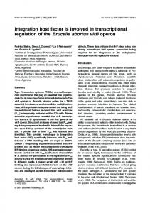
to the consensus occur approximately once in every 13 000–14 000 bp in chlamydiae. The C. trachomatis genome encodes only one IHF-like protein (Stephens et al., 1998). In most bacteria, IHF binds to an asymmetric DNA site as a heterodimer, with the aand b-subunits sharing only about 25–30% identity. Homodimers of E. coli IHF are capable of binding to an IHF site, but with considerably lower affinity than heterodimers (Zulianello et al., 1994). The crystal structure of an E. coli IHF –DNA complex has been reported (Rice et al., 1996). The b-sheet arm of the a-subunit is responsible for sitespecific recognition of DNA, with key roles played by P65a, which forms a kink in the DNA, and R60a and R63a, which interact with the most highly conserved bases in the 50 part of the consensus (WATCAA). P66, K61 and R64 in the chlamydial IHF probably serve a similar function (Fig. 12). The second portion of the consensus site (TTR) is recognized by the body of the b-subunit. R46b extends into the minor groove and makes direct or water-mediated hydrogen bonds to all three conserved bases. Positioning is thought to be enforced by a chain of salt bridges
Fig. 7. Binding of 23 h pi RB extract and rIHF to long (2285 to 115) and short (2136 to 290) upstream CRP operon probes, as determined by gel mobility shift assay.
Q 2001 Blackwell Science Ltd, Molecular Microbiology, 41, 451–462
IHF binding in Chlamydia
Fig. 8. Identification of CRP operon upstream DNA sequence bound by RB extract and rIHF determined by DNase I protection assay. The amount of RB extract (prepared at 23 h pi) and rIHF protein is indicated at the top of the lanes. The primer used to generate the probe (top strand, Fig. 1) was also used for the DNA sequence determination shown in the far left lanes. The protected region (2135 to 290) was identical for RB extract and rIHF.
between E44b and R42b and R46b (Rice et al., 1996). These equivalent functions may be provided by residues E46, R44 and R48 in chlamydial IHF (Fig. 12). We detected only a slight stimulation of in vitro transcription of the CRP operon (Fig. 11). The lack of a large effect of rIHF on CRP operon transcription may have resulted from the presence of native IHF or the absence of an IHFdependent positive transcription factor in the crude RNA polymerase preparation we obtained at 23 h pi. However, the large differences in CRP operon expression between mid-cycle (before 16–18 h pi) and later times (Hatch et al., 1986; Newhall, 1987; Fahr et al., 1995) is difficult to explain simply on the basis of enhancement by IHF on transcription from what appears to be a well-recognized major sigma factor promoter. It is more likely that the operon is negatively controlled by a repressor during the growth phases of the cycle. When we examined RB extracts for a protein binding to the 2285 to 115 probe at 17 h pi, we noted only a single retarded protein –DNA, which could be accounted for by the binding of IHF to the probe (data not shown). When we used a shorter probe (274 to 1121) that did not include the IHF binding site, we failed to detect a retarded band (data not shown). It is possible that a repressor is present in extracts at early times after infection but in amounts that cannot be detected by gel mobility shift assay. IHF has been shown to affect transcription by several mechanisms, the best understood of which is its role in bringing an upstream DNA-bound activator protein into the proximity of the s54 holoenzyme of RNA polymerase by DNA looping (Hoover et al., 1990). Although Mathews and Q 2001 Blackwell Science Ltd, Molecular Microbiology, 41, 451–462
457
Timms (2000) have identified two E. coli consensus s54 promoters in C. trachomatis, a s54 consensus recognition sequence, even allowing one mismatch, is not present within several thousand basepairs of the IHF binding site. Also, we found no evidence by gel shift or DNase I protection assays for a DNA binding site other than the IHF site within the CRP operon–ORF 444.1 intergenic region. It is possible that the chlamydial s54 is promiscuous in its sequence recognition, as has been shown for the major chlamydial sigma factor s66 (Mathews and Sriprakash, 1994; Douglas and Hatch, 1996; Tan and Engel, 1996; Tan et al., 1998; Shaumburg and Tan, 2000), and that activator binding was below the sensitivity of our assays. However, the demonstration that in vitro transcription of the CRP operon is dependent on s66 (Fahr et al., 1995) and the presence of a s66 promoter immediately upstream of the CRP operon transcription start site (Lambden et al., 1990; Fahr et al., 1995; Watson et al., 1995) suggests that s54 plays no direct role in the expression of the CRP operon. There are several ways by which IHF can alter gene expression from major sigma factor promoters (reviewed by Goosen and van de Putte, 1995). For example, in E. coli, expression of the nir operon is co-dependent on the binding upstream of FNR, the anaerobic transcription
Fig. 9. Bending of CRP operon DNA by rIHF, as determined by circular permutation assay. A. pBend-ihf was digested with eight restriction enzymes to create linear probes of about 171 bp, with the relative location of the 46 bp IHF binding site (2135 to 290) indicated by the filled boxes. B. pBend assay. The left eight lanes show the migration of unbound probes, and the next eight lanes show the retardation of probes in the presence of rIHF. The two far right lanes show migration of the multiple cloning site (MCS) of the pBend2 vector, produced by digesting the empty vector with Eco RI and HindIII in the presence (1) and absence (– ) of rIHF.
458 J. Zhong, A. L. Douglas and T. P. Hatch
Fig. 10. Binding activity to the 2136 to 290 probe in extracts prepared at different times during the developmental cycle. A. Immunoblot of the extracts reacted with anti-major outer membrane protein (MOMP). The amount of extract analysed in each lane was adjusted to provide a constant signal to the MOMP, which is present in RBs at all times after infection and in EBs. B. Gel mobility shift assay. The amount of extract tested was proportional to the amount examined in the immunoblot for all times after infection.
factor, and NarL or NarP, members of the nitrogen response regulator family. The effects of these factors on transcription initiation is modulated by a nucleoprotein complex, with IHF playing the central role (Browning et al., 2000). Browning et al. (2000) proposed a model in which binding of IHF near position 299 diverts upstream sequence away from RNA polymerase, suppressing the positive effect of FNR, which binds near position 241. In the presence of nitrite or nitrate ions in the medium, NarL and NarP are phosphorylated and bind near position 269, counteracting the effect of IHF, possibly by displacing IHF from its nearby, overlapping binding site. Our failure to detect binding of transcription factors other than IHF does not support a similar model for IHF regulation of ORF CT444.1/CRP operon expression. IHF also stimulates transcription from at least three s70 promoters in the absence of other activator proteins: bacteriophage Mu Pe (Krause and Higgins, 1986), E. coli ilvPG 2 (Parekh and Hatfield, 1996) and phage l PL1 (Giladi et al., 1998). The binding of IHF upstream of the PL promoter of phage l activates transcription either by looping an UP element to the C-terminal domain of the asubunit (aCTD) of RNA polymerase or by looping DNAbound IHF so that it makes a stimulatory contact with the aCTD (Giladi et al., 1998). Tan et al. (1998) demonstrated that the AT-rich sequence upstream of the 235 hexanucleotide of the chlamydial P1 promoter of the rRNA operon is recognized as an UP element by the aCTD of E. coli RNA polymerase, and the seven amino acid residues in the aCTD of E. coli polymerase that are most critical for DNA interaction and UP element function are conserved in the aCTD of chlamydial RNA polymerase. Thus, the role of IHF may be to loop AT-rich sequence
in the intergenic region to allow contact with RNA polymerase bound to the ORF CT444.1 or CRP promoters. Parekh and Hatfield (1996) proposed that IHF activates transcription of the ilvGMEDA operon by promoting open complex formation by a mechanism that does not require DNA looping and contact of IHF or upstream sequence with RNA polymerase. A supercoilingdependent, DNA structural transmission mechanism, in which the formation of a supercoiling-induced DNA duplex destabilized (SIDD) structure in an AT-rich region just upstream of the IHF binding site is inhibited by the binding of IHF and is transferred downstream to the promoter region, was subsequently proposed to explain the activation (Sheridan et al., 1998). The presence of ATrich sequence within the ORF CT444.1 and CRP intergenic regions is consistent with IHF enhancement of transcription by a DNA structural transmission mechanism. The orientation of the IHF binding site in all three operons described above is the same, with the consensus WATCAANNNNTTR located 50 of the affected promoter. The orientation of the proposed chlamydial IHF site is towards the ORF CT444.1 gene, suggesting that IHF may affect transcription of the putative small lipoproteinencoding gene rather than the CRP operon. However, the mechanism by which IHF stimulates transcription from ilvPG2 is independent of the orientation of the IHF binding site (Parekh and Hatfield, 1996), and Engelhorn and Geiselmann (1998) found that the substitution of an IHF site for the catabolite repressor protein binding site upstream of the malT promoter stimulated transcription in either orientation. Thus, it is not possible to predict whether ORF CT444.1 or CRP operon transcription is affected by IHF binding, simply on the basis of the orientation of the IHF binding site. Assigning a physiological role in transcription for IHF in bacteria is difficult because the number of IHF molecules
Fig. 11. Effect of rIHF on in vitro transcription of the CRP operon. rIHF was added to the reaction mix at the final concentrations indicated. The transcripts were visualized and quantified by a computer-assisted phosphorimaging system.
Q 2001 Blackwell Science Ltd, Molecular Microbiology, 41, 451–462
IHF binding in Chlamydia
Fig. 12. Comparison of the DNA contact regions of a- and b-subunits of E. coli IHF with the homologous region of C. trachomatis IHF. Amino acid residues referred to in the text are underlined.
per cell is large, ranging in E. coli from about 12 000 in exponential growth phase cells to about 55 000 in early stationary phase cells (Talukder et al., 1999), and probably exceeds the number of binding sites at all times during the cell cycle. We have not determined the number of molecules of IHF in chlamydiae; however, in contrast to E. coli, the relative amount of IHF varies dramatically during the cycle, peaking early in the late phase and dropping to undetectable levels in EBs. The binding of IHF to a site between two late stage genes suggests that it may play a role in late gene expression. An intriguing possibility is that IHF somehow overcomes the general silencing effect of histones on gene transcription, allowing the transcription of the CRP operon and/or ORF 444.1 during the early and middle phases of the late stage of the developmental cycle. Chlamydial histone binding affects the degree of negative superhelicity of DNA (Barry et al., 1993; Pedersen et al., 1994), and IHF may counter the effects of histones by a DNA structural transmission
mechanism similar to that proposed for the enhancement of transcription from ilvPG 2 by IHF in E. coli (Sheridan et al., 1998). In support of IHF playing a role in the expression of at least a subset of late genes, a conserved, perfect match E. coli IHF consensus sequence is located upstream from the hctA gene promoter in C. trachomatis serovar D and C. pneumoniae. In addition, several consensus sequences with one or two mismatches are located upstream from ltuB in both species. However, sequences with two or fewer mismatches to the consensus are not found upstream from ltuA in either C. trachomatis D or C. pneumoniae, and consensus-like sequences are found upstream of hctB only in C. trachomatis D.
Experimental procedures Strains and plasmids Chlamydia trachomatis L2 was grown in suspension cultures of L929 cells. RBs and EBs were harvested and purified on Hypaque gradients (Sanofi Winthrop Pharmaceuticals) as described previously (Hatch et al., 1986). E. coli JM107 (Stratagene) was used for the maintenance and transformation of plasmids. The E. coli host lysogen BL21(DE3)/pLysE, with a prophage carrying the T7 RNA polymerase gene, was used for IHF expression with the expression vector pET28A(1) (Novagen). pBlueScript KS1 (Stratagene) was used to generate ORF CT444.1 RNA and rRNA.
Table 1. Oligonucleotides used in the study. Namea
Sequence
934 (274) 971 (255) 972 (2266) 973 (115) 974 (2155) 975 (2115) 976 (279) Bend-1 Bend-2 Bend-seq2 IV-1 IV-2 L12K1 (2405) L12K2 (2285) L12K4 (2248) L12K5 (2210) L12K6 (2154) L12K7 (2114) L12K8 (2139) Mike G (1121) rRNA-1 rRNA-2 Su-u42 Su-t30 BS-1 (2136 to 290) BS-2 (2136 to 290)
TAATTACCTGTTATTAGAC CGTCTAATAACAGGTAATTAGC TATTGTAGCTACTGCTGCCC GAAACTAGCCCTCATAGAC GATAGAATAAATAAACTAAAATC ATTCAAAGAGATAAGTTTGC AGCAAAGCAAACAAAATAAAATAA pCTAGAAGGCAAACTTATCTCTTTGAATCCGAGCTGTTTATTATTTTATTTTGT pCTAGACAAAATAAAATAATAAACAGCTCGGATTCAAAGAGATAAGTTTGCCTT CTACCGCATTAAAGCTTGG pATAGACAATATTACATTATAAAATAAAAAT CAGCGAATTCCTTCAAGACTTGATACACAGG CAAATACTGCTTGAGCTAGAG GGGCAGCAGTAGCTACAATA CATTTTTTCATCCCTCCG GTCTGTTTGTTATCTATAGTTTAG GAAGTTTGTTTTGTTTGTAGGC CCGAGCTGTTTATTATTTTAT TGTAGGCAAACTTATCTCTTT ATTTCGTCGCTCATTTCGTC GACAGGATCCGAATAGCCTAAACCGAGCTG GATTGGATCCGTCTCCGACCGCTTGTAAG GATTGGATCCATGAAAAGATTTTTCCTTTTTATTGTAGCTAC GAACATCGATGACAAGCTTACGTAGGATCCCTAATTTTGAGACAATTC AGGCAAACTTATCTCTTTGAATCCGAGCTGTTTATTATTTTATTTTGG GATCCCAAAATAAAATAATAAACAGCTCGGATTCAAAGAGATAAGTTTGCCT
a . The numbers in parentheses indicate the 50 ends relative to the CRP operon transcription start site.
Q 2001 Blackwell Science Ltd, Molecular Microbiology, 41, 451–462
459
460 J. Zhong, A. L. Douglas and T. P. Hatch Ribonuclease protection assay RNA was isolated from uninfected L cells and infected cells at different times after infection with TRIzol reagent (Life Technologies), quantified spectrophotometrically by UV absorbency at 260 nm and stored at 2708C. ORF CT444.1 DNA was cloned into the Bam HI site of pBlueScript KS by polymerase chain reaction (PCR) amplification of C. trachomatis L2 genomic DNA, using the primers Su-u42 and Su-t30, to form pBS-KS-ORF444.1. Similarly, part of one of the chlamydial 23S rRNA genes was cloned by PCR with primers rRNA-1 and rRNA-2 to form pBS-KS-rRNA. The ORF CT444.1 and rRNA plasmids were digested with Eco RI and Acc I, respectively, to make the in vitro transcription templates, and [32P]-CTP RNA probes were generated from the phage T7 promoter with the Riboprobe system (Promega). The RPAIII hybridization ribonuclease protection kit (Ambion) was used to detect and quantify mRNA levels of the ORF CT444.1 and rRNA genes. Samples treated with RNase A and T1 were electrophoretically separated through a 6% polyacrylamide26 M urea gel. The hybridization signals were quantified by a computer-assisted phosphorimaging system, and samples were refractionated, adjusting volumes so that the rRNA signal (relatively constant during the developmental cycle) was the same at all times after infection.
Gel retardation assays Purified RBs and EBs were lysed by 1% Nonidet P-40 (Sigma) in buffer H [25 mM HEPES, pH 7.5, 10 mM EDTA, 5 mM dithiothreitol (DTT), 1 mM MgCl2, 500 mM NaCl, 25% glycerol]. After sonication, cellular debris was removed by centrifugation at 14 000 g in a microcentrifuge for 10 min. DNA fragments in the promoter region of the CRP operon were generated by PCR from C. trachomatis L2 genomic DNA with the oligonucleotide primers shown in Table 1. Gel retardation reactions were carried out as described by Cary (1991) in a volume of 20 ml containing DNA-binding buffer [25 mM TrisHCl, pH 7.5, 70 mM KCl, 1 mM EDTA, 7% glycerol, 50 mg ml21 bovine serum albumin (BSA), 3 mM CaCl2, 1 mM DTT], 1 ng of 32 P end-labelled probe, 100 ng of poly-(dI –dC) and RB extract or rIHF. After incubation at room temperature for 30 min, protein –DNA complexes were separated from the free probes by electrophoresis on 6% non-denaturing polyacrylamide gels and visualized by autoradiography. In competition assays, unlabelled competitor DNA was added to the reaction in 100-fold molar excess of the 32P-labelled probe.
DNase I protection (footprint) assay DNase I footprinting of DNA fragment 2285 to 115 was carried out essentially as described by Sasse-Dwight and Gralla (1991). The binding reaction was performed in a total volume of 50 ml containing serial dilutions of RB extract (prepared as described for the gel retardation assay) or rIHF, 2.5 ng of end-labelled DNA, 250 ng of poly-(dI–dC) and 10 ml of 5 � DNA binding buffer. After incubation at room temperature for 30 min, protein – DNA complexes were digested with 0.75 U of DNase I for 1 min at room temperature. DNA fragments were then separated on a 6% polyacrylamide DNA sequencing gel and visualized by autoradiography. DNA
sequencing reactions, carried out with the same primers used to generate the labelled probes, were used to localize the position of the protected regions.
Fractionation of binding activity in RB extracts Chlamydia trachomatis L2 whole-cell RB extract (23 h pi) was fractionated on a 10 –20% gradient SDS– polyacrylamide gel, and seven slices were cut from the gel with a razor blade, according to the positions of prestained molecular weight standards (84– 103 kDa, 52 –84 kDa, 36 – 52 kDa, 30– 36 kDa, 21.9–30 kDa, 7.4 – 21.9 kDa and , 7.4 kDa). Proteins were then eluted from gel slices in a buffer containing 0.05 M TrisHCl (pH 7.9), 0.1 mM EDTA, 5 mM DTT and 0.15 M NaCl, precipitated with cold acetone, denatured with 6 M guanidine hydrochloride and slowly renatured overnight in dilution buffer (0.05 M Tris-HCl, pH 7.9, 20% glycerol, 0.1 mM EDTA, 5 mM DTT and 0.15 M NaCl) (Hager and Burgess, 1980). After DNA binding activity was detected in the 7.4 –21.9 kDa slice, the procedure was repeated, except that 3 mm gel slices were cut within the 7.4 –21.9 kDa range.
Circular permutation (bending) assay Assessment of the ability of rIHF to bend DNA at its binding site was performed as described by Wu and Crothers (1984). Two complementary oligonucleotides containing the IHF binding site, Bend-1 and Bend-2, were annealed and cloned into the Xba I site of pBEND2 (Kim et al., 1989). The insert in the resulting plasmid, pBend-ihf, was sequenced to confirm the construction. Permuted DNA fragments were generated from pBend-ihf by digestion with the eight enzymes listed in Fig. 12, and the 171 bp fragments, containing the 46 bp IHF binding site, were gel purified using QIAEX (Qiagen). A standard gel retardation assay was then performed on each fragment with 20 ng of rIHF, except that complexes were fractionated on an 8% non-denaturing polyacrylamide gel.
In vitro transcription In vitro transcription reactions were carried out essentially as described by Schaumburg and Tan (2000). A DNA fragment (2455 to 12), containing the CRP operon promoter, the IHF binding site, upstream sequence and the first 2 bp of the operon transcript, was generated by PCR with primers IV-1 and IV-2 and cloned into the Eco RI–EcoRV sites in the in vitro transcription vector pMT504 (Tan and Engel, 1996) to form pMT504-CRP. pMT504 contains a G-less cassette of 153 bp from the Eco RV site followed by CCCC on the template strand, so that transcripts initiated from the cloned start point will terminate at the poly(C) site when the reaction is carried out in the absence of GTP. In the case of pMT504-CRP, a transcript of about 155 bases is generated. The vector also contains the P1 promoter of the chlamydial rRNA operon in the opposite orientation to the cloned promoter site, such that a control transcript of 132 bases is generated in the in vitro reactions. Transcription reactions were carried out in 10 ml of reaction mixture {25 nM supercoiled plasmid DNA, 50 mM Tris acetate, pH 8.0, 50 mM potassium acetate, 8.1 mM magnesium acetate, Q 2001 Blackwell Science Ltd, Molecular Microbiology, 41, 451–462
IHF binding in Chlamydia 27 mM ammonium acetate, 2 mM DTT, 400 mM ATP, 400 mM UTP, 1.2 mM CTP, 0.21 mM [a-32P]-CTP (Amersham; 3000 Ci mmol21), 100 mM 30 -O-methyl-guanosine 50 -triphosphate sodium salt (Amersham), 18 U of rRNasin and 10% glycerol}. Dilutions of rIHF in 1 ml were preincubated with pMT504-CRP for 10 min at room temperature, and the reaction was initiated by adding 1 ml of crude RNA polymerase prepared from RBs harvested at 23 h pi (Douglas and Hatch, 1996). After incubation at 378C for 10 min, the reactions were terminated by the addition of 10 ml of stop solution (95% formamide, 20 mM EDTA, 0.05% bromophenol blue, 0.05% xylene cyanol). A 7 ml aliquot of the incubation mixture was electrophoresed on a 6% polyacrylamide26 M urea gel, and the transcripts were visualized and quantified by a computerassisted phosphorimaging system.
Acknowledgements We thank Dr Steven Goodman for his helpful suggestions and reading of the manuscript. This work was supported by Public Health Service grant AI 19570.
References Barry, C.E., III, Hayes, S.F., and Hackstadt, T. (1992) Nucleoid condensation in Escherichia coli that express a chlamydial histone homolog. Science 256: 377 –379. Barry, C.E., III, Brickman, T.J., and Hackstadt, T. (1993) Hc1-mediated effects on DNA structure: a potential regulator of chlamydial development. Mol Microbiol 9: 273– 283. Browning, D.F., Cole, J.A., and Busby, S.J. (2000) Suppression of FNR-dependent transcription activation at the Escherichia coli nir promoter by fis, IHF and H-NS: modulation of transcription initiation by a complex nucleoprotein assembly. Mol Microbiol 37: 1258 –1269. Cary, J. (1991) Gel retardation. Methods Enzymol 208: 103– 117. Craig, N.L., and Nash, H.A. (1984) E. coli integration host factor binds to specific sites in DNA. Cell 39: 707–716. Douglas, A.L., and Hatch, T.P. (1995) Functional analysis of the major outer membrane protein gene promoters of Chlamydia trachomatis. J Bacteriol 177: 6286 –6289. Douglas, A.L., and Hatch, T.P. (1996) Mutagenesis of the P2 promoter of the major outer membrane protein gene of Chlamydia trachomatis. J Bacteriol 178: 5573 –5578. Douglas, A.L., and Hatch, T.P. (2000) Expression of the transcripts of the sigma factors and putative sigma factor regulators of Chlamydia trachomatis L2. Gene 247: 209– 214. Engelhorn, M., and Geiselmann, J. (1998) Maximal transcriptional activation by the IHF protein of Escherichia coli depends on optimal DNA bending by the activator. Mol Microbiol 30: 431– 441. Everett, K.D., Desiderio, D.M., and Hatch, T.P. (1994) Characterization of lipoprotein EnvA in Chlamydia psittaci 6BC. J Bacteriol 176: 6082– 6087. Fahr, M.J., Douglas, A.L., Xia, W., and Hatch, T.P. (1995) Characterization of late gene promoters of Chlamydia trachomatis. J Bacteriol 177: 4252 – 4260. Giladi, H., Koby, S., Prag, G., Engelhorn, M., Geiselmann, J., Q 2001 Blackwell Science Ltd, Molecular Microbiology, 41, 451–462
461
and Oppenheim, A.B. (1998) Participation of IHF and a distant UP element in the stimulation of the phage lambda PL promoter. Mol Microbiol 30: 443 –451. Goodman, S.D., Velten, N.J., Gao, Q., Robinson, S., and Segall, A.M. (1999) In vitro selection of integration host factor binding sites. J Bacteriol 181: 3246 –3255. Goodrich, J.A., Schwartz, M.L., and McClure, W.R. (1990) Searching for and predicting the activity of sites for DNA binding proteins: compilation and analysis of the binding sites for Escherichia coli integration host factor (IHF). Nucleic Acids Res 18: 4993 –5000. Goosen, N., and van de Putte, P. (1995) The regulation of transcription initiation by integration host factor. Mol Microbiol 16: 1 –7. Gourse, R.L., Ross, W., and Gaal, T. (2000) UPs and downs in bacterial transcription: the role of the alpha subunit of RNA polymerase in promoter recognition. Mol Microbiol 37: 687 –695. Hackstadt, T., Brickman, T.J., Barry, C.E., III, and Sager, J. (1993) Diversity in the Chlamydia trachomatis histone homologue Hc2. Gene 132: 137– 141. Hager, D.A., and Burgess, R.R. (1980) Elution of proteins from sodium dodecyl sulfate-polyacrylamide gels, removal of sodium dodecyl sulfate, and reconstitution of enzymatic activity: results with sigma subunit of Escherichia coli RNA polymerase, wheat germ DNA topoisomerase, and other enzymes. Anal Biochem 109: 76 –86. Hales, L.M., Gumport, R.I., and Gardner, J.F. (1994) Determining the DNA sequence elements required for binding integration host factor to two different target sites. J Bacteriol 176: 2999– 3006. Hatch, T.P. (1996) Disulfide cross-linked envelope proteins: the functional equivalent of peptidoglycan in chlamydiae? J Bacteriol 178: 1–5. Hatch, T.P., Miceli, M., and Sublett, J.E. (1986) Synthesis of disulfide-bonded outer membrane proteins during the developmental cycle of Chlamydia psittaci and Chlamydia trachomatis. J Bacteriol 165: 379– 385. Hoover, T.R., Santero, E., Porter, S., and Kustu, S. (1990) The integration host factor stimulates interaction of RNA polymerase with NIFA, the transcriptional activator for nitrogen fixation operons. Cell 63: 11 –22. Kalman, S., Mitchell, W., Marathe, R., Lammel, C., Fan, J., Hyman, R.W., et al. (1999) Comparative genomes of Chlamydia pneumoniae and C. trachomatis. Nature Genet 21: 385– 389. Kaul, R., Hoang, A., Yau, P., Bradbury, E.M., and Wenman, W.M. (1997) The chlamydial EUO gene encodes a histone H1-specific protease. J Bacteriol 179: 5928 – 5934. Kim, J., Zwieb, C., Wu, C., and Adhya, S. (1989) Bending of DNA by gene regulatory proteins: construction and use of a DNA bending vector. Gene 85: 15 – 23. Krause, H.M., and Higgins, N.P. (1986) Positive and negative regulation of the Mu operator by Mu repressor and Escherichia coli integration host factor. J Biol Chem 261: 3744 –3752. Lambden, P.R., Everson, J.S., Ward, M.E., and Clarke, I.N. (1990) Sulfur-rich proteins of Chlamydia trachomatis: developmentally regulated transcription of polycistronic mRNA from tandem promoters. Gene 87: 105– 112. Mathews, S.A., and Sriprakash, K.S. (1994) The RNA
462 J. Zhong, A. L. Douglas and T. P. Hatch polymerase of Chlamydia trachomatis has a flexible sequence requirement at the 210 and 235 boxes of its promoters. J Bacteriol 176: 3785 –3789. Mathews, S.A., and Timms, P. (2000) Identification and mapping of sigma-54 promoters in Chlamydia trachomatis. J Bacteriol 182: 6239 –6242. Mathews, S.A., Volp. K.M., and Timms, P. (1999) Development of a quantitative gene expression assay for Chlamydia trachomatis identified temporal expression of s factors. FEBS Lett 458: 354– 358. Newhall, W.J.V. (1987) Biosynthesis and disulfide crosslinking of outer membrane components during the growth cycle of Chlamydia trachomatis. Infect Immun 55: 162–168. Parekh, B.S., and Hatfield, G.W. (1996) Transcriptional activation by protein-induced DNA bending: evidence for a DNA structural transmission model. Proc Natl Acad Sci USA 93: 1173 –1177. Pedersen, L.B., Birkelund, S., and Christiansen, G. (1994) Interaction of the Chlamydia trachomatis histone H1-like protein (Hc1) with DNA and RNA causes repression of transcription and translation in vitro. Mol Microbiol 11: 1085– 1098. Read, T.D., Brunham, R.C., Shen, C., Gill, S.R., Heidelberg, J.F., White, O., et al. (2000) Genome sequences of Chlamydia trachomatis MoPn and Chlamydia pneumoniae AR39. Nucleic Acids Res 28: 1397 – 1406. Remacha, M., Kaul, R., Sherburne, R., and Wenman, W.M. (1996) Functional domains of chlamydial histone H1-like protein. Biochem J 315: 481 –486. Rice, P.A., Yang, S., Mizuuchi, K., and Nash, H.A. (1996) Crystal structure of an IHF-DNA complex: a protein-induced DNA U-turn. Cell 87: 1295 – 1306. van Rijn, P.A., van de Putte, P., and Goosen, N. (1991) Analysis of the IHF binding site in the regulatory region of bacteriophage Mu. Nucleic Acids Res 19: 2825 –2834. Sasse-Dwight, S., and Gralla, J.D. (1991) Footprinting of promoter-DNA complexes in vivo. Methods Enzymol 208: 146–147. Schaumburg, C.S., and Tan, M. (2000) A positive cis-acting DNA element is required for high-level transcription in Chlamydia. J Bacteriol 182: 5167 –5171. Schmid, M.B. (1990) More than just ‘histone-like’ proteins. Cell 63: 451 –453. Shaw, E.I., Dooley, C.A., Fischer, E.R., Scidmore, M.A., Fields, K.A., and Hackstadt, T. (2000) Three temporal classes of gene expression during the Chlamydia trachomatis developmental cycle. Mol Microbiol 37: 913–925. Shen, L., Shi, Y., Douglas, A.M., Hatch, T.P., O’Connell, C.M.C., Chen, J.M., and Zhang, Y.X. (2000) Identification and characterization of promoters regulating tuf expression
in Chlamydia trachomatis serovar F. Arch Bichem Biophys 379: 46 – 56. Sheridan, S.D., Benham, C.J., and Hatfield, G.W. (1998) Activation of gene expression by a novel DNA structural transmission mechanism that requires supercoilinginduced DNA duplex destabilization in an upstream activating sequence. J Biol Chem 273: 21298 –21308. Stephens, R.S., Kalman, S., Lammel, C., Fan, J., Marathe, R., Aravind, L., et al. (1998) Genome sequence of an obligate intracellular pathogen of humans Chlamydia trachomatis. Science 282: 754 –759. Talukder, A.A., Iwata, A., Nishimura, A., Ueda, S., and Ishihama, A. (1999) Growth phase-dependent variation in protein composition of the Escherichia coli nucleoid. J Bacteriol 181: 6361 – 6370. Tan, M., and Engel, J.N. (1996) Identification of sequences necessary for transcription in vitro from the Chlamydia trachomatis rRNA P1 promoter. J Bacteriol 178: 6975 –6982. Tan, M., Gaal, T., Gourse, R.L., and Engel, J.N. (1998) Mutational analysis of the Chlamydia trachomatis rRNA P1 promoter defines four regions important for transcription in vitro. J Bacteriol 180: 2359 –2366. Taylor, M., Buttler, R., Chambers, S., Casimiro, M., Badii, F., and Merrick, M. (1996) The RpoN-box motif of the RNA polymerase sigma factor sigma N plays a role in promoter recognition. Mol Microbiol 22: 1045 –1054. Thompson, J.F., and Landy, A. (1988) Empirical estimation of protein-induced DNA bending angles: applications to 1 sitespecific recombination processes. Nucleic Acid Res 16: 9687 –9705. Wagar, E.A., and Stephens, R.S. (1988) Developmentalform-specific DNA-binding proteins in Chlamydia spp. Infect Immun 56: 1678 –1684. Watson, M.W., Clarke, I.N., Everson, J.S., and Lambden, P.R. (1995) The CrP operon of Chlamydia psittaci and Chlamydia pneumoniae. Microbiology 141: 2489 –2497. Wu, H.M., and Crothers, D.M. (1984) The locus of sequencedirected and protein-induced DNA bending. Nature 308: 509– 513. Zhang, L., Douglas, A.L., and Hatch, T.P. (1998) Characterization of a Chlamydia psittaci DNA binding protein (EUO) synthesized during the early and middle phases of the developmental cycle. Infect Immun 66: 1167 – 1173. Zhang, L., Howe, M.M., and Hatch, T.P. (2000) Characterization of in vitro DNA binding sites of the EUO protein of Chlamydia psittaci. Infect Immun 68: 1337 – 1349. Zulianello, L., de la Gorgue de Rosny, E., van Ulsen, P., van de Putte, P., and Goosen, N. (1994) The HimA and HimD subunits of integration host factor can specifically bind to DNA as homodimers. EMBO J 13: 1534 –1540.
Q 2001 Blackwell Science Ltd, Molecular Microbiology, 41, 451–462