Dec 15, 2014 - They are excited by a laser (Laser pointer 100mW, ArmLaser,. San Francisco ... June 26-29, 2013, Sunriver, Oregon, USA. SBC2013-14800.
Proceedings of the ASME 2013 Summer Bioengineering Conference SBC2013 June 26-29, 2013, Sunriver, Oregon, USA
SBC2013-14800 CHARACTERIZATION OF MICROSCALE PARTICLES USING A MICROFLUIDIC FLOW CYTOMETER EQUIPPED WITH A MULTI-PLEX PHOTON COUNTER
Pouya Asrar and Nastaran Hashemi
Mechanical Engineering Department Iowa State University Ames, Iowa, 50010 USA
(PDMS). The sample flow is compressed by two sheath flows. The sample and sheath flows are introduced into the channel using a bidirectional syringe pump (Cole-Parmer, Vernon Hills, IL) at 10 µL/min and 200 µL/min, respectively. The height and width of the channel are 130 and 390 , respectively. The grooves are designed and fabricated as chevrons at the top and bottom of the channel. The dimensions of the chevrons are 100 µm (width) and 65 (height) [1]. The microchannel is consisted of four arrays of chevrons for sheathing and four reversed chevrons for unsheathing (recycling) process [2]. The microspheres are Fluorescent Sky Blue particles (Spherotech Inc., Lake Forest, IL) with excitation peak of 635 nm. They are excited by a laser (Laser pointer 100mW, ArmLaser, San Francisco, California) at 635 nm in wavelength. Multimode optical fibers were inserted in the channel to collect scattered light. The fibers are perpendicular to the excitation fiber and carry the scattered light from microspheres to MPPC (ceramic S10362-11100C, Hamamatsu, Japan). A data acquisition (DAQ) unit (NI USB6351, National Instrument, Austin, TX) is used to receive the data which are obtained by the MPPC module (Ceramic type, Hamamatsu, Japan). Analyzing the data provides information about the microspheres such as size and fluorescent intensity of particles. The
ABSTRACT We have shown the design and fabrication of a microfluidic flow cytometer. The microfluidic flow cytometer has been used to characterize microspheres of different sizes. The device is consisted of a microchannel, electronics, and integrated optics. The microchannel has three inlets. Two inlets are used to introduce sheath flows and one middle inlet is assigned as sample inlet. The sample flow is hydrodynamically focused at the center of the microchannel by two side streams (sheath flows). Also arrays of four chevron grooves compress the sample flow from the top and bottom of the microchannel. The core flow contains microspheres at a certain concentration. Detection of the microspheres at the interrogation region of the channel is performed by integrated optics and electronics. The scattered light emitted from the microspheres is collected by a multi-plex photo diode (MPPC). The results are recorded using data acquisition (DAQ) unit. The MPPCs employed in the setup is the new generation of photon counter devices with an excellent detection limit, a compact size, and capability of recording data at high gain compared to previous generation of photodetectors such as photomultipliers or avalanche photon diodes. The flow cytometer was sensitive enough to collect data from 3 µm microspheres using such mentioned sensitive photon counting unit. We have also used COMSOL Multiphysics software to investigate velocity and pressure distribution as well as concentration distribution along the microchannel. The average voltage collected by MPPC was 1.9 V for 10.2 µm and 1.6 V for 3.2 µm microsphere.
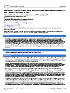
sample flow contains microspheres which are purchased). The inlet and outlet entries and the microchannel are shown in Fig. 1. The figure shows a close-up of the sheathing chevrons by which the microspheres are focused at the interrogation region. The same feature in reverse direction is designed in the second half of the design to perform recycling of the sample and sheath flows.
Experimental Setup Applying photolithography techniques and soft baking, the mold is fabricated. The microchannel is made in polydimethylsiloxane
1
Downloaded From: http://proceedings.asmedigitalcollection.asme.org/ on 12/15/2014 Terms of Use: http://asme.org/terms
Copyright © 2013 by ASME
maximum compression in vertical direction occurred right after the last chevron (d). The concentration of the core and sheath flow flows were assumed to be 1 and 0 , respectively. The quadratic solution was selected as the method of solving for fluid dynamics part of the simulation to improve the quality of the results.
FIG. 1. The geometry of the whole channel including chevrons on the top and bottom of the channel. A close-up of the chevrons’ section is also shown.
Results and Discussion The sheath flow rate was 200 µL/min and the sample (core) flow rate was 10 µL/min. The sample flow contained microspheres with diameter of 3.2 and 10.2 . The data collected by MPPC (Fig. 2) showed that for larger microsphers, both height and width of the signal output were larger. The concentration of the microsphres was 50 beads/ . The background noise was also monitored regularly to make sure it remained constant over the course of the experiment. The sampling rate of DAQ was samples per second.
FIG. 3. The concentration distribution along the channel for the cross section at: a) before the chevrons, b) after the first chevron, c) after the third chevron and d) after the last chevron.
Fig. 4. shows the average and standard deviation of collected data points for each microsphere. The average voltage for 3.2 and 10.2 microspheres was 1.6 V and 1.9 V and the standard deviation was 0.039 and 0.009, respectively.
Fig. 4. Statistical approach for Average of signal output regarded to two sizes of microspheres is shown.
Fig. 2. The output signal from MPPC module for (a) 3.2 10.2 microspheres.
Conclusion It was shown that for two high intensity florescence microspheres of different sizes, the output signal was larger for the 10 µm microspheres. The results showed that for 10.2 µm microspheres the width of the signal was more expanded compared to 3.2 µm microspheres which mean larger microspheres have passed through the interrogation region in a longer period of time. The results for COMSOL simulation showed that the sample flow compresses by passing through more chevron grooves.
and (b)
Fig. 3 shows the results of COMSOL simulations for concentration distribution along the channel. The simulation was performed for quarter of the channel because of symmetry characteristics of the whole microchannel. This also helped in reducing the computational time. The velocity profile was adjusted as fully developed at the entrance of the channel. Additionally, looking at the concentration simulation along the channel (Fig. 3), as the core (sample) flow entered the channel (red region), it was compresses horizontally by sheath flows on both sides (b) and once the sample flow passed the chevrons, it was compressed vertically on the top and bottom. The
References [1] N. Hashemi, et al., 2011. “Microflow Cytometer for optical analysis of phytoplankton.” Biosensors and Bioelectronics, 4263-4269 [2] Hashemi, et al., 2010. “Dynamic reversibility of hydrodynamic focusing for recycling sheath fluid” Lab on a Chip, 1952-1959
2
Downloaded From: http://proceedings.asmedigitalcollection.asme.org/ on 12/15/2014 Terms of Use: http://asme.org/terms
Copyright © 2013 by ASME