C. Using mRNA differential display and screening of the cDNA libraries, we have cloned from tomato fruit a full-length HCT1 cDNA (heat induced/chilling ...
885
Plant Molecular Biology 36: 885–895, 1998. c 1998 Kluwer Academic Publishers. Printed in Belgium.
Molecular cloning of a novel heat induced/chilling tolerance related cDNA1 in tomato fruit by use of mRNA differential display2 Dina K. Kadyrzhanova, Konstantinos E. Vlachonasios, Philippos Ververidis� & David R. Dilley�� Department of Horticulture, Plant and Soil Sciences Building, Michigan State University, East Lansing, MI 48824, USA; � (current address: Institute of Molecular Biology and Biotechnology, Foundation for Research and Technology, P.O. Box 1527, Heraklion 711 10, Crete, Hellas, Greece; �� author for correspondence) Received 19 February 1997; accepted in revised form 25 November 1997
Key words: chilling injury, heat shock protein, tomato
Abstract Chilling injury was circumvented by heat-treating mature green tomatoes (Lycopersicon esculentum, cv. Mountain Springs) at 42 � C for two days prior to storing them at 2 � C for one or two weeks, whereas fruits stored at 2 � C without preheating developed typical chilling injury symptoms and failed to ripen at 20 � C. Using mRNA differential display and screening of the cDNA libraries, we have cloned from tomato fruit a full-length HCT1 cDNA (heat induced/chilling tolerance related). The protein (17.6 kDa) predicted from coding region of HCT1 cDNA has high identity with class II cytosolic small HSPs. The gene corresponding to HCT1 cDNA was termed as LeHSP 17.6. Southern-blot hybridization indicates that LeHSP 17.6 belongs to a two-member gene family. Northern blot analysis indicates the heat-induced transcript of the LeHSP 17.6 remains up-regulated during subsequent exposure of the fruit to chilling temperatures for at least one week and upon transfer to ripening temperatures for one day. Fruits which were only chilled show a low level of expression of the LeHSP 17.6 transcript. We hypothesize that LeHSP 17.6 may be involved in protecting the cell from metabolic dysfunctions leading to ripening failure caused by chilling injury. This is the first report of a class II cytosolic smHSPs encoding gene in tomato. Introduction The useful postharvest life of many fruits of tropical or subtropical origin is limited because they must be stored above 10–12 � C to avoid chilling injury [37]. For example, if mature green tomatoes are stored for only a few days at temperatures below 10 � C and subsequently returned to a normally permissive ripening temperature, they fail to ripen and become susceptible to microbial spoilage [18]. Prestorage heat-treatment has been found to increase the chilling tolerance of tomato and other subtropical fruits [17, 36, 38, 56]. 1 GenBank Accession No. of the LeHSP 17.6 cDNA sequence is U72396. 2 This research was partially funded by the United States–Israel Binational Agricultural Research and Development Fund (BARD No. 15–2175–92R) and the Michigan Agricultural Experiment Station.
Increased tolerance to chilling injury by high temperature treatment has been related to the accumulation of heat shock proteins (HSPs) [27, 36]. Plant HSPs consist of a few high molecular weight classes 60 kD, 70 kD, 90 kD, 100 kD and a complex group of low molecular weight proteins with molecular sizes ranging from �17 to 30 kD [23, 43, 65]. The small HSPs are structurally related and are encoded by six discrete gene families [67]. Classes I and II encode cytosolic proteins while classes III and IV encode chloroplast and endoplasmic reticulum localized proteins [16, 65]. A fifth class encodes the mitochondrial proteins [8, 32]. For class VI, which is represented by a single 22.3 kD HSP from soybean, the intracellular location is proposed to be the endoplasmic reticulum [26]. Cytosolic small HSPs are particularly abundant in plants subjected to stress conditions but their specific functions are not known. Recently, in vitro studies demonstrated that
886 small HSPs as well as high molecular weight HSPs display elements suggesting molecular chaperone activities [29]. While they ostensibly function in the development of thermotolerance [30, 31, 71], some plant HSPs are expressed when the plant is subjected to other stresses such as water stress [2], heavy-metal toxicity [40] and cold stress [5, 25, 41, 62]. To gain insight about the molecular mechanisms by which heat-treatment affords subsequent protection against chilling injury in tomato fruits, we employed the mRNA differential display techniques. The mRNA differential display method [35] is proving to be a good tool for detecting specifically expressed genes in plant tissues [13, 20, 46, 58, 61, 63, 68]. We herein report the identification and cloning of a full-length HCT1 cDNA (heat-induced/chilling tolerance related). HCT1 cDNA encodes a putative 17.6 kD protein which has high identity with class II cytosolic small HSPs of other plants. The gene corresponding to HCT1 cDNA was termed as LeHSP 17.6. The transcript of LeHSP 17.6 is induced by heat-treatment at 42 � C for 2 days and remains up-regulated during a subsequent one week exposure to chilling temperatures of 2 � C. The heattreated fruits ripened normally following the low temperature storage while non-heated fruits showed typical chilling injury symptoms. We hypothesize that the translation product of LeHSP 17.6 may be involved in protecting the cell from the metabolic dysfunctions leading to ripening failure caused by chilling injury.
taining: 1% triisopropylnaphthalene sulfonate, 6% paminosalicylate, 5% (v/v) Tris-saturated phenol and 50 mM Tris-HCl, pH 8.0). The homogenate was extracted 4 times with an equal volume of phenol:chloroform (1:1) and once with chloroform. RNA was precipitated at ,20 � C by addition of 0.1 vol 3M sodium acetate pH 5.6 and 2.5 vols of ethanol in two steps: first, 1 vol of ethanol was added and if a carbohydrate and DNA precipitate formed, this was removed and the remaining 1.5 vol of ethanol was added. RNA was precipitated with 4 M lithium chloride and the pellet was dissolved in 250 mM potassium acetate pH 7.0. The RNA was precipitated with 3 vols of ethanol, the pellet washed with 80% (v/v) ethanol, dried and dissolved in diethylpyrocarbonate (DEPC)-treated water. RNA concentration and purity were determined spectrophotometrically. Integrity of RNA was evaluated by fractionation of an aliquot on 1.2% agarose/formaldehyde gel. Poly (A)+ mRNA was obtained from total RNA using the PolyAT-tractR mRNA isolation kit (Promega, Madison, WI, USA). Concentration of poly(A)+ mRNA was measured using the DNA DipStickTM kit (Invitrogen, San Diego, CA, USA). Genomic DNA was isolated from tomato leaves as described by Doyle and Doyle [9] except that SDS replaced CTAB in the extraction buffer. DNA concentration, purity and integrity was determined spectrophotometrically and by running an aliquot on 0.8% agarose gel.
Materials and methods
Differential display. Differential mRNA display [35] was performed using the RNA mapTM kit (GenHunter, Brookline, MA, USA) according to the manufacturer’s recommendation. Total RNA samples were DNAse I treated as described by Liang et al. [34] using RNAsefree DNAse I and human placental ribonuclease inhibitor from Boehringer Mannheim (Indianapolis, IN, USA). DNAse-free total RNA samples (0.2 �g) were used for the first-strand cDNA synthesis. T12 MN anchor primers (where M is degenerate A, C, G and N=A, C, G or T) were used in four reverse transcription (RT) reactions. PCR amplification of one-tenth of the first-strand synthesis cDNA products was done in the presence of [33 P] dATP [4]. Five decamers (AP1 to AP5 ) were used in combination with the respective T12 MN. AmpliTaq polymerase from Perkin-Elmer Cetus (Norwalk, CT, USA) was used for PCR reactions. Control reactions were performed in the absence of reverse transcriptase. Amplified cDNAs were electrophoresed through a 6% denaturing polyacrylam-
Plant material and temperature treatments. Tomato fruits were harvested at the mature green stage and subjected to different temperature treatments: heattreatment at 42 � C for 2 days (designated as H); heattreatment at 42 � C for 2 days then cold storage at 2 � C for 7 days plus 1 day at 20 � C (designated as HC); and cold storage at 2 � C for 7 days then 1 day at 20 � C (designated as C). As a control for all of the treatments, mature green tomato fruits (designated as MG) were used. Fruit pericarp tissue from each temperature treatment and control was frozen in liquid nitrogen and stored at ,80 � C. Nucleic acid isolation. Total RNA was extracted and purified according to Grierson and Covey [14] and Fray and Grierson [11] with some modifications. Frozen fruit pericarp was ground in liquid nitrogen with a motor and pestle and extracted with buffer con-
887 ide gel, containing 7 M urea. The 32 P-end labeled �X174/Hae III (Gibco BRL, Life Technologies, Gaithersburg, MD, USA) was used as the molecular weight markers. Electrophoresis was done at constant 60 W for 3–4 h followed by drying the gels on Whatman 3MM without fixing. Isolation of cDNA bands. Autoradiograms and gels were aligned and appropriate bands identified on the autoradiographs were excised. DNA was recovered according to the manufacturer’s instructions. Each gel was reexposed to X-ray film to verify that the band of interest had been excised [73]. cDNAs were then reamplified using the same PCR conditions and primers as before. PCR products were analyzed on a 1.2% agarose gel and cDNAs were purified from agarose slices using the QIAEXIITM kit (Qiagen, Chatsworth, CA, USA). Subsequently, eluted cDNAs were labeled with [�-32 P]-dCTP (DuPont/NEN Products, Boston, MA, USA) in the presence of the respective T12 MN oligomer by using the Random Primer Labeling System (Gibco BRL, Life Technologies, Gaithersburg, MD, USA) for use in RNA gel blot analysis. RNA and DNA gel blot analysis. Total RNA samples were denatured and electrophoresed (15 �g per lane) in denaturing 1.2% agarose/2.2 M formaldehyde gels for RNA gel blot hybridization. RNA size markers (Gibco BRL, Life Technologies, Gaithersburg, MD, USA) were used as length standards. Genomic DNA was digested with appropriate restriction enzymes. Restriction fragments (20 �g per lane) were separated by electrophoresis on 0.8% agarose gel for DNA gel blot hybridization. The gels were blotted onto HybondN nylon membranes (Amersham, Aylesbury, UK). RNA and DNA were fixed to the membranes using a microwave oven for 2 min at full setting (700 W) [3] and hybridization was performed according to standard protocols [55]. RNA blots were prehybridized for 3 h and DNA blots for 24 h in hybridization buffer containing 50% formamide, 5�SSC, 25 mM potassium phosphate, pH 7.4, 5�Denhardt’s solution, 50 �g/ml denatured salmon sperm DNA. After alkaline denaturation 32 P-labeled probe was added to hybridization buffer with dextran sulfate in 5% (w/v) final concentration and blots were hybridized for 16 h at 42 � C. RNA blots were washed twice with 1�SSC and 0.1% SDS at room temperature for 15 min and one time with 0.2�SSC and 0.1% SDS at 60 � C for 30 min and autoradiographed. DNA blots were washed three
times with 2�SSC and 0.1% SDS at room temperature for 15 min and twice with 0.1�SSC and 0.5% SDS at 42 � C for 15 min and autoradiographed 24 h with double intensifying screens at ,80 � C. The cloning of cDNA bands. Following RNA blot hybridization cDNA bands were recovered using the Northern-blot affinity capturing method [33]. A piece of the nylon membrane containing the captured probe was cut out, using the autoradiogram for band localization. The membrane pieces were stripped by boiling in water for 5 min and the eluted probes were precipitated with 0.3M sodium acetate and ethanol using glycogen as carrier. The reconstituted probe was reamplified by PCR using the same conditions as described for the differential display step with no radioactive labeled nucleotide. The PCR product was ligated and subcloned in pCRII vector of the TA-Cloning kit (Invitrogen, San Diego, CA, USA). Multiple plasmid preparations were performed for each clone using the standard alkaline lysis method [55] and analyzed by digestion with EcoRI for presence and size of the cDNA insert. cDNA band inserts were purified from agarose slices, random-radiolabeled and used in RNA blot analysis. Those detecting differentially expressed mRNA were sequenced. Library construction and screening. Amplified cDNA libraries was constructed from heat-treated (H) and heat and cold-treated (HC) tomato fruit poly (A)+ mRNA in �gt 11 vector by using CapFinderTM PCR cDNA Library Construction kit (Clontech, Palo Alto, CA, USA) according to manufacturer’s instructions. Screening of the (H) and (HC) cDNA libraries was done by the Long-Distance (LD)-PCR based method [1]. In 50 �l reaction mixture 106 plaques from (H) or (HC) cDNA libraries were screened by amplification with 50 ng of the 30 -gene specific primer (30mer) derived from a sequence near the poly (A) tail of HCT1 cDNA band and 50 ng of the 50 -PCR primer (22 -mer) supplied in CapFinderTM PCR cDNA Library Construction kit from Clontech. PCR buffer and LD-polymerase from the same kit from Clontech were used. The LD-PCR band was eluted from agarose gel and cloned into pCRII vector by using TA-cloning. Sequencing and sequencing analysis. Doublestranded DNA sequencing was performed on an Applied Biosystems 373A (Foster City, CA, USA) at Michigan State University Instrumentation Facility, East Lansing, MI, USA. DNA sequence of tomato
888
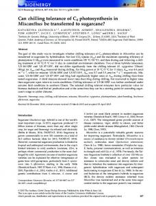
Figure 1. Differential display of total RNA from four different treatments of tomato fruit. (1) RNA was isolated from control fruits at the mature green stage (MG), (2) heat-treated at 42� C for 2 days (H), (3) heat-treated at 42� C for 2 days following by storage at 2� C for one week and then transferred at 20� C for 1 day (HC) and (4) fruits that were stored at 2� C for one week following by one day at 20� C (C). Total RNA was reverse transcribed with the anchored primer T12 MC. The anchor primer and the arbitrary primer (AP1 , AP2 , AP3 , AP4 , AP5 ) were used for the amplification step of differential display. The 32 P-end labeled �X174/Hae III (Gibco BRL) was used as molecular weight marker.
LeHSP 17.6 is available from the GenBank as Accession number U72396. Sequences were compared to the National Center of Biotechnology Information nonredundant sequence database using the Fast A and BLAST programs. Alignment of sequences was done using the MEGALIGN program of DNA STAR (DNA STAR, Madison, WI, USA).
Results Differential display. The effect of heat and/or cold treatment on tomato fruit gene expression at the tran-
scriptional level was studied using the mRNA differential display [35]. Twenty RT-PCR reactions for each total RNA treatment have been conducted by combining five 50 arbitrary decamers (AP1 -AP5 ) and four 30 anchor primers (T12 MG, T12 MC, T12 MA, T12 MT). An example of the differential display pattern is presented in Figure 1. About 40–80 amplified bands ranging from 100 to 600 bp in size were visible for each primer set. The anchor primers T12 MA and T12 MT showed lower specificity and selectivity than the T12 MG and T12 MC primers. From a total of about 1200 cDNA bands, 142 showed an increased pattern during heat shock (H), 41 showed high intensity upon heat shock
889
and subsequently cold temperatures (HC), while 18 were up-regulated during cold treatment (C) (Table 1). Twenty nine cDNAs were specifically expressed on both H and HC treatments and selected for further evaluation. After elution from the gel, these bands were reamplified with the same set of primers used for the differential display under the same PCR conditions with no radioactive labeled nucleotide. The size of the fragments were verified on 1.2% agarose by electrophoresis. Other differential display bands derived from the same sets of primer combinations in RT-PCR reactions were detected but have not been completely characterized.
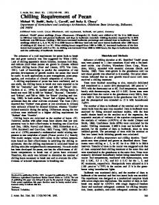
Cloning and sequence analysis of the specific cDNA bands. The cDNA bands confirmed by RNA blot analysis were recovered using the Northern blot affinity capturing method [33], and cloned into the pCRII plasmid vector by using the TA-Cloning kit from Invitrogen. The subcloned cDNA fragments were released from the pCRII vector by digestion with EcoRI enzyme and random prime labeled for use as a probe in RNA blot hybridization. Northern blot analysis resulted in an identical autoradiogram (Figure 2) to that obtained previously. The partial cDNA clones were termed as HCT (heat-induced/chilling tolerance related). Herein we present one representative named as HCT1. The HCT1 nucleotide sequence contained the sequences of the decamer and the oligo (dT) primers used for the differential display as well as part of the open reading frame (ORF) (data not shown). Database searches revealed the following similarities. At the nucleotide level, the 273 bp HCT1 fragment showed 79% similarity with the class II cytosolic small HSP from soybean [50], and pea [28]. The carboxyl-end terminus (40 amino acids) of the ORF presented in the HCT1 encoding protein had 92% identity to the soybean cytosolic class II small HSP and contained the conserved GVL motif that is present in all small heat shock proteins [67]. The 30 non-coding region of the HCT1, contained a putative polyadenylation signal AATAAA 104 bp upstream of the poly (A) tail [21].
Expression pattern of the specific cDNA bands corresponding mRNAs during heat-shock and subsequent chilling temperatures. To verify the authenticity of the differential display bands the reamplified cDNAs were used as a probes for RNA blot hybridizations. Upon Northern analysis, twenty-one cDNAs showed levels of transcript induction that confirm the differential display pattern (Figure 2). The cDNAs did not reveal any transcripts in mature green tomato fruit, while they detected remarkably up-regulated transcripts at 42 � C. Interestingly, the level of transcript induction remained elevated during the subsequent period of 7 days at 2 � C followed by one day at room temperature. However, the transcripts homologous to the selected cDNAs were detected at a lower level of induction on chilled treated fruits. The length of the transcripts was approximately 0.8 kb. Northern blot analysis could detect but not confirm the specific pattern for some cDNA fragments, while other cDNA fragments did not reveal any detectable hybridization signals.
Construction, screening of the cDNA libraries and sequence analysis of a full-length HCT1 cDNA. The cDNA libraries produced in �gt11 with poly (A)+ mRNA from heated (H) and heated and chilled (HC) tomato fruits contained about 106 recombinants with inserts ranged in size from 0.5 to 6 kb. The LD-PCR based screening of 106 plaques of amplified (H) and (HC) cDNA libraries resulted in isolation of a fulllength HCT1 cDNA 738 bp in size we have further designated as LeHSP 17.6. The computer analysis of the entire sequence of HCT1 cDNA revealed an uninterrupted reading frame of 477 nucleotides starting at the first ATG 108 and ending with a TGA 154 bases upstream of the poly (A) tail. The 30 untranslated region includes a putative polyadenylation signal. The deduced amino acid sequence of the LeHSP 17.6 corresponds to a polypeptide of 158 amino acids with an expected molecular mass of 17.6 kD and a pI of 6.63. The computer search through the nonredundant protein database showed that LeHSP 17.6 protein is highly similar (>70%) over its entire length to
Figure 2. RNA blot hybridization analysis of total RNA from tomato fruit. Approximately 15 �g of total RNA from control mature green tomato fruit (MG), heat-treated fruit at 42 � C for 2 days (H), heattreated fruit as (H) followed by storage at 2 � C for one week and then placed at 20 � C for one day (HC) and non heat-treated fruit stored at 2 � C for one week and transferred at 20 � C for one day (C) was hybridized with 32 P random primed labeled reamplified HCT1 cDNA. The mRNA molecular weight is indicated.
890 Table 1. Analysis of differential display gels showing cDNA bands corresponding to H, HC, C and MG tomato fruit RNAs Primer combinations in RT-PCR reactions
+
T12 MG
+
T12 MC
+
T12 MA
T12 MT
+
Number of cDNA bands H HC C
Total (including MG)
AP1 AP2 AP3 AP4 AP5
8 9 15 13 5
2 1 5 2 1
4 1 0 1 0
58 53 54 57 41
AP1 AP2 AP3 AP4 AP5
16 7 11 10 8
5 5 1 3 2
10 0 1 1 0
69 64 73 61 65
AP1 AP2 AP3 AP4 AP5
4 1 1 5 3
2 4 1 1 1
0 0 0 0 0
77 69 67 57 41
AP1 AP2 AP3 AP4 AP5
7 4 7 2 6
1 1 1 1 1
0 0 0 0 0
54 59 64 39 56
142
41
18
1196
Total
cytosolic class II small heat shock proteins [65]. The highest identity (75%) was found with the 17.7 kD HSP from pea [28], 74% with the 17.9 kD HSP from soybean [50], 73% with the 17.9 kD HSP from parsley [10] and 70% with small HSPs from alfalfa [22] as well as with the 17.2 kD HSP from Pharbitis nil [24]. Thus based on DNA sequence and deduced protein sequence analysis, the LeHSP 17.6 corresponding to the HCT1 cDNA can be assigned to the class II cytosolic smHSP encoding gene in tomato. Southern blot analysis. To estimate the LeHSP 17.6 gene copy number, Southern blot analysis of genomic tomato DNA digested with three different restriction enzymes (EcoRI, BamHI, HindIII) was conducted, using the entire LeHSP 17.6 cDNA without its poly(A) tail as the probe under low-stringency hybridization and washing conditions. Two bands were detected in each restriction digest, indicating that the LeHSP 17.6 transcript is encoded by a two-member gene family in the tomato genome (data not shown).
Discussion Employing the mRNA differential display techniques and screening the cDNA libraries, we have identified and cloned a full-length HCT1 cDNA (heat/induced/chilling tolerance related) whose expression pattern changes during heat-treatment and subsequent cold storage of tomato fruits. Northern blots confirmed the specific expression of the HCT1 cDNA. Only transcripts from heated and heated-and-chilled tomato tissues were preferentially expressed; although transcripts were detected in fruits which were only chilled, they were at a much lower level. These data show that HCT1 cDNA originates from a gene(s) whose transcription is activated by heat-treatment and maintained up-regulated during subsequent cold storage. The induction of the same transcripts at both high and low temperatures suggests, in part, that the 50 and 30 untranslated regions of the corresponding gene(s) might contain the elements controlling induction and
891 selective stabilization of the mRNAs induced by both stresses [62]. The protein (17.6 kD) predicted from the coding region of HCT1 cDNA has high identity with class II cytosolic small HSPs from other plants so we have designated it as LeHSP 17.6. This is the first report of a class II cytosolic smHSP encoding gene in tomato. The optimal multiple sequence alignment in Figure 3 identifies several conserved amino acids among the LeHSP 17.6 and the other plant cytosolic class II smHSPs. The carboxyl end terminus LeHSP 17.6 contains the conserved ‘heat shock’ domain. The conserved domain consists of two subdomains I and II separated by a variable length hydrophilic region [67]. The subdomain I contains the motif P-X14-GVL (where X is any amino acid) and the subdomain II consists of a similar motif P-X14 -N-V/L/I-V/L/I. The significance of these conserved carboxyl-terminus domains for the structure and function of smHSP has not been determined [67]. Contrary to the carboxyl end terminal, the amino terminal of the LeHSP 17.6 differs from the other smHSPs. This may serve to confer some functional specificity to the protein. Interestingly, the LeHSP 17.6 includes a putative protein kinase C phosphorylation site S28 DK at the N-terminal where the serine residue is preferentially phosphorylated by protein kinase C [70] and a putative cAMP-dependent protein kinase phosphorylation site RKFS85 [72] at the C-terminus, indicated that phosphorylation of the LeHSP 17.6 might occur. However, in early studies Nover and Scharf [44] failed to detect phosphorylation of the smHSP in tomato culture cells. Similarly, Susuki et al. [60] could not find phosphorylation of the cytosolic class I and II smHSPs. Waters et al. [67] suggested that the lack of the consensus RXXS phosphorylation motif is a possible explanation of the insufficient smHSP phosphorylation. In the first box of the alignment a conserved basic amino acid sequence is present (RKR) and corresponds to a putative Xenopus type nuclear localization signal (NLS) [53]. According to that motif, 2 basic amino acids are followed by 10 residues and the next 5 residues contain at least 3 basic residues. The two basic regions cooperate in binding, whereas the spacer may facilitate their cooperative interaction [49]. Alternatively, the LeHSP 17.6 at the C-terminus (K148 KKPK), contains a putative SV40 large T-antigen nuclear targeting signal [12]. Recent studies by Wollgiehn et al. [69] have suggested that the smHSPs move between the nucleus and the cytoplasm in a stress-dependent fash-
ion. Small proteins, like the 13.8 kD heat shock protein from yeast [39], have been shown to have an NLS capable of redirecting a reporter protein. By histochemical analysis it has been shown that the SV40 sequence can function as an NLS in transgenic tobacco [64]. However, not all the sequences similar to SV40 NLS are recognized by the plant nuclear import machinery [59]. Perhaps, these putative nuclear localization signals are responsible for the shuttling of the smHSPs from cytoplasm to the nucleus during stress. Finally, the carboxyl end of the cytosolic class II smHSPs contains a polyproline motif PPPEPKKP. This motif, particularly the diproline sequence PxxP, is recognized by proteins with Src homology 3 (SH3) domains [51, 52]. In a number of cell types, the SH3 domains function to regulate cellular events such as protein localization, enzyme activity and substrate requirement [6]. The sequences of these proteins determine specific signal transduction pathways [48]. It remains to be elucidated if the smHSPs bind to proteins with SH3 domains. Genomic Southern-blot hybridization indicates that LeHSP 17.6 belongs to a two-member gene family. In agreement, Schoffl and Key [57] reported that the soybean cytosolic class II smHSP is encoded by single gene. In contrast, the Pharbitis nil class II smHSPs are encoded by a multigene family, with at least four representatives [24]. The transcription of the LeHSP 17.6 gene is heatinduced and is maintained up-regulated during subsequent exposure to chilling temperature and this correlated with tolerance to chilling injury. The mechanism remains to be determined and the function of the putative LeHSP 17.6 is unknown. However, our data is in good agreement with results by Lurie and colleagues [54]. They found that protection of tomatoes from chilling injury afforded by prestorage heat-treatment is correlated with the induction of transcription of HSP 17 and HSP 70 mRNAs and with translation of HSP 17 and HSP 23 proteins which persist during subsequent storage of the fruit at chilling temperature. Other heatinduced transcripts may be involved as well. Recently, Collins et al. [7] reported that heat shock of mung bean hypocotyls induced synthesis of several HSPs and only de novo synthesized HSP 79 and HSP 70 remained at significantly higher levels in tissue during a subsequent chilling period. These data together with our results suggest that the synthesis and action of HSPs attained by heat-treatment may be involved in protecting the fruit and other parts of the plant from heat and chilling stress damage. One of the possible models
892
Figure 3. LeHSP 17.6 is a cytosolic class II small HSP. The amino acid sequence alignment of the LeHSP 17.6 tomato protein with cytosolic class II smHSPs from pea (M33901), soybean (X07159), parsley (X95716), Pharbitis nil (M99429) and alfalfa (X98617). Consensus sequence appears below the alignment typed in bold. A putative nuclear localization signal is indicated in box I. Box II indicates a potential polyproline motif. The underlined sequences suggest a putative protein kinase C phosphorylation sites. Asterisks define the important residues within the heat shock domain. Gaps within the alignment were introduced to optimize the alignment.
for the mechanism by which heat-treatment attenuate heat and chilling injury may be attributed to ‘molecular chaperone’ activities of HSPs [65]. Molecular chaperone are a group of intracellular proteins that control correct folding, oligomeric assembly, transport across membranes or disposal by degradation of other conformer unstable proteins by binding to them and release of them. Additionally, molecular chaperones prevent incorrect interaction within and between non-native polypeptides which result in their irreversible aggregation [15]. Recently, the in vitro evidence for molecular chaperone activity of plant smHSPs was demonstrated
by Vierling and colleagues [29]. They observed that recombinant HSP 18.1 and HSP 17.7 representing class I and class II cytosolic smHSPs from pea were able to enhance the refolding of chemically denatured model substrate citrate synthase and lactate dehydrogenase and prevented their aggregation and irreversible inactivation. Plant smHSPs can assemble into multimeric units and form soluble high molecular weight complexes between 200–400 kDa [29, 67]. Lin and colleagues [19] observed that the isolated 280 kDa smHSP complex from soybean was able to protect up to 75% of
893 the total soluble proteins of the cell from heat denaturation in vitro. These smHSP complexes can associate into insoluble larger cytoplasmic aggregates termed as ‘heat shock granules’ [43]. It has been suggested that these HS granules are transient sites for non-heat shock mRNA, preventing its degradation during heat stress [42]. These large structures may be common to all smHSPs [47] and their formation may be reversible and occur mainly at highly stressing temperatures [67]. Another role of plant HSPs is the protection of some proteins from and the targeting of other proteins to proteolysis [66]. This putative function of HSPs may be involved in the protection of plants from different stress damages such as heat shock and chilling injury. We plan to study the LeHSP 17.6 in transgenic plants to test the in vivo role of its protein product in attenuation of chilling injury in tomatoes.
12. 13.
14. 15. 16.
17.
18. 19.
20.
References 1. Ali-Osman F, Akande O: Amplification of the human glutathione s-transferase-pi cDNA from a �gt 11 cDNA library with the ExpandTM Long Template PCR system. Biochemica 4: 28 (1995). 2. Almoguera C, Coca MA, Jordano J: Tissue-specific expression of sunflower heat shock proteins in response to water stress. Plant J 4: 947–958 (1993). 3. Angeletti E, Battiloro E, Pascale E, D’Ambrosio E: Southern and Northern blot fixing by microwave oven. Nucl Acids Res 23: 879–880 (1995). 4. Bauer D, Muller H, Reich J, Riedel H, Ahrenkiel V, Warthoe P, Strauss M: Identification ofdifferential expressed mRNA species by an improved display technique (DDRT-PCR). Nucl Acids Res 21: 4272–4280 (1993). 5. Cabane M, Calvet P, Vincens P, Boudet AM: Characterization of chilling-acclimation-related proteins in soybean and identification of one as a member of the heat shock protein (HSP 70) family. Planta 190: 346–353 (1993). 6. Cohen GB, Ren R, Baltimore D: Modular binding domains in signal transduction proteins. Cell 80: 237–248 (1995). 7. Collins GG, Nie X, Saltviet ME: Heat shock proteins and chilling sensitivity of mung bean hypocotyls. J Exp Bot 46: 795–802 (1995). 8. Dong J-Z, Dunstan DI: Characterization of three heat-shockprotein genes and their developmental regulation during somatic empryogenesis in white spruce. Planta 200: 85–91 (1996). 9. Doyle JJ, Doyle JL: A rapid DNA isolation procedure for small quantitites of fresh leaf tissues. Phytochem Bull 19: 11–15 (1987). 10. Eckey-Kaltenbach H, Kiefer E, Grosskopf E, Ernst D, Sandermann H: Differential transcript induction of parsley pathogenesis. Plant Mol Biol 33: 343–350 (1997). 11. Fray RG, Grierson D: Identification and genetic analysis of normal and mutant phytoene synthase genes of tomato by sequencing, complementation and co-suppression. Plant Mol Biol 22: 589–602 (1993).
21.
22.
23. 24.
25.
26.
27.
28.
29.
30.
31.
32.
Garcia-Bustos J, Heitman J, Hall MN: Nuclear protein localization. Biochem Biophys Acta 1071: 83–101 (1991). Goormachtig S, Valerio-Lepiniec M, Szczyglowski K, Van Montagu M, Holsters M, de Bruijn FJ: Use of differential display to identify novel Sesbania rostrata genes enhanced by Azorhizobium caulinodans infection. Mol Plant-Microbe Interact 8: 816–824 (1995). Grierson D, Covey S: The properties and function of rapidlylabeled nuclear RNA. Planta 130: 317–321 (1976). Hartl FU: Molecular chaperones in cellular protein folding. Nature 381: 571–580 (1996). Helm KW, Lafayette PR, Nagao RT, Key JL, Vierling E: Localization of small heat-shock proteins to the higher plant endomembrane system. Mol Cell Biol 13: 238–247 (1993). Hirose T: Effect of pre- and interposed warming on chilling injury, respiratory rate and membrane permeability of cucumber fruits during cold storage. J Jpn Soc Hort Sci 53: 459–466 (1985). Hobson GE: Low-temperature injury and the storage of ripening tomatoes. J Hort Sci 62: 55–62 (1987). Jinn TL, Chen YM, Lin CY: Characterization and physiological function of class I low-molecular-mass, heat shock protein complex in soybean. Plant Physiol 108: 693–701 (1995). Johnson RR, Cranston HJ, Chaverra ME, Dyer WE: Characterization of cDNA clones for differentially expressed genes in embryos of dormant and nondormant Avena fatua L. caryopses. Plant Mol Biol 28: 113–122 (1995). Joshi CP: Putative polyadenylation signals in nuclear genes of higher plants: a compilation and analysis. Nucl Acid Res 15: 9627–9640 (1987). Keleman Z, Dudits D, Gyorgyey J: New member of alfalfa small heat shock proteins is also expressed in somatic embryos. Accession number X98617 (unpublished). Key J, Lin CY, Chen TM: Heat shock proteins of higher plants. Proc Natl Acad Sci USA 76: 3526–3530 (1981). Krishna P, Felsheim RF, Larkin JC, Das A: Structure and lightinduced expression of a small heat-shock protein gene of Pharbitis nil. Plant Physiol 100: 1772–1779 (1992). Krishna P, Sacco M, Cherutti JF, Hill S: Cold-induced accumulation of HSP90 transcripts in Brassica napus. Plant Physiol 107: 915–923 (1995). LaFayette PR, Nagao RT, O’Grady K, Vierling E, Key JL: Molecular characterization of cDNAs encoding lowmolecular-weight heat shock proteins of soybean. Plant Mol Biol 30: 159–169 (1996). Lafuente MT, Belver A, Guye MG, Saltveit ME: Effect of temperature conditioning on chilling injury of cucumber cotyledons. Plant Physiol 95: 443–449 (1991). Lauzon LM, Helm KW, Vierling E: A cDNA clone from Pisum sativum encoding a low molecular weight heat shock protein. Nucl Acids Res 18: 4274 (1990). Lee GJ, Pokala N, Vierling E: Structure and in vitro molecular chaperone activity of cytosolic small heat shock proteins from pea. J Biol Chem 270: 10432–10438 (1995). Lee JH, Hubel A, Schoffl F: Derepression of the activity of genetically engineered heat shock factor causes constitutive synthesis of heat shock proteins and increased thermotolerance in transgenic Arabidopsis. Plant J 8: 603–612 (1995). Lee, Y-RL, Nagao RT, Key JL: A soybean 101-kD heat shock protein complements a yeast HSP104 deletion mutant in acquiring thermoterance. Plant Cell 6: 1889–1897 (1994). Lenne C, Douce R: A low molecular mass heat-shock proteins is localized to higher plant mitochondria. Plant Physiol 105: 1255–1261 (1994).
894 33. Li F, Barrnathan ES, Kariko K: Rapid method for screening and cloning cDNAs generated in differential mRNA display: application of Northern blot for affinity capturing of cDNAs. Nucl Acids Res 22: 1764–1765 (1994). 34. Liang P, Averboukh L, Pardee AB: Distribution and cloning of eukaryotic mRNA by means of differential display: refinements and optimization. Nucleic Acids Res 21: 3269–3275 (1993). 35. Liang P, Pardee AB: Differential display of eukaryotic mRNA by means of the polymerase chain reaction. Science 257: 967– 971 (1992). 36. Lurie S and Klein JD: Acquisition of low-temperature tolerance in tomatoes by exposure to high-temperature stress. J Amer Soc Hort Sci 116: 1007–1012 (1991). 37. Lyons JM: Chilling injury in plants. Ann Rev Plant Physiol 24: 445–466 (1973). 38. McCollum TG, D’Aquino S, McDonald RE: Heat treatment inhibits mango chilling injury. HortScience 28: 197–198 (1993). 39. Moreland RB, Langevin GL, Singer RH, Garcea RL, Hereford LM: Amino acid sequences that determine the nuclear localization of yeast histone 2B. Mol Cell Biol 7: 4048–4057 (1987). 40. Neumann D, Lichtenberger O, Gunther D, Tschiersch K, Nover L: Heat shock proteins induce heavy-metal tolerance in higher plants. Planta 194: 360–367 (1994). 41. Neven LG, Haskell DW, Guy CL, Denslow N, Klein PA, Green LG, Silverman A: Association of 70-kilodalton heat shock cognate proteins with acclimation to cold. Plant Physiol 99: 1362–1369 (1992). 42. Nover L: Heat shock response. CRC Press, Boca Raton (1991). 43. Nover L, Neumann D, Scharf KD: Heat Shock and Other Stress Response Systems of Plants. Springer-Verlag, Berlin (1990). 44. Nover L, Scharf K: Synthesis, modifications and structural binding of heat shock granules in tomato cell cultures. Eur J Biochem 139: 303–313 (1984). 45. Nover L, Scharf KD, Neumann D: Cytoplasmic heat shock granules are formed from precursor particles and are associated with a special set of mRNA. Mol Cell Biol 9: 1298–1308 (1989). 46. Oh BJ, Balint DE, Giovannoni JJ: A modified procedure for PCR-based differential display and demonstration of use in plants for isolation of genes related to fruit ripening. Plant Mol Biol Rep 13: 70–81 (1995). 47. Osteryoung KW, Vierling E: Dynamics of small heat shock protein distribution within the chloroplasts of higher plants. J Biol Chem 269: 28676–28682 (1994). 48. Pawson T: Protein module and signaling networks. Nature 373: 573–560 (1995). 49. Raikhel N: Nuclear targeting in plants. Plant Physiol 100: 1627–1632 (1992). 50. Raschke E, Baumann G, Schoffl F: Nucleotide sequence analysis of soybean small heat shock protein genes belonging to two different multigene families. J Mol Biol 199: 549–557 (1988). 51. Ren R, Mayer BJ, Cicchett P, Baltimore D: Identification of a ten-amino acid proline-rich SH3 binding site. Science 259: 1157–1161 (1993). 52. Rickles RJ, Botfield MC, Zhou XM, Henry PA, Brugge JS, Zoller MJ: Phage display selection of ligand residues important for Src homology 3 domain binding specificity. Proc Natl Acad Sci USA 92: 10909–10913 (1995). 53. Robbins J, Dilworth SM, Laskey RA, Dingwall C: Two independent basic domains in nucleoplasmin nuclear targeting
54.
55.
56.
57.
58.
59. 60.
61.
62.
63.
64.
65. 66. 67.
68.
69.
70.
71.
sequence: Identification of a class of biparticle nuclear targeting sequence. Cell 64: 615–623 (1991). Sabehat A, Weiss D, Lurie S: The correlation between heatshock protein accumulation and persistence and chilling in tomato fruit. Plant Physiol 110: 531–537 (1996). Sambrook J, Fritsch EF, Maniatis TA: Molecular cloning: A laboratory manual. 2nd Edition, Cold Spring Harbor Laboratory, Cold Spring Harbor, New York (1989). Sanxter SS, Nishijima KA, Chan HT: Heat-treating ‘Sharwil’ avocado for cold tolerance in quarantine cold treatments. HortScience 29: 1166–1168 (1994). Schoffl F, Key JL: Identification of a multigene family for small heat shock proteins in soybean and physical characterization of one individual gene coding region. Plant Mol Biol 2: 269–278 (1983). Sharma YK, Davis KR: Isolation of a novel Arabidopsis ozoneinduced cDNA by differential display. Plant Mol Biol 29: 91– 98 (1995). Silver PA: How proteins enter the nucleus. Cell 64: 489–497 (1991). Susuki T, Krawitz D, Vierling E: A chloroplast small heat shock protein forms a large homo-oligomer and is not phosphorylated. In Molecular Chaperones and the Heat Shock Response Metting 5/1-5/5, 1996, pp. 288. Cold Spring Harbor Lab, Cold Spring Harbor, NY (1996). Tieman DM, Handa AK: Molecular cloning and characterization of genes expressed during early tomato (Lycopersicon esculentum Mill) fruit development by mRNA differential display. J Amer Soc Hort Sci 121: 52–56 (1996). Van Berkel J, Salamini F, Gebhardt C: Transcripts accumulating during cold storage of potato (Solanum tuberosum L.) tubers are sequence related to stress-responsive genes. Plant Physiol 104: 445–452 (1994). Van der Knaap E, Kende H: Identification of a gibberellininduced gene in deep-water rice using differential display of mRNA. Plant Mol Biol 28: 589–592 (1995). Van der Krol AR, Chua NH: The basic domain of plant BZIP proteins facilitates import of a reporter protein into plant nuclei. Plant Cell 3: 667–675 (1991). Vierling E: The role of heat shock proteins in plants. Ann Rev Plant Physiol Mol Biol 42: 579–620 (1991). Vierstra RD: Protein degradation in plants. Annu Rev Plant Physiol Mol Biol 44: 385–410 (1993). Waters EL, Lee GJ, Vierling E: Evolution, structure and function of the small heat shock proteins in plants. J Exp Bot 296: 325–338 (1996). Wilkinson JQ, Lanahan MB, Conner TW, Klee HJ: Identification of mRNAs with enhanced expression in ripening strawberry fruit using polymerase chain reaction differential display. Plant Mol Biol 27: 1097–1108 (1995). Wollgiehn R, Neumann D, Nieden UZ, Musch A, Scharf KD, Nover L: Intracellular distribution of small heat stress proteins in culture cells of Lycopersicon peruvianum. J Plant Physiol 144: 491–499 (1994). Woodgett JR, Hunter T, Gould KL: Protein kinase C and its role in cell growth. In Elson EL, Frazier WA, Graser L (eds). Cell Membranes: Methods and Reviews, 3, p. 215. Plenum, New York (1986). Yeh CH, Yeh KW, Wu SH, Chang PFL, Chen YM, Lin CY: A recombinant rice 16.9 kDa heat shock protein can provide thermoprotection in vitro. Plant Cell Physiol 36: 1341–1348 (1995).
895 72. Zetterqvist OZ, Ragnarsson U, Engstrom LT: Substrate specificity of cyclic AMP-dependent protein kinase. In Kemp BE (ed). Peptides and Protein Phosphorylation, p. 43. CRC Press, Boca Raton (1990).
73.
Zimmerman JW, Schultz RM: Analysis of gene expression in the preimplantation mouse embryo use of mRNA differential display. Proc Natl Acad Sci USA 91: 5456–5460 (1994).