[Downloaded from www.aece.ro on Monday, December 18, 2017 at 15:52:59 (UTC) by 191.101.66.57. Redistribution subject to AECE license or copyright.]
Advances in Electrical and Computer Engineering
Volume 15, Number 3, 2015
Classification of Parameters Extracted from Cardiotocographic Signals for Early Detection of Metabolic Acidemia in Newborns Cristian ROTARIU1, Hariton COSTIN1,2, Alexandru PĂSĂRICĂ3, Dragoş NEMESCU1 1 Grigore T. Popa University of Medicine and Pharmacy, Iaşi,700115, Romania 2 Institute of Computer Science, Romanian Academy, Iaşi, 700054, Romania 3 Gheorghe Asachi Technical University, Iaşi, 700506, Romania
[email protected] 1
Abstract—Fetal acidosis is reflected by the values of umbilical cord pH and base deficit (BDecf): normal recordings (pH over 7.2 and BDecf under 8 mmol/l) and abnormal recordings (pH under 7.2 and BDecf over 8 mmol/l). The purpose of this paper is to present the implementation of an automated system for detecting fetal acidosis in cardiotocographic recordings. The method uses spectral analysis of medium (0.07-0.13 Hz) and high (0.13-1 Hz) frequency spectrum. We implement the algorithm for segments of the recordings without signal loss for better classification. We determined the normalized medium and high frequency components and mid to high frequency ratio. The recordings in the database are divided into a control group (100 normal recordings) and a test group (431 normal or abnormal recordings). A t-test with the p value under 0.05 between the two groups is used to classify the test group. The classification is improved by including the presence of late and prolonged decelerations in the classification process, obtaining the final results, which are comparable to the best ones in current literature. Index Terms—cardiotocographic signals, fetal heart rate monitoring, metabolic acidemia detection, pattern classification, spectral analysis.
I. INTRODUCTION The fetus is normally equipped with physiological defense mechanisms that prevent the long term onset of fetal oxygen deficiency during labor, but in some cases these mechanisms may be weakened, leading to fetal distress. The lack of oxygen that reaches his blood stream through the umbilical cord determines an adaptation of the metabolic processes from normal aerobic metabolism to anaerobic metabolism. The latter has toxic byproducts such as organic acids (lactic acid) that can’t be eliminated easily [1]. This may lead to metabolic imbalances (metabolic acidosis) and further in time to neurological dysfunctions, cerebral palsy or encephalopathy [2]. Metabolic acidosis is reflected by the clinical parameters umbilical pH value and base deficit value (BDecf). The method commonly used to determine these values is the fetal scalp blood sampling, an invasive procedure that is time consuming, making it impossible to be used multiple times during labor, and also presents a high risk for the fetus (it requires to take blood samples from the fetus, with possible complications such as hemorrhages, infections or early labor) [3]. In obstetrics, cardiotocography (CTG) is a method of
recording the fetal heart rate (FHR) and the uterine contractions (UC) during pregnancy. These signals reflect changes in fetal behavior and are important parameters used in most fetal status assessment techniques. Spectral analysis of FHR and the identification of FHR decelerations coupled with UC is widely used to monitor the autonomic nervous system of the fetus that may cause changes in FHR during oxygen deficiency [4]. FHR monitoring is one of the most used methods that help the physician to diagnose possible abnormalities, and recognize the pathologic conditions during pregnancy stages [5]. The analysis of CTG signals is an area of interest for monitoring maternal and fetal physiological parameters. The literature contains a series of articles that have the main focus of identifying and classifying the CTG signals, in order to determine the state of the fetus at birth. The primary disadvantage of implementing such methods is the inconsistency of the databases used in the evaluation of presented methods. These vary from small local databases to large ones that have signals selected based on clinical and technical criteria. Table I presents a series of noteworthy articles from 2009 to 2015 on the subject of CTG signals analysis and the results obtained. The main purpose of these papers was to classify the signal in two classes: normal or abnormal (acidemic, adverse or pathological). The methods are different in each paper, but the comparison between results is done by means of sensitivity (SE) and specificity (SP) of the method [6]. TABLE I. ARTICLES ON THE SUBJECT OF CTG SIGNAL ANALYSIS Article Criteria Method Classes SE(%) SP(%) Gmed(%) Costa et al. Multiscalar Normal/ pH12 57 97 74,36 2010 [8] CTG abnormal Siira et al. Normal/ pH 8mmol/L). This condition allows the identification of false positive and false negative recordings, increasing the overall performance of the classification to 98.38% sensitivity and 78.24% specificity, with the class distribution shown in Table VI. TABLE VI. TEST GROUP CLASSIFICATION AFTER INTRODUCING FHR DECELERATION CONDITION
Record type Abnormal Normal Total
Determined records TP FN 61 1 TN FP 289 80 431
Reference records 62 369 431
FHR (bpm)
FHR signal
Max count 44 26 7
1.03 x 10
4
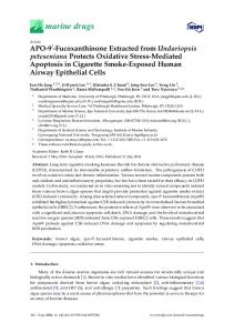
Figure 11. Representation of late decelerations of the FHR signal
200
FHR Prolonged deceleration Baseline
150 100 50 1.16
Amplitude (mmHg)
Min count 5 0 0
4
40
GROUP
Deceleration type Early decelerations Late decelerations Prolonged decelerations
1.03
x 10 Uterine contraction Contraction peak
1.18
1.2
1.22 1.24 Samples Uterine contractions
1.26 x 10
4
Uterine contraction Contraction peak
80 60 40 20 0 1.16
1.18
1.2
1.22 Samples
1.24
1.26 x 10
4
Figure 12. Representation of a prolonged deceleration
IV. DISCUSSIONS Due to high values of signal loss, i.e. an average of 18.6% across the entire database with a maximum value of 53.5%, it is necessary to identify a method that allows the improvement of spectral analysis results. The most used method is the cubic spline interpolation of missing signal, but this method modifies the values of frequency domain metrics (nMF, nHF and MFHF). In consequence, we implemented a method that improves these results. This consists of identifying the signal segments without loss and computing the FFT for these segments. The global value for each record is determined by a weighted average, with the weight represented by the ratio of the segment length to the signal length. 165
[Downloaded from www.aece.ro on Monday, December 18, 2017 at 15:52:59 (UTC) by 191.101.66.57. Redistribution subject to AECE license or copyright.]
Advances in Electrical and Computer Engineering We approached this study by taking into consideration two clinical outcomes that are indicative to the fetal status at birth: pH value and base deficit value. Based on these indicators we defined two classes: normal recordings (pH ≥ 7.2 and BDecf ≤ 8 mmol/L) and abnormal recordings (pH < 7.2 and BDecf > 8 mmol/L). The database was divided into two groups: a control group of 100 normal recordings and a test group with the remaining 431 both normal and abnormal recordings. This allowed us to perform a t-test between the two groups in order to classify the test group recordings into normal and abnormal based on frequency domain metrics. The obtained results point towards a good separation of classes based on these metrics. We initially obtained a sensitivity value of 98.38% and a specificity of 67.47%. To improve the result we introduced a new condition that removes some of the false positive and false negative recordings, which consists in the presence of late and prolonged decelerations associated with uterine contractions in the FHR tracing. Although the methodology presented in this study is widely used, the overall aim was novel. V. CONCLUSIONS Spectral parameters may be used to establish significant differences between normal and pathological fetal outcomes based on pH and base deficit values. Also they can be used to predict the fetal outcome problems that may occur. The overall performance of the classification method is improved when introducing the criteria of late and prolonged decelerations associated with uterine contractions, to 98.38% sensitivity and 78.24% specificity. The presence of early decelerations indicates a normal recording and allows us to identify false positive recordings, and the presence of late and prolonged decelerations indicates an abnormal recording and allows us to remove false negative classifications. Future work on the identification of fetal acidosis can be done and improved by using data collected from patients that are not in labor, in order to minimize the amount of signal loss induced by fetal and maternal movement and intense uterine contractions. This can be realized by longterm CTG monitoring with a non-invasive device during pregnancy (starting with the 25-28th week of pregnancy). REFERENCES [1]
[2]
[3]
[4]
166
A. C. Gjerris, J. Staer-Jensen, J. S. Jorgensen, T. Bergholt, C. Nickelsen,”Umbilical cord blood lactate: A valuable tool in the assessement of fetal blood acidosis,” European Journal of Obstretics & Gynecology and Reproductive Biology, vol. 139, Issue 1, pp. 1620, Jan. 2008. [Online]. Available: http://dx.doi.org/10.1016/j.ejogrb.2007.10.004 E. Soncini, S. Paganelli, C. Vezzani, G. Gargano, G. Battista, “Inatrapartum fetal heart rate monitoring: evaluation of standardized system of interpretation for prediction of metabolic acidosis at delivery and neonatal neurological morbidity,” The Journal of Maternal-Fetal & Neonatal Medicine, vol. 27, no. 14, pp. 1465-1469, Sept. 2014. [Online]. Available: http://dx.doi.org/ 10.3109/14767058.2013.858690 E. Chandraharan, “Fetal scalp blood sampling during labor: is it a useful diagnostic test or a historical test that no longer has a place in modern clinical obstretics?,” BJOG: An international Journal of Obstreticts & Gynaecology, vol. 121, Issue 9, pp. 1056-1062, Aug. 2014. [Online]. Available: http://dx.doi.org/10.1111/1471-0528.12614 J.Y.Kwon, I. Y. Park, J.C. Shin, J. Song, R. Tafreshi, J. Lim,”Specific changes in spectral power of fetal heart rate variability related to fetal acidemia during labor: Comparison between preterm and term
Volume 15, Number 3, 2015
[5] [6] [7]
[8]
[9] [10]
[11]
[12] [13]
[14]
[15] [16]
[17]
[18] [19]
[20]
[21]
[22]
[23]
[24]
fetuses,” Early Human Development, vol. 88, Issue 4, pp. 203-207, April 2012. [Online]. Available: http://dx.doi.org/10.1016/j.earlhumdev.2011.08.007 M.P. Nageotte, “Featl heart rate monitoring,” Seminars in Fetal & Neonatal Medicine, vol. 20, pp. 1-5, Mar. 2015. [Online]. Available: http://dx.doi.org/10.1016/j.siny.2015.02.002 A. Indrayan, “Medical Biostatistics, Third Edition”, Chapman & Hall/CRC Press, USA, pp. 280-283, 2012 A. Costa, D. Ayres-de-Campos, F. Costa, C. Santos, J. Bernardes ”Prediction of neonatal acidemia by computer analysis of fetal heart rate and ST event signals”, American Journal of Obstetrics and Gynecology, vol. 201, pp. 464-452, Nov. 2009. [Online]. Available: http://dx.doi.org/10.1016/j.ajog.2009.04.033 C. Elliott, P. Warrick, E. Graham, E. Hamilton, “Graded classification of fetal heart rate tracings: association with neonatal metabolic acidosis and neurologic morbidity,” American Journal of Obstetrics and Gynecology, vol. 202, no. 3, pp. 258.e1-258.e8, Mar. 2010. [Online]. Available: http://dx.doi.org/10.1016/j.ajog.2009.06.026 S. Siira, “Intrapartum hypoxia and power spectral analysis of fetal heart rate variability,” Uniprint Suomen Yliopistopaino Oy - Oulu, Finland, pp. 33-42, 2012 J. Spilka, V. Chudacek, M. Koucky, M. Huptych, P. Janku, G. Georgoulas, C. Stylios, “Using nonlinear features for fetal heart rate classification,” Biomedical Signal Processing and Control, vol. 7, Issue 4, pp. 350-357, July 2012. [Online]. Available: http://dx.doi.org/10.1016/j.bspc.2011.06.008 A. Georgieva, S. J. Payne, M. Moulden, C. W. G. Redman. “Artificial neural networks applied to fetal monitoring in labour,”. Neural Computing and Applications, vol. 22, pp. :85–93, Jan. 2013. [Online]. Available: http://dx.doi.org/10.1007/s00521-011-0743-y V. Chudáček, J. Spilka, M. Burša, et al. , “Open access intrapartum CTG database”, BMC Pregnancy and Childbirth, pp. 14:16, Jan. 2014. [Online]. Available: http://dx.doi.org/10.1186/1471-2393-14-16 G.S. Dawes, M. Lobb, M. Moulden, C.W. Redman, T. Wheeler, “Antenatal cardiotocogram quality and interpretation using computers,” BJOG: An International Journal of Obstretics & Gynaecology, vol. 99, Issue 10, pp. 791-797, Aug. 2005. [Online]. Available: http://dx.doi.org/10.1111/j.1471-0528.1992.tb14408.x P.A. Warrick, E.Ff Hamilton, D. Precup, R. Kearney, “Classification of normal and hypoxic fetuses from systems modeling of intrapartum cardiotocography,” IEEE Transactions on Biomedical Engineering, vol. 57, Issue 4, pp. 771–779, April 2010. [Online]. Available: http://dx.doi.org/10.1109/TBME.2009.2035818 E.M. Graatsma, “Monitoring of Fetal Heart Rate and Uterine Activity”, Ridderprint BV, Amsterdam, Holland, pp. 39-55, 2010 C-Y. Chen, C. Yu, C-C. Chang, C-W. Lin, “Comparison of a Novel Computerized Analysis Program and Visual Interpretation of Cardiotocography,” PLoS ONE, vol. 9, Issue 12, Dec. 2014. [Online]. Available: http://dx.doi.org/10.1371/journal.pone.0112296 U. Schneider, E. Schleussner, A. Friedler, S. Jaekel, .M. Liehr, J. Haueisen, D. Hoyer, “Fetal heart rate variability reveals defferential dynamics in the intrauterine development of the sympathetic and parasympathetic branches of the autonomic nervous system,” Physiologcal Measurements, vol. 30, no. 2, pp. 215-226, Jan. 2009. [Online]. Available: http://dx.doi.org/10.1088/0967-3334/30/2/008 V. Munteanu, D. Tarniceriu, “Estimation theory and optimal filtering,” Ed. Technopress, Iasi, Romania, pp. 306-310, 2005 V. Maier, S. G. Pavel, C. D. Maier, I. Birou, “Correct Application of the Discrete Fourier Transform in Harmonics,” Advances in Electrical and Computer Engineering, vol. 8, no. 1, pp. 26-30, 2008, doi:10.4316/AECE.2008.01005 M. Jezewski, R. Czabanski, J. Wrobel, K. Horoba, “Analysis of extracted cardiotocographic signal features to improve automated prediction of fetal outcome”, Biocybernetics and Biomedical Cardiology, vol. 30, no.4, pp. 29-47, Feb. 2010. A.G. Cahill, K. A. Roehl, A. O. Odibo, G. A. Macones, “Association and prediction of neonatal acidemia,” American Journal of Obstretics and Gynecology, vol. 207, Issue 3, pp. 206.e1-206.e8, Sept. 2012. [Online]. Available: http://dx.doi.org/10.1016/j.ajog.2012.06.046 Y. Hatakeyama, H. Kataoka, N. Nakajima, T. Watabe, Y. Okuhara, “Level evaluation system for cardiotocography,” 15th International Symposium on Soft Computing and Intelligence Systems, pp. 265269, Dec. 2014. [Online]. Available: http://dx.doi.org/10.1109/SCISISIS.2014.7044686 L. Jimenez, R. Gonzalez, M.J. Gaitan, S. Carrasco, C. Vargas, “Computerized algorithm for baseline estimation of fetal heart rate,” Computers in Cardiology, vol. 29, pp. 477-480, Sept. 2002. [Online]. Available: http://dx.doi.org/10.1109/CIC.2002.1166813 H. J. Seltman, “Experimental design and analysis,” Carnegie Melon University, Chapter 6, pp. 141-161, Nov. 2014