International Journal of Advanced Information Science and Technology (IJAIST) ISSN: 2319:2682 Vol.6, No.4, April 2017 DOI:10.15693/ijaist/2017.v6i4.40-45
CLASSIFICATION SCHEME BASED ON COMMON NEURAL NETWORK FOR BRAIN TUMOR DETECTION 1
M.Vasuki, 2N.Geethapriya, 3M.Brindha 1, 2 PG Scholar, 3Assistant professor 1 Computer Science and Engineering, 2Communication Systems, 3Department of CSE 1, 2, 3 Sri Ramakrishna Institute of Technology Coimbatore Mail id:
[email protected],
[email protected]
Abstract: Brain tumour (tumour- British English, tumor-American English) is a group of cell that grows abnormally in the cell, nerves and other parts of the brain. Methods such as X-Ray, CT-Scan, MRI is available to detect the brain tumour. Many researchers have found that people die due to inaccurate detection of the affected brain tumour part. It is necessary to find the accurate part of the affected area of the brain tumor. Brain tumor is one of the major causes of death among people. The chances of the death in brain tumor are more as compared to other diseases. To avoid the chances of death in the early detection of brain tumor is necessary. Nowadays Brain tumor detection and classification is one of the most active research areas in medical image processing field. The segmentation of brain tumors in magnetic resonance images (MRI) is a challenging and difficult task because of the variety of their possible shapes, locations, image intensities. Detection of brain tumor manually by doctors is a very difficult and time consuming process. To avoid the misclassification and to minimize the time, automatically brain tumor detection and classification is necessary. So, in this work, a common Reflex Fuzzy Min-Max (RFMM) neural network classification scheme is proposed for brain tumor detection. Initially, the input image is pre-processed to remove poison noise as well as active contour is used for edge detection to improve the detection result. Then, tumor region are segmented by Glow-worm Swarm based Clustering (GSC) scheme. After that, the geometric and texture features are extracted. Finally, the RFMM neural network is classified the MRI (DICOM) image into normal or abnormal image. The simulation results shows that RFMM neural network obtained gives better accuracy when compared to the existing Support Vector Machine (SVM) algorithm. Keywords: Brain tumor, MRI (DICOM) image, texture, geometric, neural network, classification. I.INTRODUCTION Brain tumour is the collection or growth of abnormal cells in the brain. Brain tumour is classified into two main types namely cancerous and non cancerous. The cancerous brain tumour is called as Malignant. It is the cancerous type of tumour which occurs in the brain due to the abnormal growth of cells. It is danger compared to non cancerous brain tumour. The other type is the non cancerous brain tumor which is called as Benign. It is the normal brain tumour present in the brain. It is less risk compared to the malignant tumour since it is the initial stage of tumour. The symptoms of the brain tumour are mostly common for both malignant and benign. The common symptoms are headache, vomiting, blurred vision, difficulty in walking, etc. The nonstandard growth of cells inside the skull, it may be cancerous or noncancerous. The tumor is a one type of cancer. Cancer begins in cells, a building block that forms tissues. Tissues make up the organs of the body. Normally, cells grow and divide to form new cells as the body needs them. When cells grow old, they die, and new cells take their place. Sometimes this orderly process goes wrong. New cells form when the body does not need
them, and old cells do not die when they should (Louis et al 2007). These extra cells can form a mass of tissue called a growth or tumor. A lot of tumors can spread into the brain; the most regular ones are lung cancer, breast cancer, melanoma, kidney cancer, bladder cancer, certain sarcomas, testicular and germ cell tumors, and a number of others. Some types of cancers only spread to the brain infrequently, such as colon cancer, or very rarely, such as prostate cancer. Brain tumors destroy brain cells or it in some way damage cells by producing inflammation, compressing other parts of the brain as the tumor grows, inducing brain swelling and causes increased pressure within the skull. Brain Tumor Detection Using Neural Network brain tumor segmentation in magnetic resonance imaging (MRI) has turn into an emergent Research area in the field of medical imaging system. Brain tumor detection helps in finding the exact size and position of tumor. Brain tumors are a dissimilar group of central nervous system neoplasm’s that arise within or adjacent to the brain. Some are curable by surgical resection, but many cannot be wipe out by current treatments, and, when they are, disabling neurological injury often occur. Brain tumor is one of the major
40
International Journal of Advanced Information Science and Technology (IJAIST) ISSN: 2319:2682 Vol.6, No.4, April 2017 DOI:10.15693/ijaist/2017.v6i4.40-45 elements for the increase in Mortality among children and adults. A tumor is a group of tissue that grows out of control of the normal forces that regulate growth. (Selvanayaki et al 2010). The terrible brain tumors can be distributed into two generic categories depending on the tumors root, their growth pattern and malignancy. Primary brain tumors are tumors that begin from cells in the brain or from the covering of the brain. A secondary or metastatic brain tumor arises when cancer cells spreading to the brain from a primary cancer in parts inside of the body. The approximately brain tumors may be of any size, have a array of shapes, may appear at any location and may appear in different image intensities (Marcel et al 2004). 1.1. Categories of Brain Tumor: a) Primary brain tumor: Primary brain tumors starts with brain tissue . b) Secondary brain tumor: Secondary brain tumors are more ordinary. These types of cancers start somewhere else in the body and travel to the brain. Lung, breast, kidney, colon, and skin cancers are among the most common cancers that can spread to the brain.
scanning is placed on a moveable bed which can pass through the magnet. The magnet creates a strong magnetic field that aligns the protons of hydrogen atoms, which then exposed to a beam of radio waves. This spins the various protons of the body, and they produce a faint signal that is detected by the receiver portion of the MRI scanner. The receiver information is been processed by a computer, and then an image is produced. The image and resolution produced by MRI is quite detailed and can detect tiny changes of structures within the body. A contrast agent like gadolinium can be used to increase the accuracy of the images (Joseph 2013). An MRI scan can be used as an extremely accurate method of disease detection throughout the body. In the beginning, trauma to the brain can be seen as bleeding or swelling. Other abnormalities often found include brain aneurysms, stroke, tumors of the brain, as well as tumors or inflammation of the spine. MRI scanners can produce 1500 images per second. Intraoperative MR imaging can acquire high contrast images of soft tissue anatomy. It can also be acquired individually in as little as half a second per image. a)
c) Benign brain tumors: Benign brain tumors don't have cancer cells. They grow gradually, often be removed, not often spread towards the brain tissue. They can cause problems if they press on certain areas of the brain, though. Depending on where they are located in the brain, they can be life-threatening. d) Malignant brain tumors: Malignant brain tumors have cancer cells. The rates of growth vary, but cells can invade healthy brain tissue nearby. Malignant tumors infrequently spread beyond the brain or spinal cord. 1.2. Magnetic Resonance Image: Currently most of the medical imaging studies and detection are conducted by means of MRI, Positron Emission Tomography (PET) and Computed Tomography (CT) scan. An MRI scan is one among radiology method includes magnetism, radio waves, and a computer to produce images of human body structure. The MRI scanner is a tube surrounded by a massive circular magnet. The patient who needs the
Advantages: MRI is mainly useful for scanning and detection of abnormalities in soft tissue structures in the body and soft organs like the brain or the heart. There is no contribution of any kind of radiations in the MRI, so it is safe for the people who can be in danger to the effects of radiations such as pregnant women or babies. MRI scan provides sufficient information about the blood circulation throughout the body and blood vessels and also enabling the detection of problems related to the blood circulation.
b) Disadvantages:
·MRI scan is done in an covered space, so the people who are fearful of being in a closely enclosed surface, are facing problems with MRI to be done. MRI scans engage really loud noises while processing because they absorb a really high amount of electric current supply. II. EXISITING SYSTEM
Support Vector Machine (SVM) is an algorithm that was developed for pattern classification. It is applied to various optimization problems such as regression and the data classification. The data points are identified as being positive or negative, and the problem is to find
41
International Journal of Advanced Information Science and Technology (IJAIST) ISSN: 2319:2682 Vol.6, No.4, April 2017 DOI:10.15693/ijaist/2017.v6i4.40-45 a hyper-plane that separates the data points by a maximal margin. The statistical features from the images are collected and supplied to SVM feature space. As the Tumor images presents a distinct cluster in the feature space a linear SVM classifier is sufficient can easily generate a completely separable spaces. At the detection stage the features of the input images are plotted on the hyper plane and Euclidian distance is calculated from each group. The smallest distance is considered as the detected class. Thus the image can be classified as either a tumor image or a nontumor image. SVM is a binary classifier. SVM is a useful technique for data classification. It is easier to classify the data and it involves testing for some data instances. The objective o f SVM is to produce a model which predicts target value of data instances in the testing set which are given only the attributes. The steps within SVM classification involves the identification as if familiarly connected to the identified classes called as feature selection or feature extraction. The objective is to find classifier with largest margin between closest positive and negatively labeled support vectors. Normal vector for optimal separating hyper plane wopt is found using a quadratic optimization procedure.
In existing system, Support Vector Machine (SVM) based classifier with Berkeley Wavelet Transformation (BWT) based brain tumor segmentation has been presented. The experimental results achieved accuracy, specificity, and sensitivity. Disadvantage: The classification accuracy was not good and it has taken high training time for process the image. So, the performance was not good.
Most common disadvantage of nonparametric techniques such as SVMs is the lack of transparency of results. SVMs could not represent the score of all companies as a simple parametric function of the financial ratios, since its dimension perhaps very high. It cans a linear combination of single financial ratios or it may have an additional simple functional form. The weights of the financial ratios are not constant. Thus the marginal contribution of each financial ratio to the score is changeable. Using Gaussian kernel, every company has its own weights according to the difference between the value of their own financial ratios and those of the support vectors of the training data sample.
III.PROPOSED SYSTEM
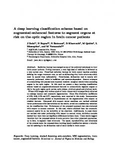
Figure 1. Proposed block diagram representation a) PRE-PROCESSING Pre-processing using bilateral filter to remove the poison noises without reduces the sharpness of the images. Then, the edge has been deducted by using active contour scheme to improve the segmentation process. Here, the level set active contour method has been used. The level set method is a numerical technique that is used to track the interfaces and shapes. In level set method, the contours are set as zero level set of higher dimensional function, known as level set function. Initially, geometrical shaped contours are set and after a number of iteration the contours spread over the vision of interest or the edges of the object boundaries. b) EDGE DETECTION USING ACTIVE CONTOUR First, active contour models can be easily formulated under an energy minimization framework and allow incorporation of various prior knowledge, such as shape and intensity distribution, for robust image segmentation. Second, they can provide smooth and closed contours as segmentation results, which are necessary and can be readily used for further applications, such as shape analysis and recognition. Algorithm for edge detection: 1. Initialize T ← I while convergence/maximum iterations not reached 2. Compute E1 (T) and E2 (T) 3. Compute α∗ 4. Update T end while 5. Do erosion operation on thresholded image 6. Subtract image after erosion from threshold image 7. Pixels greater than zero gives boundary of image.
42
International Journal of Advanced Information Science and Technology (IJAIST) ISSN: 2319:2682 Vol.6, No.4, April 2017 DOI:10.15693/ijaist/2017.v6i4.40-45 c)
GLOW-WORM SWARM BASED CLUSTERING (GSC) SCHEME BASED SEGMENTATION
To separate the tumor region from the image the segmentation process has been carried out. Here, the clustering concept has been implemented for separation process. Two classes are considered such as normal region and tumor region. The Glowworm Swarm Optimization (GSO) scheme has been implemented for this clustering process to separate the regions. The algorithm for GSC is given as follows: 1: Input: Dataset M D of N unified multimedia documents, max iterations 2:
Output: Clusters of multimedia documents
3:
iterations ← 0
4:
repeat
5:
for each glowworm agent GAi ∈ M D do
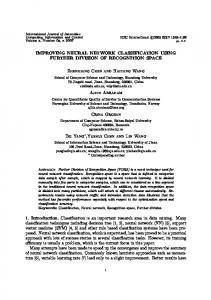
We base our texture feature extraction on the spatial gray level co-occurrence-matrix (GLCM). The GLCM method considers the spatial relationship between pixels of different gray levels. The method calculates a GLCM by calculating how often a pixel with a certain intensity i, occurs in relation with another pixel j, at a certain distance d, and orientation θ. For instance, if the value of a pixel is 1 the method looks, for instance, the number of times this pixel has 2 in the right side. Each element (i, j) in the GLCM is the sum of the number of times that the pixel with value i, occurred in the specified relationship to a pixel with value j, in the raw image. e)
6: Initialize the number of objects present in the cluster of GAi as its luciferin value Li 7:
given the geometrical information and it contains the features like centroid, Eccentricity, area, perimeter, circularity, shape index, solidity, orientation and euler number. The texture feature has been the given Grey Level Co-occurrence Matrix (GLCM) information's. The energy, entropy, covariance, mean, homogeneity features has been extracted.
end for
8: Arrange the glowworms with respect to their luciferin (L) value in ascending order 9:
for each glowworm agent GAi ∈ M D do
10:
Find the neighbors Ni using the rule (1)
11:
Calculate the probability for neighbor GAj ∈
Ni using the formula 12: Select the best neighbor GAj having the minimum probability 13:
Move GAi towards the cluster of GAj .
14:
end for
15:
iterations ← iterations + 1
CLASSIFICATION BASED ON REFLEX FUZZY MIN-MAX NEURAL NETWORK
Classification is the procedure for classifying the input pattern into analogous classes. When the input data set is represented by its class membership, it is called supervised learning. It employs two phases of processing- training phase and testing phase. For training segment, properties of image characteristics were isolated and an unique explanation of each classification category is been produced. During testing phase these features space partitions are used to classify image features. Fuzzy min max (FMM) model is a combination of both fuzzy set and neural network for arrangement. The extracted features are given as input to the RFMM neural network model. FMNN is a concept of merging the fuzzy logic with neural network for pattern classification. Based on this concept the proposed scheme has been introduced. Here, the reflex concept has been introduced to reduce the misclassification problem in FMNN. The reflex is inspired from the reflex system of human brain which takes over the control in hazardous conditions unconsciously. IV. EVALUATION AND RESULTS
16: until no glowworms remaining to merge or iterations ≤ maxiterations d) FEATURE EXTRACTION From the segmented region, the geometric and texture features are extracted to improve the DICOM image classification. The shape has been
For the implementation of this proposed work we use the Image Processing Toolbox under MATLAB Software. The proposed algorithms were tested on DICOM image sample sets for evaluation in brain tumour diagnosis. The experimental aims to verify the performance improvement of SVM and proposed RFMMNN in learning granular data, study
43
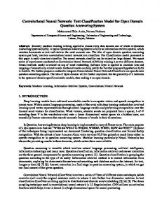
International Journal of Advanced Information Science and Technology (IJAIST) ISSN: 2319:2682 Vol.6, No.4, April 2017 DOI:10.15693/ijaist/2017.v6i4.40-45 the effect of variation of granularity of test data on classification performance. The input brain images are collected from dataset. It is controlled initially by the pre processing stage using bilateral filter. Bilateral filter is a non-linear and noniterative filter. It prevents averaging across image edges, it is edge-preserving. It is shown in the figure 4.1. The pre-processed image is moved to the next level for segmenting. Here the Glowworm Swarm Optimization (GSO) is used. As shown in the figure 4.2, the segmented image is extracted with the help of the artificial neural network. The best classification algorithm is considered for processing the extracted image; it is shown in the figure 4.4.
Fig 4.4: Neural network based classification V.CONCLUSION AND FUTURE WORK 5.1 Conclusion Figure 4.1 Pre-processed and edge detected images
Fig 4.2: Segmented images
The research work offered with a bi-lateral filter based pre-processing stage; Glow-worm Swarm Optimization based segmentation, a novel algorithm RFMMNN for classification of brain tumors is proposed. Here, pre-processing was done by bilateral filter since it was optimal in flat parts of the image. The de-noised images were given as the input to edge detection. Texture features are contrast, correlation, homogeneity and energy were extracted by means of gray level co-occurrence matrix method. The extracted features are then given to the RFMMNN classifier. Using this algorithm one could segment the brain tumors accurately from an MR brain image. Finally, the classification done and the experimental results shows the proposed RFMMNN attained better accuracy, precision and recall compared than existing SVM. 5.2 Future work The proposed segmentation algorithm focuses mainly on two dimensional anatomical structures. It can also be extended to three dimensional volumetric structures. As the intensity and orientation based segmentation method is capable of extracting the features of the medical images accurately, it can also be applied to the fusion image. As the online service is used for the detection of a brain cancer type, the research can be extended to other brain cancer types and location also.
Fig 4.3: Feature extracted images
44
International Journal of Advanced Information Science and Technology (IJAIST) ISSN: 2319:2682 Vol.6, No.4, April 2017 DOI:10.15693/ijaist/2017.v6i4.40-45 REFERENCES [1] Al-Tamimi, M. S. H., & Sulong, G. (2014). Tumor brain detection through MR images: a review of literature. Journal of Theoretical and Applied Information Technology, 62(2), 387-403. [2] Karnan, M., & Logheshwari, T. (2010, December). Improved implementation of brain MRI image segmentation using ant colony system. In Computational Intelligence and Computing Research (ICCIC), 2010 IEEE International Conference on (pp. 1-4). IEEE. [3] Wang, H., Das, S. R., Suh, J. W., Altinay, M., Pluta, J., Craige, C., ... & Alzheimer's Disease Neuroimaging Initiative. (2011). A learning-based wrapper method to correct systematic errors in automatic image segmentation: consistently improved performance in hippocampus, cortex and brain segmentation. NeuroImage, 55(3), 968-985. [4] Li, W., Song, S., & Qian, X. (2011). Active contours with selective local or global segmentation property for multiobject image. Optical Engineering, 50(6), 067009-067009. [5] Zhang, K., Zhang, L., Song, H., & Zhou, W. (2010). Active contours with selective local or global segmentation: a new formulation and level set method. Image and Vision computing, 28(4), 668676. [6] Lin, S. W., Ying, K. C., Chen, S. C., & Lee, Z. J. (2008). Particle swarm optimization for parameter determination and feature selection of support vector machines. Expert systems with applications, 35(4), 1817-1824. [7] Kai xiao,A.Lei Liang, Hai Bing Guan, Aboul Ella Hassanien ,”Extraction and Application of Deformation Based Feature in Medical Images”,ELSEVIER Neuro computing 2013. [8] Padma Nanda Gopal &R.Sukanesh,” wavelet statistical feature based segmentation and classification of brain computed tomography images”IET Image Prosess, Vol7 pp 25-32, 2013.
[12] Rao V, Sarabi M S, Jaiswal A.” Brain tumor segmentation with deep learning. MICCAI Multimodal Brain Tumor Segmentation Challenge” (BraTS),56–59,2015. BIOGRAPHIES Ms.M.Vasuki received her B.E degree in Information Technology from Sri Ramakrishna Institute of Technology, Coimbatore,Anna university, Chennai, in 2015.Currently,she is a Post Graduate Student,ME in Computer Science and Engineering,Sri Ramakrishna Institute of Technology, Coimbatore, Anna University, Chennai.Her current research interests includes Digital Image Processing. Ms.N.Geethapriya received her B.E degree in Electronics and Communication Engineering from Avinashilingam University, Coimbatore, in 2015. Currently,she is a Post Graduate Student,ME in Communication Systems,Sri Ramakrishna Institute of Technology, Coimbatore, Anna University, Chennai. She has published her papers in International Journals. Her current research interests include Wireless communication, Internet of Things,Digital Image Processing.
Ms. M.Brindhacompleted her M.E. degree in Computer Science and Engineering at Sri Ramakrishna Engineering College, Anna University, Chennai in the year 2016. She received her B.E. degree in Electronics and Communication Engineering from KTVR Knowledge park for Engineering and Technology, Coimbatore, Anna University, Chennai in the year 2014. Currently she is working as Assistant Professor in CSE Department at Sri Ramakrishna Institute of Technology. She is having 1 year of teaching experience. She has published papers in International Journals.Her area of interest is Image mining, Data mining.
[10] Hashem Kalbkhani, Mahrokh G Shayesteh, Behrooz Zali-vargahan “Robust algorithm for Brain Magnetic Resonance Image Classification based on GARCH variances Series”, ELSEVIER Biomedical Signal Processing and Control 8,909-919,2013. [11] Richa Aggarwal, Amanpreet Kaur,”Comparative Analysis of Different Algorithms For Brain Tumor Detection”, International Journal of Science and Research (IJSR) ISSN (Online): 2319-7064 Impact Factor (2012): 3.358, Volume 3 Issue 6, June 2014.
45