of this study were to use kinematic MR imaging to assess the dynamic changes of the cervical spine in ..... reliable diagnostic indicator of the genesis of spondy-.
AJNR Am J Neuroradiol 19:1763–1771, October 1998
Classification System Based on Kinematic MR Imaging in Cervical Spondylitic Myelopathy Claus Muhle, Jo ¨rg Metzner, Dieter Weinert, Axel Falliner, Gisbert Brinkmann, Maximillian H. Mehdorn, Martin Heller, and Donald Resnick
BACKGROUND AND PURPOSE: Functional myelographic studies are often used to evaluate the dynamic changes of the cervical spinal canal during flexion and extension. The purposes of this study were to use kinematic MR imaging to assess the dynamic changes of the cervical spine in patients at different stages of degenerative disease and to describe a classification system based on static and dynamic factors in the pathogenesis of cervical spondylitic myelopathy. METHODS: Eighty-one patients with different stages (I–IV) of degenerative disease of the cervical spine were examined with MR imaging. In the neutral position (0°) and at maximum flexion and extension, spinal stenosis was classified for each segment according to the following grading system: 0 5 normal, 1 5 partial obliteration of the anterior or posterior subarachnoid space, 2 5 complete obliteration of the anterior or posterior subarachnoid space, and 3 5 cervical cord compression or displacement. RESULTS: At flexion and extension, the prevalence of spinal stenosis and cervical cord impingement increased as the stage of degenerative disease progressed. With regard to a pincer effect (anterior and posterior cord impingement) and cord encroachment at multiple segments, statistically significant differences were observed at stages III and IV as compared with stages I and II. Significant increase in cord impingement was seen in 22 (27%) of 81 patients at extension, as compared with four (5%) of 81 patients at flexion. CONCLUSION: Regardless of the stage of degenerative disease and grade of spinal stenosis at the neutral position (0°), cervical spinal motion may contribute to the development of cervical spondylitic myelopathy. Although cervical spondylitic myelopathy is the most common disease of the spinal cord that occurs during and after middle age, the pathophysiology remains unclear (1– 4). Besides static factors that lead to canal encroachment, including congenitally narrowed canal, disk bulging, and spondylitic bars, experimental studies have indicated that vascular compromise contributes to the development of cervical spondylitic myelopathy (1– 4). In addition, dynamic factors, such as cervical spinal motion, have been described by various authors in myelographic investigations as further reducing the functional diameter of the spinal canal (5–9). Recently, kinematic MR imaging studies
were performed to define the physiological changes that occur in the subarachnoid space and cervical cord at different positions of flexion and extension of the neck (10). To our knowledge, however, no in vivo study has documented the kinematic characteristics of the cervical spine in patients at different stages of degenerative disease. The objectives of this study were to assess the dynamic changes of the cervical spine in patients at different stages of degenerative disease and to describe a classification system based on static and dynamic factors that may play a role in the pathogenesis of cervical spondylitic myelopathy.
Received February 3, 1998; accepted after revision June 15. From the Departments of Diagnostic Radiology (C.M., J.M., G.B., M.H.), Neurosurgery (D.W., M.H.M.), and Orthopedics (A.F.), Christian-Albrechts-University Kiel, Germany; and the Department of Radiology, Veterans Affairs Medical Center, San Diego, CA (D.R.). Address reprint requests to Claus Muhle, MD, Department of Diagnostic Radiology, Christian-Albrechts-University Kiel, Arnold-Heller-Str. 9, 24105 Kiel, Germany.
Methods Patients Eighty-one patients (46 men and 35 women; 30 to 74 years old; mean age, 58 years) with degenerative disease of the cervical spine were studied. All patients were referred for kinematic MR imaging studies because of their clinical findings. Sixteen patients had reported dizziness, 19 had cervical neck pain, 29 had cervical radiculopathy, and 20 had cervical myelopathy. In addition to kinematic MR imaging, conven-
© American Society of Neuroradiology 1763
1764
MUHLE
AJNR: 19, October 1998 tional radiography of the cervical spine was performed in 54 patients, flexion and extension radiography in 34, myelography in six, and CT-myelography in 10.
Patient Positioning All patients were examined on a 1.5-T MR unit in the supine position inside a device consisting of a movable support for the head and a stationary frame attached to the patient’s table (11, 12) (Fig 1). This allowed examination of the cervical spine in positions ranging from 50° of flexion to 30° of extension by increments of 5° to 10°. For signal reception, a 16-cm circular flexible receive surface coil was placed on the posterior surface of the patient’s neck so that it encircled the cervical spine, allowing free and unrestricted motion at flexion and extension.
FIG 1. Patient positioning inside the device at the neutral position (0°). The positioning device consists of a stationary support for the neck, which is attached to the table, and a movable support (curved arrow) for the head. Owing to lengthening of the radius of the cervical spine with flexion and shortening with extension, the support for the head slides on two parallel rods, which are connected by a common axis of rotation (white arrow) with two other rods, which are attached to the stationary frame. Depending on the position on the scale (straight back arrow), MR images can be obtained from 50° of flexion to 30° of extension.
Imaging Protocol The kinematic MR imaging protocol consisted of a sagittal T1-weighted spin-echo sequence with parameters of 600/20/2 (TR/TE/excitations), a 256 3 192 matrix, and a 25-cm field of view; a sagittal turbo spin-echo T2-weighted sequence with parameters of 4700/112/2 (TR/TEeff/excitations), a 256 3 192 matrix, and a 25-cm field of view; an axial T2*-weighted gradient-echo sequence in the neutral position (0°) with parameters of 630/15/2 with a 15° flip angle, a 205 3 256 matrix, and a 16-cm field of view; and a sagittal T2-weighted turbo spinecho sequence at maximum flexion and extension of the neck with parameters of 4700/112/2 (TR/TEeff/excitations) a 256 3 192 matrix, and a 25-cm field of view. A 3-mm section thickness and 0.5-mm intersection gap were used for all MR images. The T2-weighted turbo spin-echo sequence was adopted for kinematic MR imaging studies because of prior documentation that this sequence is useful both in the assessment of intramedullary lesions and in differentiating the cervical spinal canal, the subarachnoid space, and the cervical cord (13–15).
Evaluation All radiologic images of the cervical spine were evaluated independently by two observers experienced in spine MR imaging. Stage of Degenerative Disease.—To evaluate the specific degenerative stage for each patient, the clinical data and all available images were categorized according to a classification
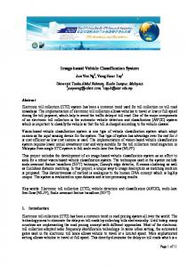
FIG 2. Classification system of cervical spondylitic myelopathy. Contribution of static factors (degenerative stage) and dynamic factors (flexion and extension) in the pathogenesis of cervical spondylitic myelopathy on kinematic MR images. At stage 0, physiological alignment of the cervical spine can be observed. At stage I, in patients with disk herniation or protrusion and preserved integrity of the cervical spine, subarachnoid space narrowing can be seen with no cord impingement with flexion and extension; in patients with cervical spondylosis (stage II), only anterior cord impingement can be seen with flexion and extension. At this stage, no posterior cord encroachment or pincer effect is evident. At stage III, in patients with cervical spondylosis and restricted motion of the involved segment, anterior or posterior cord impingement and different kinds of pincer effects (see also Fig 6 and Table 5) can be observed, because of secondary instability of adjacent levels. At stage IV, in patients with cervical spondylitic myelopathy, cord impingement can be diagnosed from the anterior aspect, the posterior aspect, or both anterior and posterior aspects (pincer effects) at one or multiple levels, adjacent or apart from the primarily involved segment (see also Fig 6 and Table 5). If only anterior cord compression (top row) or no cervical cord compression (bottom row) is seen at the level of the intramedullary lesion at the neutral position, cord impingement might be obvious with extension of the cervical neck. Note oval intramedullary lesion at stage IV relates to cystic degeneration (ie, cervical spondylitic myelopathy).
AJNR: 19, October 1998
CERVICAL SPONDYLITIC MYELOPATHY
1765
Results Grade of Spinal Stenosis at the Neutral Position (0°) during Flexion and Extension According to the Stage of Degenerative Disease
FIG 3. Evaluation of spinal stenosis at the neutral position (0°) in extension and flexion using grading system (11). Grade 0, normal width of the spinal canal, no signs of anterior and posterior subarachnoid space narrowing; grade 1, partial obliteration of the anterior or posterior subarachnoid space or of both; grade 2, complete obliteration of the anterior or posterior subarachnoid space or of both; grade 3, anterior or posterior cord impingement or both (pincer effect).
system based on pathoanatomic observations described by Kirkaldy-Willis (16) and by Handal et al (17). This classification system (“degenerative cascade”) describes different stages of degenerative disease, as follows (Fig 2): stage I, diskogenic phase, characterized by disk degeneration, disk protrusion, and disk herniation without osteophytic formation; stage II, spondylosis, with disk degeneration, disk protrusion, and disk herniation as well as initial osteophytic formation; and stage III, stabilization, with bridging osteophytes and immobilization of the involved segment. In addition to these stages, stage 0, representing normal findings of the cervical spine based on previously published data of kinematic MR imaging studies of healthy subjects, and stage IV, representing cervical spondylitic myelopathy, were added to the classification system (10). In this system, degenerative changes of the joints of Luschka and the facet joints were not evaluated and did not contribute to the stage of degenerative disease. Grade of Spinal Stenosis at the Neutral Position (0°) during Flexion and Extension on Kinematic MR Imaging.—To ascertain the dynamic changes of spinal stenosis during flexion and extension relative to the neutral position (0°), the extent of spinal stenosis for each patient was determined for each segment at the neutral position (0°) and at maximum flexion and extension, according to the grading system (11) described in Figure 3.
Statistical Analysis Student’s t-test was used to evaluate statistically significant differences with regard to changes in spinal stenosis evident among the different stages of degenerative disease. Significant results were assumed on the 5% level (P # .05). Inter- and intraobserver agreement was expressed for the stage of degenerative disease and for the grade of spinal stenosis at the neutral position (0°) and at maximum flexion and extension using the k statistic. Agreement was said to be almost perfect when k was greater than .80, good when k 5 .80 to .61, moderate when k 5 .60 to .41, fair when k 5 .40 to .21, and poor when k was less than .20 (18).
Thirteen patients had stage I disease, 42 patients had stage II disease, 11 patients had stage III disease, and 15 patients had stage IV disease. Stage I.—At the neutral position (0°), degenerative changes were observed in 31 segments, with cervical compression seen in six patients (46%) and seven segments (22%) (Table 1). In this group, anterior cervical cord compression was noted in seven segments, with cord impingement at one segment in five patients and at two segments in one patient (Fig 4). In none of the patients was functional cord impingement noted at flexion and extension (Table 2). In three (23%) of 13 patients, subarachnoid space narrowing was diagnosed at flexion and extension, with no change in spinal stenosis in 10 (77%) of 13 patients. A decrease of spinal stenosis was found in five patients at flexion, with cervical cord decompression seen in one patient (Table 3). In comparison with flexion, at extension, widening of the cervical canal was observed in only one patient. Stage II.—Of 42 patients with stage II disease, at the neutral position (0°), anterior cervical compression was diagnosed in 11 patients (26%) and 11 segments (9%) (Table 1, Fig 5). During flexion and extension MR imaging, no change in spinal stenosis was found in 15 patients (36%), and an increase of subarachnoid space narrowing was observed in 22 patients (52%). In addition to cervical cord compression at the neutral position (0°), anterior cervical cord impingement was diagnosed in five patients (12%), occurring in four patients (9%) at extension and in one patient (2%) at flexion (Table 2). At this stage, no functional pincer effect or posterior cord compression was observed. Widening of the cervical canal was observed at flexion in 26 patients (62%) and at extension in five patients (12%) (Table 3). In five patients, anterior cervical cord decompression was seen at flexion. Stage III.—Of 11 patients with stage III disease, at the neutral position (0°), cervical cord compression was diagnosed in six patients (54%) either from the anterior aspect (n 5 5) or from both the anterior and posterior aspects (n 5 3) at one (n 5 4) or two (n 5 2) segments (Table 1). In all patients, restricted motion of the involved cervical segment was accompanied by instability of one or more adjacent segments on kinematic MR imaging. In addition to static cord compression (n 5 6), functional cord impingement was observed at flexion in one (9%) of 11 patients and at extension in eight (73%) of 11 patients at one or multiple segments (Table 2). With regard to the site of functional cord compression, in five patients, cervical cord impingement resulted from the anterior aspect, the posterior aspect, or both the anterior and posterior aspects (pincer effect). At stages III and IV, different
1766
MUHLE
AJNR: 19, October 1998
TABLE 1: Number of patients with degenerative changes according to the stage of degenerative disease at neutral position (0°)
Degenerative Stage (n 5 81)
No. (%) of patients with Grade 3 Cord Compression
I (n 5 13) II (n 5 42) III (n 5 11) IV (n 5 15)
No. of Patients with Grade 3 Cord Compression at Segment:
No. of Segments with Grade 3 Cord Compression from:
Anterior Aspect
Posterior Aspect
Anterior and Posterior Aspects
1
2
3
7 11 5 7
... ... ... 5
... ... 3 7
5 11 4 8
1 ... 2 4
... ... ... 1
6 (46) 11 (26) 6 (54) 13 (87)
FIG 4. 26-year-old woman with disk herniation at C5 and small disk protrusion at C6 (stage I). At the neutral position (0°) and at 30° of extension, cervical cord compression (curved arrow) can be seen. At flexion, there is increased compression and stretching on the cervical cord (straight arrow) owing to narrowing of the anterior subarachnoid space and anterior shifting of the cervical cord. There was no change in the patient’s management with this additional information obtained at kinematic MR imaging.
TABLE 2: Number of patients and segments with cervical cord impingement at flexion and extension, from anterior, posterior, or both anterior and posterior (pincer effect) aspects at flexion and extension, and at one or multiple segments at flexion and extension according to the stage of degenerative disease No. (%) of patients with Grade 3 Cord Impingement at: Degenerative Stage (n 5 81)
I (n 5 13) II (n 5 42) III (n 5 11) IV (n 5 15)
Flexion from: Flexion Extension
1 (2) 1 (9) 2 (13)
4 (9) 8 (73) 12 (80)
No. of Patients with Grade 3 Cord Impingement at:
No. of Segments with Grade 3 Cord Impingement at:
Anterior Posterior Aspect Aspect
1 1 3
... ... ...
Flexion at Segment:
Extension from:
Anterior and Anterior and Anterior Posterior Posterior 1 Posterior Aspect Aspect Aspects Aspects ... ... ...
4 5 1
... 5 8
... 5 14
2
3
Extension at Segment: 4
1 2
3
4
1 ... ... ... 3 1 ... ... 1 ... ... ... 2 5 1 ... 1 1 ... ... 3 5 3 1
AJNR: 19, October 1998
CERVICAL SPONDYLITIC MYELOPATHY
1767
TABLE 3: Number of patients and segments with decrease of spinal stenosis at flexion and extension according to the stage of degenerative disease No. (%) of Patients with Decrease of Spinal Stenosis at: Degenerative Stage (n 5 81)
I (n 5 13) II (n 5 42) III (n 5 11) IV (n 5 15)
No. of Segments with Cord Decompression at: Flexion from:
Flexion
5 26 7 13
Extension
38 62 64 87
1 5 1 ...
8 12 9 ...
Extension from:
Anterior Aspect
Posterior Aspect
Anterior and Posterior Aspects
Anterior Aspect
Posterior Aspect
Anterior and Posterior Aspects
1 5 1 2
... ... 3 6
... ... ... 2
... ... ... ...
... ... ... ...
... ... ... ...
FIG 5. 28-year-old patient with cervical spondylosis (stage II). On kinematic MR images at the neutral position (0°), partial obliteration of the anterior subarachnoid space at C6 –C7 is noted. At flexion, there is widening of the posterior subarachnoid space (arrowheads), with anterior cord compression (curved arrow), caused by narrowing of the subarachnoid space and anterior shifting of the cervical cord. At extension, no changes are evident relative to the neutral position (0°).
kinds of pincer effects were observed (Fig 6). In contrast, a decrease of spinal stenosis was observed at flexion in seven patients (64%) as compared with only one patient (9%) at extension (Table 3). Stage IV.—Among patients with stage IV disease, at the neutral position (0°), cervical cord impingement was observed in 13 patients (87%) in 19 segments, from the anterior aspect in seven segments, from the posterior aspect in five segments, and from both anterior and posterior aspects (pincer effect) in seven segments (Table 1). In addition to cervical cord compression seen in 13 patients at the neutral position (0°), functional cord impingement was found in 12 patients (80%) at extension and in two patients (13%) at flexion (Table 2). In this group, at extension, a pincer effect was observed in 14 segments, posterior cervical cord encroachment in eight segments, and anterior cervical
cord compression in one segment (Figs 7 and 8). In contrast, at flexion, cervical cord impingement was diagnosed only from the anterior aspect in three segments. With regard to the location of the intramedullary lesions, kinematic MR imaging showed functional cord impingement in four of six patients, whereas no cervical cord compression was seen at the neutral position (0°). In addition, a pincer effect was observed at extension in four of five patients with cervical cord compression seen only from the anterior or posterior aspect at the neutral position (0°) (Fig 7). Furthermore, functional cord impingement was observed either adjacent to the level of cervical cord compression at static examination (n 5 4) or at levels more than one segment apart (n 5 5) (Fig 8). In contrast, widening of the subarachnoid space was found at flexion in 13 patients (87%), with cervical cord decompres-
1768
MUHLE
AJNR: 19, October 1998
good agreement in comparisons of the grade of cervical spinal stenosis at positions of maximum flexion and extension relative to the neutral position (0°) and stage of degenerative disease (Table 4).
Discussion
FIG 6. Pincer effects observed at flexion and extension using kinematic MR imaging. At flexion, compared with the neutral position (0°), cervical cord impingement can be observed from the posterior aspect, owing to anterolisthesis of the superior vertebra relative to the adjacent vertebra below. At extension, different kinds of pincer effects can be noted. a), As compared with the neutral position (0°), cord compression from the posterior margin of one vertebral body (or osteophyte, or disk) and the lamina or ligamentum flavum of the adjacent inferior segment is noted above the anterior abnormality. b), As compared with the neutral position (0°), cord compression from the posterior margin of one vertebral body (or osteophyte, or disk) and the lamina or ligamentum flavum of the adjacent inferior segment is noted at the same level as the anterior abnormality. c), At extension, cord compression can be observed from the posterior margin of one vertebral body (or osteophyte, or disk) and the lamina or ligamentum flavum of the next inferior segment below the level of the anterior abnormality. d), Multiple pincer effects: cord impingement can be seen from the posterior margins of two or more vertebral bodies (or osteophytes, or disks) and the laminae or ligamenta flava of the adjacent levels.
sion seen from an anterior aspect, a posterior aspect, or both, in 10 of 19 involved segments (52%) (Table 3; Figs 7 and 8). In contrast, no decrease (0%) of spinal stenosis was seen at extension. Independent of the stage of degenerative disease (I–IV), in all patients except one, functional cord compression was observed between 30° of flexion and 30° of extension. The number of patients with functional cord impingement was greater at stage IV than at stage II (P # .05), at stages IV/III than at stage I (P # .001), and at stage II than at stage I (P # .05). At extension, posterior or both anterior and posterior cord impingement (pincer effect) was greater at stage IV than at stages I/II, and at stage III than at stages I/II (P # .001). In the assessment of patients with functional cord impingement at one or more segments, statistically significant differences were observed between stages IV/III and stages I/II (P # .01). There was also a statistically significant association between the decrease of spinal stenosis at flexion as compared with extension at all degenerative stages (I–IV) (P # .01). Intra- and interobserver tests (k statistic) showed
Cervical spondylitic myelopathy results from segmental or generalized degenerative disease of the cervical spine. It has been proposed that a congenitally narrowed cervical spinal canal, progressive spondylosis, mechanical spinal cord compression, or alterations in vascularity of the spinal cord contribute to the development of cervical spondylitic myelopathy (19). As our results suggest, these factors become increasingly important in the development or deterioration of cervical spondylitic myelopathy during cervical spinal motion. Considering that the cervical spine is a dynamic, mobile structure, our studies show that in addition to cervical cord compression at the neutral position (0°), in a number of patients, functional cord impingement also can be observed in early stages of degenerative disease (Tables 2 and 5). Because of the preserved flexibility and integrity of the cervical spine in the initial stages of disk degeneration, functional cord impingement was not observed at stage I at flexion and extension. In patients with herniated disks and cord compression at the neutral position (0°), however, our studies showed increased compression and stretching of the cord at flexion, as was described previously in cadaveric and myelographic investigations (8, 20 –22). With loss of disk height and osteophytic formation, segmental instability may occur during cervical spinal motion (17, 19). At this stage, as was demonstrated with kinematic MR imaging, cervical cord impingement can be seen only from an anterior aspect at flexion and extension (Table 5). There are several reasons for this. At flexion, the cervical cord follows a direct path through the cervical canal (20 –22). Physiologically, this may result in up to 43% narrowing of the anterior subarachnoid space at flexion with simultaneous widening in up to 40% to 90% of the posterior subarachnoid space (10). In the presence of osteophytes and increased intersegmental motion, these structures can bulge into the cervical cord at flexion and cause neurologic damage (8, 21, 23). At extension, as has been described by different authors, backward gliding of the vertebrae may result in narrowing of the anterior subarachnoid space or even cervical cord encroachment at one or more cervical segments, independent of the grade of anterior subarachnoid space narrowing at the neutral position (8, 10, 20 –22). At stage III, osteophytes and protruding disks can lead to restricted motion of the involved cervical segment with secondary instability of the adjacent cervical levels. To increase joint stability, secondary changes, such as facet joint arthrosis, laminar hypertrophy, and thickening of the ligamentum flavum, may occur (17). Since these structures are positioned centrally, the posterior space available for the cervical
AJNR: 19, October 1998
CERVICAL SPONDYLITIC MYELOPATHY
1769
FIG 7. 72-year-old patient with spondylitic cervical myelopathy (stage IV). T2-weighted turbo spin-echo kinematic MR images show cervical cord atrophy with pathologic signal changes at C4 with anterior cervical cord compression at the neutral position (0°) (thick straight arrow). At flexion, functional cord impingement with increased stretching of the cervical cord is noted at C5 (curved arrow). At extension, a pincer effect is observed at C4, corresponding to the intramedullary lesion (thin arrows). Note also posterior cord encroachment due to a thickened ligamentum flavum at C7 (arrowhead) and posterior subarachnoid space narrowing at C7. Because of the kinematic MR imaging findings, the patient underwent laminectomy with anterior fusion rather than anterior decompression surgery with fusion.
FIG 8. 56-year-old patient with cervical spondylitic myelopathy and restricted motion at C6 and C7 (stage IV). At the neutral position (0°), cervical cord compression with intramedullary signal abnormalities is seen at the level of C4. At the neutral position (0°), spinal stenosis is recognized from C3 to T2, with cord compression at multiple levels. At flexion, a widening of the posterior subarachnoid space is noted. At extension, multiple pincer effects (straight arrows) can be observed at C4, C5, and C6. Note also increased spinal stenosis due to a thickened ligamentum flavum (curved arrow) at T2. Because of increased spinal stenosis at extension with pincer effects observed at multiple levels, the patient underwent laminectomy rather than anterior decompression surgery.
1770
MUHLE
AJNR: 19, October 1998
TABLE 4: Comparison of the grade of cervical spinal stenosis at maximum flexion and extension with neutral position (0°) according to the stage of degenerative disease: k value of intra and interobserver variability
k Value Evaluation
Intraobserver Variability
Interobserver Variability
Stage of degenerative disease Grade of spinal stenosis
.723 .823
.682 .756
aging studies are needed to verify the potential of each pincer effect to cause cervical cord damage. In contrast, at flexion, a pincer effect was observed only in one patient at C3–C4, and this was due to anterior degenerative spondylolisthesis. This observation, as well as the finding that in a large number of patients a decrease in spinal stenosis with even cord decompression was observed at flexion, may indicate a minor role of flexion in contributing to cervical spondylitic myelopathy (24).
TABLE 5: Classification system of cervical spondylotic myelopathy: contribution of static factors (degenerative stage) and dynamic factors (flexion and extension) to pathogenesis of disease as determined with use of kinematic MR imaging Degenerative Stage
Description Degenerative Stage (Static Factors)*
Flexion and Extension (Dynamic Factors)
0
Physiological disk height, no evidence of disk degeneration or osteophyte formation
Physiological alignment of the cervical spine at flexion and extension; narrowing of the anterior space at flexion and widening at extension; widening of the posterior subarachnoid space at flexion with narrowing at extension; cervical cord lengthening and narrowing at flexion with shortening and widening at extension (10)
I
Disk degeneration, disk protrusion, disk herniation; no evidence of osteophyte formation; anterior cord compression possible
Kyphotic malalignment of the involved cervical segment at neutral position (0°) possible; subarachnoid space narrowing at flexion and extension possible; no functional cord impingement at flexion and extension
II
Cervical spondylosis; intervertebral osteochondrosis; anterior cord compression possible
Segmental instability with possibility of anterior cord impingement at flexion and extension; no posterior cord impingement or pincer effect
III
Cervical spondylosis; invertebral osteochondrosis with bridging osteophytes; anterior or anterior and posterior cord impingement possible
Segmental immobility or restricted motion, with secondary instability of the adjacent segments with possibility of anterior, posterior, or anterior and posterior cord impingement (functional pincer effect); different kinds of pincer effects at stages III and IV
IV
Cervical spondylotic myelopathy; anterior, posterior, or anterior and posterior cord encroachment possible
If no cord compression at the level of the intramedullary lesion is obvious, cord impingement can be seen in a number of patients at extension; additionally, cord impingement can be diagnosed from anterior, posterior, or anterior and posterior (functional pincer effect) at one or multiple levels, adjacent or apart from the primarily involved segment
* Note.—Conventional radiography, functional radiography, CT, CT-myelography, and static and kinematic MR imaging of the cervical spine can contribute to the definition of the degenerative stage.
cord decreases. Compared with functional cord encroachment, observed at extension in fewer than 10% of patients with stage II disease, functional cord impingement, in addition to static cervical cord compression, was observed at extension in 73% to 87% of patients with stage III or IV disease, mostly from the posterior aspect or the anterior and posterior aspects of the cervical spine. These pincer effects included the original descriptions recorded by Penning and Van der Zwaag (8) and other pincer effects that, to our knowledge, have not been described previously. Although we believe that the pincer effect contributes significantly to the development of cervical spondylitic myelopathy, further follow-up kinematic MR im-
The best evidence that cervical spinal motion may contribute to the pathogenesis of cervical spondylitic myelopathy was found in patients with stage IV degenerative disease (25, 26). On the assumption that a reliable diagnostic indicator of the genesis of spondylitic myelopathy is the demonstration of cord compression at the site of a cord lesion, kinematic MR imaging showed functional cord impingement in four of six patients, whereas no cervical cord compression was seen at the neutral position. In addition, a pincer effect was seen at extension in the location of intramedullary lesions in four of five patients with cervical cord compression only from the anterior or posterior aspect at the neutral position (8). Therefore, it
AJNR: 19, October 1998
CERVICAL SPONDYLITIC MYELOPATHY
can be assumed that patients with stages III or IV disease may have a higher risk of initial or additional cord damage due to an increased prevalence of static and dynamic cervical cord encroachment than do patients with stage I or II changes, who have no or less static and functional cervical cord compression. Our study has several limitations. The patients were positioned supine inside a device, and the results may not be identical when patients are examined in the upright position; however, previous comparative studies of functional radiographs obtained with the patient in the upright position and kinematic MR images have demonstrated no significant differences in segmental motion of the cervical vertebrae (10). Second, despite the fact that a comparative study of functional myelography and kinematic MR imaging was not possible, our preliminary myelographic results in six patients showed a similar grade of spinal stenosis as that seen on kinematic MR images. Third, although the grade of spinal stenosis at the neutral position (0°) was verified in all patients who underwent decompression surgery, no verification of functional cervical spinal stenosis could be obtained at operation.
Conclusion Kinematic MR imaging showed an increasing prevalence of functional cord impingement from the posterior aspect and from both anterior and posterior aspects (pincer effect) during extension of the neck in patients at increasing stages of degenerative disease of the cervical spine. In contrast, at flexion, cervical cord decompression was observed in a large number of patients. Our observations suggest that, in addition to static factors, cervical spinal motion contributes to cervical cord compression and to the pathogenesis of cervical spondylitic myelopathy (27). The extent to which kinematic MR imaging of the cervical spine might influence the evaluation of cervical spondylosis is a subject for further investigation.
References 1. Tarlov IM, Klinger H, Vitale S. Spinal cord compression studies, I: experimental techniques to produce acute and gradual compression. Arch Neurol Psychiatry 1953;70:813– 819 2. Tarlov IM, Klinger H. Spinal cord compression studies, II: time limits for recovery after acute compression in dogs. Arch Neurol Psychiatry 1954;71:271–290 3. Taylor AR. Mechanism and treatment of spinal-cord disorders associated with cervical spondylosis. Lancet 1953;1:717–723 4. Gooding MR, Wilson CB, Hoff JT. Experimental cervical myelopathy: effects of ischemia and compression of the canine cervical
1771
spinal cord. J Neurosurg 1975;43:9 –17 5. Adams CBT, Logue V. Studies in cervical spondylitic myelopathy, I: movement of the cervical roots, dura and cord, and their relation to the course of the extrathecal roots. Brain 1971;94:557–568 6. Guidetti B, Fortuna A. Long-term results of surgical treatment of myelopathy due to cervical spondylosis. J Neurosurg 1969;30:714 – 721 7. Olsson SE. The dynamic factor in spinal cord compression: a study on dogs with special reference to cervical disc protrusion. J Neurosurg 1958;15:308 –321 8. Penning L, Van der Zwaag P. Biomechanical aspects of spondylitic myelopathy. Acta Radiol 1966;5:1090 –1103 9. Chen IH, Vasavada A, Panjabi MM. Kinematics of the cervical spine canal: changes with sagittal plane loads. J Spinal Disord 1994;7:93–101 10. Muhle C, Wiskirchen J, Weinert D, et al. Biomechanical aspects of the subarachnoid space and cervical cord in healthy subjects using kinematic MR imaging. Spine 1998;23: 11. Muhle C, Wiskirchen J, Brinkmann G, et al. Kinematische MRT bei degenerativen Halswirbelsa ¨ulenvera ¨nderungen (abstract in English). Fortschr Rontgenstr 1995;163:148 –154 12. Muhle C, Melchert UH, Brossmann J, Schro ¨der C, Wiskirchen J, Heller M. Positionsgestell zur kinematischen Magnetresonanztomographie der Halswirbelsa ¨ule (abstract in English). Fortschr Rontgenstr 1995;162:252–254 13. Sze G, Kawamura Y, Negishi C, et al. Fast spin-echo MR imaging of the cervical spine: influence of echo train length and echo spacing on image contrast and quality. AJNR Am J Neuroradiol 1993;14:1203–1213 14. Constable RT, Smith RC, Gore JC. Signal-to-noise and contrast in fast spin echo (FSE) and inversion recovery FSE imaging. J Comput Assist Tomogr 1992;16:41– 47 15. Jones KM, Mulkern RV, Schwartz RB, Oshio K, Barnes PD, Jolesz FA. Fast spin-echo MR imaging of the brain and spine: current concepts. AJR Am J Roentgenol 1992;158:1313–1320 16. Kirkaldy-Willis WH. Managing Low Back Pain. 2nd ed. New York: Churchill Livingstone; 1988:117–143 17. Handal JA, Knapp J, Poletti S. The structural degenerative cascade: the cervical spine. In: White AH, ed. Spine Care: Diagnosis and Conservative Treatment. St Louis: Mosby; 1995:16 –26 18. Davies M, Fleiss JL. Measuring agreement for multinominal data. Biometrics 1982;38:1047–1051 19. Abdu WA. Pathophysiology of cervical spondylitic myelopathy. In: Saunders RL, Bernini PM, eds. Cervical Spondylitic Myelopathy. Cambridge, MA: Blackwell; 1992:18 –28 20. Breig A. Biomechanics of the Central Nervous System. Stockholm: Almquist & Wiksell; 1961 21. Breig A, Turnbull I, Hassler O. Effects of mechanical stresses on the spinal cord in cervical spondylosis. J Neurosurg 1966;25:45–56 22. Breig A. Overstretching and circumscribed pathological tension in the spinal cord: a basic cause of symptoms in cord disorders. J Biomech 1970;3:7–9 23. Waltz TA. Physical factors in the production of myelopathy of cervical spondylosis. Brain 1967;90:395– 404 24. Boulos AS, Lovely TJ. Degenerative cervical spondylolisthesis: diagnosis and management in five cases. J Spinal Disord 1996;9: 241–245 25. Bohlmann HH. Cervical spondylosis with moderate to severe myelopathy: a report of 17 cases treated by Robinson anterior cervical discectomy and fusion. Spine 1977;2:151–162 26. Hayashi H, Okada K, Hashimoto J, Tada K, Ueno R. Cervical spondylitic myelopathy in the aged patient: a radiographic evaluation of the aging changes in the cervical spine and etiologic factors of myelopathy. Spine 1983;3:618 – 625 27. White AW, Panjabi MM. Clinical Biomechanics of the Spine. 2nd ed. Philadelphia: Lippincott; 1990:666
Please see the Editorial on page 1594 in this issue.