Clinical Notes Active pulmonary tuberculosis in a patient with Langerhans’ cell histiocytosis Oguzhan Okutan, MD, Zafer Kartaloglu, MD, Ahmet Ilvan, MD, Omer Deniz, MD, Emir Silit, MD, Rauf Gorur, MD.
L
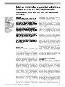
angerhansʼ cell histiocytosis (LCH) or pulmonary histiocytosis is an uncommon parenchymal lung disease of unknown origin. The lesions of LCH contain single of multifocal proliferation of histiocytes similar in phenotype to dendritic Langerhansʼcell.1 The natural history is extremely a variable. The disorder ranges in clinical severity from a solitary eosinophilic granuloma of bone to a generalized disease with multiple organ involvement. Rarely, LCH has been reported to be associated with some disorders such as pulmonary tuberculosis.2 Here, we report a cases of a 21-year-old male patient with LCH and active pulmonary tuberculosis. A 21-year-old male patient was hospitalized due to complaints of progressive dyspnea in the last 6 months, occasional cough and night sweats. There was nothing marked in the medical history of the patient who was working in tourism industry. He had 9 years smoking history. Physical examination of respiratory system and other systems were normal. Routine hematological studies were normal; there was no peripheral eosinophilia, the erythrocyte sedimentation rate was 35 mm/h. Other hematological and biochemical data on admission were within normal ranges. Postero-anterior (PA) chest x-ray revealed extended heterogeneous reticulonodular infiltration in both lung fields. Thorax high resolution computed tomography (HRCT) revealed a reticulonodular infiltration involving all lung areas in which there were thin walled cavitary lesions among those the biggest one was 10 mm in diameter (Figure 1). Nonspecific culture of sputum revealed normal oral flora. Tuberculin skin test was negative. Acid-fast bacilli (AFB) was not detected in the sputum, in gastric lavage, and in bronchial washing fluid under microscopic examination. A fiber optic bronchoscopy was performed. There was no endobronchial pathology. Brochoalveolar lavage fluid examination and cell distribution were nondiagnostic. Transbronchial biopsy specimens showed normal lung mucosa and parenchyma. Pulmonary function tests (PFT) revealed restrictive disease. The PFT presented the following values: Vital capacity (VC), 3.45 L (62% pred); forced vital capacity (FVC), 2.85 L (56% pred); FEV1, 2.79 (78% pred);
Figure 1 - Thorax high resolution computed tomography scan revealed thin walled cavitary lesions in the upper lung fields among those the biggest one was 10 mm in diameter and micronodular densities.
and peak expiratory flow rate, 6.25 L/sec (71% pred). Diffusion capacity was lowered (4.9 mmol/min/kPa, 62% pred). The patient underwent a video assist thoracic surgery for definite diagnosis. Histopathological examination of the specimen of biopsy revealed many eosinophils, histiocytes and lymphocytes all of which showed centrilobular distribution pattern consisting of dense chronic inflammation, edema, and fibrosis. Muscular hyperplasia and dilatation were detected. Immunohistochemical study of S-100 protein of biopsy specimen showed S-100 protein positive histiocytes in granulomatous lesions. In addition, moderate interstitial fibrosis was seen with trichrome staining. Histological findings were compatible with LCH. There was no clinical or laboratory finding compatible with LCH in other organs. Abdominal ultrasound was normal. During laboratory evaluation Mycobacterium tuberculosis was isolated on culture media. The final diagnosis was LCH and pulmonary tuberculosis. Antituberculosis treatment with izoniazid, rifampicin, pyrazinamide and ethambutol was started. Corticosteroids were not used for the treatment. A repeat thorax HRCT examination after 6 months from the beginning of the treatment revealed marked improvement in nodular lesions, and thin walled cystic lesions in the upper lung fields. Marked regression was detected for micro nodular opacities in both lung fields. Our patient was diagnosed with pulmonary LCH proven through lung biopsy and AFB culture positive pulmonary tuberculosis. Antituberculosis treatment resulted in an objective radiological improvement and AFB culture negativity. The clinical course of LCH ranges from spontaneous resolution to a chronic and sometimes lethal disease. The etiology of LCH remains obscure, but the vast www.smj.org.sa
Saudi Med J 2006; Vol. 27 (3)
401
Tuberculosis and Langerhansʼ cell histiocytosis
majority (up to 97%) of patients are current smokers or ex-smokers. Although, spontaneous improvement in patients with LCH has been described, the role of smoking cessation in the natural history remains controversial. After diagnosis, our patient stopped smoking. In pulmonary LCH, chest x-ray usually reveals a diffuse reticulonodular pattern with micronodules measuring 2-5 mm in diameter and pulmonary cysts that predominate in the upper lobes. However, in occasional cases chest x-ray can be normal3 and CT scan of the lung may disclose focal or diffuse abnormalities.4 In pulmonary LCH cases, it was observed that nodular opacities, thick-walled cysts, and ground-glass opacities underwent regression. Thin-walled cysts, linear opacities, and emphysematous lesions remained unchanged or progressed.3 In active pulmonary tuberculosis cases, CT scan reveals combination finding of centrilobular nodule or branching linear structure, bronchial wall thickening.5 In the present case, HRCT scan revealed a reticulonodular infiltration involving all lung areas in which there were thin-walled cavitary lesions. Due to the high incidence of spontaneous remission in untreated patients, the efficiency of different immunosuppressive therapeutic strategies for pulmonary LCH remains unknown. Since severe impairment of pulmonary function at the time of diagnosis has negative prognostic value in patients with LCH6 an early intervention may be beneficial in terms of survival. In our patient, we have shown substantial clinical and radiological improvement after antituberculosis treatment, and cessation of smoking without corticosteroid treatment. It was not revealed if the improvement were due to antituberculosis treatment or cessation of smoking. Reviewing the literature, we identified only one published LCH case with pulmonary tuberculosis.2
402
Saudi Med J 2006; Vol. 27 (3)
www.smj.org.sa
In summary, this case demonstrated that LCH could be associated with active pulmonary tuberculosis. Thus, it should be kept in mind that LCH and pulmonary tuberculosis might exist as concomitant disorders in the same patient particularly in areas where prevalence of tuberculosis is high. Received 11th September 2005. Accepted for publication in final form 31st December 2005. From the Departments of Chest Diseases (Okutan, Kartaloglu, Ilvan, Deniz), Radiology (Silit), and Thoracic Surgery (Gorur), GATA Haydarpasa Training Hospital, Istanbul, Turkey. Address correspondence and reprint requests to: Dr. Oguzhan Okutan, GATA Camlica Gogus Hastaliklari Hastanesi, 81020 Acibadem, Istanbul, Turkey. Tel. +90 (216) 3257250. Fax. +90 (216) 3257257. E-mail:
[email protected]
References 1. Willman CL, Busque L, Griffith BB, Favara BE, McClain KL, Duncan MH, et al. Langerhansʼ cell histiocytosis (histiocytosis X). A clonal proliferative diseases. N Eng J Med 1994; 331: 154-160. 2. Arai T, Inoue Y, Yamamoto S, Akira M, Uesugi H, Hayashi S, et al. Incipient stage of pulmonary Langerhans-cell histiocytosis complicated with pulmonary tuberculosis was examined by high-resolution computed tomography. Respir Med 2005; 99: 1188-1190. 3. Schonfeld N, Frank W, Wening S, Uhrmeister P, Allica E, Preussler H, et al. Clinical and radiological features, lung function and therapeutic results in pulmonary histiocytosis X. Respiration 1993; 60: 38-44. 4. Brauner MW, Grenier P, Tijani K, Battesti JP, Valeyre D. Pulmonary Langerhansʼ cell histiocytosis: evolution of lesions on CT scans. Radiology 1997: 204: 497-502. 5. Lee KS, Hwang JW, Chung MP, Kim H, Kwon OJ. Utility of CT in evaluation of pulmonary tuberculosis in patients without AIDS. Chest 1996; 110: 977-984. 6. Delobbe A, Durieu J, Duhamel A, Wallaert B. Determinants of survival in pulmonary Langerhansʼ cell granulomatosis (histiocytosis X). Eur Respir J 1996; 9: 2002-2006.