J. Microbiol. Biotechnol. (2011), 21(5), 515–518 doi: 10.4014/jmb.1010.10061 First published online 8 March 2011
Cloning and Expression of a bpr Gene Encoding Bacillopeptidase F from Bacillus amyloliquefaciens CH86-1 Kwon, Gun-Hee1, Jae-Yong Park2, Jong-Sang Kim3, Jinkyu Lim3, Cheon-Seok Park4, Dae Young Kwon5, and Jeong Hwan Kim1,6* 1
Division of Applied Life Science (BK21 Program), Graduate School, Gyeongsang National University, Jinju 660-701, Korea Department of Food Science and Nutrition, Catholic University of Daegu, Gyeongsan 712-702, Korea 3 School of Applied Biosciences, Kyungpook National University, Daegu 702-701, Korea 4 Department of Food Science and Biotechnology, Kyung Hee University, Yongin 446-701, Korea 5 Food Function Research Division, Korea Food Research Institute, Sungnam 463-746, Korea 6 Research Institute of Life Sciences, Gyeongsang National University, Jinju 660-701, Korea 2
Received: October 27, 2010 / Revised: December 30, 2010 / Accepted: February 12, 2011
A gene encoding bacillopeptidase F, bpr86-1, was cloned from B. amyloliquefaciens CH86-1 isolated from cheonggukjang. This gene could encode a preproenzyme of 1,431 amino acids. When bpr86-1 was introduced into B. subtilis WB600 via pHY300PLK, an E. coli-Bacillus shuttle vector, the transformant showed fibrinolytic activity. During growth on LB, the fibrinolytic activity of cells increased sharply when they entered the stationary phase. The highest activity (761.4 mU/mg protein) was observed at 96 h of cultivation. Keywords: Bacillus amyloliquefaciens CH86-1, bacillopeptidase F, bpr gene, fibrinolytic enzymes
Bacillus amyloliquefaciens CH86-1, which was isolated from cheonggukjang, a traditional Korean fermented soyfood, secretes several proteases with fibrinolytic activity [4]. Among these proteases, AprE86-1, a 27-kDa enzyme in its mature form, is the main fibrinolytic enzyme and is encoded by aprE86-1, a homolog of aprE from Bacillus subtilis [6, 7]. This gene shares significant homology with nattokinase genes from B. subtilis and closely related species [6]. B. amyloliquefaciens CH86-1 secretes other minor fibrinolytic proteases, the presence of which can be demonstrated by fibrin zymography of culture supernatants [4] (see also Fig. 3). Bacillopeptidase F is one of the minor proteases secreted by B. subtilis and other species [2]. Wu et al. [11] reported that bacillopeptidase F of B. subtilis is synthesized as a preproenzyme, and that small proteins are *Corresponding author Phone: +82 557721904; Fax: +82 557721909; E-mail:
[email protected]
generated through processings at both the amino and carboxyl termini [11]. Yamagata et al. [13] found that a 90-kDa serine protease, secreted by B. subtilis (natto) No. 16, might be a degradation product from the 133-kDa mature enzyme derived from a preproenzyme (1,433 amino acids). Although proteases are generally believed to help cells survive in adverse environments by providing peptides and amino acids from denatured or unnecessary proteins [8, 10], the exact role and contribution of each enzyme is largely unknown. We cloned a gene encoding bacillopeptidase F from the chromosome of B. amyloliqufaciens CH86-1 by PCR. Primers were designed based on the bpr sequence of B. amyloliquefaciens FZB42 for which the genome sequence is available. PCR was performed with Ex-Taq polymerase (Takara, Shiga, Japan) using an MJ Mini PCR system (BioRad, Hercules, CA, USA) and the primers bprF (5'-GCGGATCCATGATGCCCTCGACATT-3', BamHI site underlined) and bprR (5'-CGTCTAGACATCAGAAGC ACAATGGCTG-3', XbaI site underlined). PCR conditions were as follows: initial denaturation at 95oC for 5 min, followed by 30 cycles of 95oC for 30 s, 60oC for 30 s, and 74oC for 2 min, and a final 4 min extension at 74oC. The 4,988 bp fragment thus amplified was cloned into pHY300PLK (Takara), a Bacillus-E. coli shuttle vector, resulting in pHYbpr86-1. DNA sequencing confirmed that the fragment contained bpr, and the gene was named bpr86-1 accordingly. The ORF of bpr86-1 begins at nucleotide (nt) 327 and ends at nt 4,622. It could encode a protein of 1,431 amino acids, designated Bpr86-1, and the pI and molecular mass of which were calculated to be 5.95 and 154,512.71 Da, respectively. The first 30 amino acids seem to constitute a signal peptide, as predicted by the SignalP 3.0 Server program, and comparision with other bacillopeptidase F
516
Kwon et al.
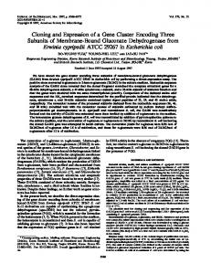
Fig. 1. Alignment of the first 320 amino acids of Bpr86-1 with other bacillopeptidase F sequences.
FJ432003 (B. amyloliquefaciens CH86-1); CP000560 (B. amyloliquefaciens FZB42); FN597644 (B. amyloliquefaciens DSM7); AL009126 (B. subtilis 168); D44498 [B. subtilis (natto) No. 16]. * indicates conserved amino acids among five sequences. Amino acids constituting the catalytic triad are marked with a rectangle. The presumed cleavage points for the pre- and pro-sequences are marked with an arrow. Other portions of the enzymes are shown (near the serine residue of the catalytic triad and the C-terminus).
sequences suggests that the next 166 amino acids might be a pro-sequence [13]. The first 320 amino acids of Bpr86-1 and portions of other regions translated from the DNA sequence are shown together with other bacillopeptidases
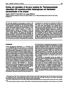
Fig. 2. Growth and fibrinolytic activities of bacilli.
in Fig. 1. Bpr86-1 shows 99%, 94%, 71%, and 70% homology to the enzymes from B. amyloliquefaciens FZB42, B. amyloliquefaciens DSM7, B. subtilis 168, and B. subtilis (natto) strain 16, respectively. The pI and the molecular
A, B. - ▲ -, B. amyloliquefaciens CH86-1; - ● -, B. subtilis WB600 [pHYbpr86-1]; - ○ -, and B. subtilis WB600 [pHY300PLK] (control). C. A fibrin plate showing fibrinolytic activity of cells. P denotes plasmin (Sigma) at 1.5 mU. Numbers indicate incubation time in h and (-) denotes the control. A standard curve was obtained at different plasmin concentrations (0.75-12 mU). Ten µl (20 µg) was spotted and the plate was incubated for 16 h at 37oC. All the measurements were repeated three times and the average values are shown.
BACILLOPEPTIDASE F GENE FROM BACILLUS AMYLOLIQUEFACIENS CH86-1
517
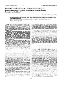
Fig. 3. SDS-PAGE (left) and fibrin zymogram (right) of culture supernatant.
M, protein size marker (DokDo-MARK, Elpisbio, Daejeon, Korea); B. amyloliquefaciens CH86-1 at 48 h (lane 1), 96 h (2), and 120 h (3); B. subtilis WB600 [pHYbpr86-1] at 48 h (4), 96 h (5), and 120 h (6); B. subtilis WB600 [pHY300PLK] at 48 h (7), 96 h (8), and 120 h (9). Fifteen µg was applied to a 10% acrylamide gel. Bands marked on the left are absent in the control.
mass of the expected mature enzyme are 5.06 and 132,658.36 Da, respectively. In most fibrinolytic serine proteases, three amino acids constituting the catalytic triad are conserved [9]. These residues (Asp33, His80, Ser258) were located in Bpr86-1, by comparision with other bacillopeptidase F sequences, and are marked in Fig. 1. The entire sequence of bpr86-1 was deposited in GenBank under the accession number FJ432003. pHYbpr86-1 was introduced into B. subtilis WB600, a strain lacking six protease activities [12], by electroporation. Bacillus competent cell preparation and electroporation procedures were performed as previously described [3]. B. subtilis TF (cells harboring pHYbpr86-1) and a B. subtilis control (cells harboring pHY300PLK) were grown in LBTc (tetracycline, 10 µg/ml) up to 120 h at 37oC with aeration (Fig. 2). B. amyloliquefaciens CH86-1 was grown in LB. At intervals, culture supernatant (70 ml) was obtained by centrifugation, and then filtered and precipitated with ammonium sulfate [80% (w/v)]. The pellet thus obtained was resuspended in 20 mM Tris-HCl (pH 8.0), dialyzed against the same buffer at 4oC overnight with four buffer changes, freeze-dried, and resuspended in a small volume of 20 mM Tris-HCl (pH 8.0). The protein concentration was determined by the Bradford method [1] and fibrinolytic activity was measured using a fibrin plate as previously described [3, 7]. The fibrinolytic activity of B. subtilis TF increased significantly during the stationary phase and reached the highest point at 96 h (Fig. 2). The control did not show any activity during the same period. TF showed a basal level of activity until 48 h, after which it increased sharply, reaching 533.8 (mU/mg protein) at 72 h, 761.4 at 96 h, and 528.9 at 120 h (Fig. 2). The control showed only 41.9 mU/mg protein at 120 h, which was less than 10% of the activity of TF. PCR-amplified brp86-1 contains its own promoter; expression of bpr86-1 seemed to occur under
the control of this promoter. The results showed that the synthesis of bacillopeptidase F was greatly induced when cells entered the stationary phase. The increase in bacillopeptidase F activity was parallel to the increase in total fibrinolytic activity of B. amyloliquefaciens CH86-1 cells during the stationary phase [7]. Thus, it is likely that the expression of all fibrinolytic proteases is induced when cells enter the stationary phase. SDS-PAGE and fibrin zymography were performed on the culture supernatant from TF and control cells as previously described [3, 5]. Bands corresponding to peptides of 90, 55, and 40 kDa, observed on an SDS-polyacrylamide gel of B. subtilis TF, were most likely derived from bacillopeptidse F (Fig. 3). On the fibrin zymogram, bands of approximately 45 and 17 kDa were observed in CH86-1 and B. subtilis TF, but were absent in the control. Larger derivatives of Bpr86-1 were difficult to locate in B. amyloliquefaciens CH86-1. This result may be due to the fact that Bpr86-1 is one of the minor proteases in B. amyloliquefaciens CH86-1 and its derivatives are unstable. It has been reported that smaller fragments are generated from bacillopeptidase F as the results of processing at either the N- or C-terminus or both [11, 13]. When 2D gel fibrin zymography was performed on the culture supernatant of B. amyloliquefaciens CH861, synthesis of proteases larger than 70 kDa (pI around 7.0) was strongly induced at the stationary phase, and the band intensity was higher at 100 h than at 67 h (results not shown). Bacillopeptidase F and its derivatives were probably responsible for enhancing activity at least partially. Enhanced bacillopeptidase F activity might be advantageous for host cells in that it enables them to adjust to adverse growth conditions in which available nutrients including proteins and peptides are limited. Detailed studies on bacillopeptidase F and its processed derivatives will be helpful in understanding its function and role in the overall fibrinolytic capability of
518
Kwon et al.
B. amyloliquefaciens CH86-1. The results will help us to develop industrial applications of bacillopeptidase F and identify producing organisms.
Acknowledgments This work was supported by a research grant from the Biofoods Research Program of the Korea Science and Engineering Foundation (KOSEF), and G.-H. Kwon was supported by the 2nd stage of the Brain Korea 21 Program of the Ministry of Education, Science and Technology (MEST, Korea). REFERENCES 1. Bradford, M. M. 1976. Rapid and sensitive methods for the quantification of microgram quantities of protein utilizing the principle of protein-dye binding. Anal. Biochem. 72: 248-254. 2. Choi, N.-S., D.-M. Chung, C. H. Ryu, K.-S. Yoon, P. J. Maeng, and S. H. Kim. 2006. Identification of three extracellular proteases from Bacillus subtilis KCTC 3014. J. Microbiol. Biotechnol. 16: 457-464. 3. Jeong, S.-J., G.-H. Kwon, J. Chun, J. S. Kim, C.-S. Park, D. Y. Kwon, and J. H. Kim. 2007. Cloning of fibrinolytic enzyme gene from Bacillus subtilis isolated from cheonggukjang and its expression in protease-deficient Bacillus subtilis strains. J. Microbiol. Biotechnol. 17: 1018-1023. 4. Kim, G. M., A. R. Lee, K. W. Lee, J.-Y. Park, J. Chun, J. Cha, Y.-S. Song, and J. H. Kim. 2009. Characterization of a 27 kDa fibrinolytic enzyme from Bacillus amyloliquefaciens CH51 isolated from cheonggukjang. J. Microbiol. Biotechnol. 19: 997-1004. 5. Kim, S.-H., N.-S. Choi, and W. Y. Lee. 1998. Fibrin zymography: A direct analysis of fibrinolytic enzymes on gels. Anal. Biochem. 263: 115-116.
6. Lee, A. R., G. M. Kim, G.-H. Kwon, K. W. Lee, J.-Y. Park, J. Chun, J. Cha, Y.-S. Song, and J. H. Kim. 2010. Cloning of aprE86-1 gene encoding a 27 kDa mature fibrinolytic enzyme from Bacillus amyloliquefaciens CH86-1. J. Microbiol. Biotechnol. 20: 370-374. 7. Lee, A. R., G. M. Kim, J.-Y. Park, H. D. Jo, J. Cha, Y.-S. Song, J. Chun, and J. H. Kim. 2010. Characterization of a 27 kDa fibrinolytic enzyme from Bacillus amyloliquefaciens CH86-1 isolated from cheonggukjang. J. Kor. Soc. Appl. Biol. Chem. 53: 56-61. 8. Park, C. H., S. J. Lee, S. G. Lee, W. S. Lee, and S. M. Byun. 2004. Hetero- and autoprocessing of the extracellular metalloprotease (Mpr) in Bacillus subtilis. J. Bacteriol. 186: 6457-6464. 9. Peng, Y., X. Yang, and Y. Zhang. 2005. Microbial fibrinolytic enzymes: An overview of source, production, properties, and thrombolytic activity in vivo. Appl. Microbiol. Biotechnol. 69: 126-132. 10. Veening, J.-W., O. A. Igoshin, R. T. Eijlander, R. Nijland, L. W. Hamoen, and O. Kuipers. 2008. Transient heterogeneity in extracellular protease production by Bacillus subtilis. Mol. Syst. Biol. 184: 1-15. 11. Wu, X. C., S. Nathoo, A. S.-H. Pang, T. Carne, and S. L. Wong. 1990. Cloning, genetic organization, and characterization of a structural gene encoding bacillopeptidase F from Bacillus subtilis. J. Biol. Chem. 265: 6845-6850. 12. Wu, X. C., W. Lee, L. Tran, and S. L. Wong. 1991. Engineering a Bacillus subtilis expression-secretion system with a strain deficient in six extracellular proteases. J. Bacteriol. 173: 49524958. 13. Yamagata, Y., R. Abe, Y. Fujita, and E. Ichishima. 1995. Molecular cloning and nucleotide sequence of the 90k serine protease gene, hspK, from Bacillus subtilis (natto) No. 16. Curr. Microbiol. 31: 340-344.