JOURNAL OF BACTERIOLOGY, Nov. 1997, p. 6566–6572 0021-9193/97/$04.0010 Copyright © 1997, American Society for Microbiology
Vol. 179, No. 21
Cloning and Expression of a Gene Cluster Encoding Three Subunits of Membrane-Bound Gluconate Dehydrogenase from Erwinia cypripedii ATCC 29267 in Escherichia coli DO-YOUNG YUM,1 YOUNG-PHIL LEE,2
AND
JAE-GU PAN1*
Bioprocess Engineering Division, Korea Research Institute of Bioscience and Biotechnology, Yusong, Taejon 305-600,1 and Biotech Research Institute, LG Chemical Ltd., Taejon 305-380,2 Korea Received 5 June 1997/Accepted 11 August 1997
We have cloned the gene cluster encoding three subunits of membrane-bound gluconate dehydrogenase (GADH) from Erwinia cypripedii ATCC 29267 in Escherichia coli by performing a direct-expression assay. The positive clone converted D-gluconate to 2-keto-D-gluconate (2KDG) in the culture medium. Nucleotide sequence analysis of the GADH clone revealed that the cloned fragment contained the complete structural genes for a 68-kDa dehydrogenase subunit, a 47-kDa cytochrome c subunit, and a 24-kDa subunit of unknown function and that the genes were clustered with the same transcriptional polarity. Comparison of the deduced amino acid sequences and the NH2-terminal sequences determined for the purified protein indicated that the dehydrogenase, cytochrome c, and 24-kDa subunits contained typical signal peptides of 22, 19, and 42 amino acids, respectively. The molecular masses of the processed subunits deduced from the nucleotide sequences (65, 45, and 20 kDa) coincided well with the molecular masses of subunits estimated by sodium dodecyl sulfatepolyacrylamide gel electrophoresis. In E. cypripedii and recombinant E. coli, the GADH was constitutively formed and the activity of GADH was enhanced more than twofold by addition of D-gluconate to the medium. The holoenzyme glucose dehydrogenase of E. coli was reconstituted by addition of pyrroloquinoline quinone to the culture medium, and the conversion of D-glucose or D-gluconate to 2KDG by recombinant E. coli harboring the cloned GADH gene was attempted in batch culture. The conversion yields for D-glucose were 0.95 mol of 2KDG/mol of D-glucose after 16 h of cultivation, and those for D-gluconate were 0.95 mol of 2KDG/mol of D-gluconate after 12 h of cultivation. in GDH activity in the absence of exogenous PQQ (13). Therefore, we tried to convert D-glucose to 2KDG via D-gluconate by using recombinant E. coli harboring the cloned GADH gene in the presence of PQQ.
The conversion of D-glucose to D-gluconate, 2-keto-D-gluconate (2KDG), and 2,5-diketo-D-gluconate (25DKG) in several species of the genera Acetobacter, Gluconobacter, and Erwinia is mediated by membrane-bound dehydrogenases linked to the cytochrome chains located in the cytoplasmic membrane of the bacterium (1, 31). Membrane-bound gluconate dehydrogenases (GADHs), which catalyze the production of 2KDG from D-gluconate, have been purified and characterized from Pseudomonas aeruginosa (16, 19), P. fluorescens (17), Klebsiella pneumoniae (17), Serratia marcescens (17), and Gluconobacter dioxyacetonicus (28). The subunit structures and enzymatic properties of these GADHs were found to be very similar (17). These GADHs consisted of three subunits; namely, flavoprotein, cytochrome c, and a third subunit whose function is unknown. Despite these findings, no further work on gene structures has been reported. In the present work, we describe the direct-expression cloning of the gene cluster encoding three subunits of membranebound GADH from Erwinia cypripedii ATCC 29267. E. coli K-12 derivatives are capable of synthesizing the apo-glucose dehydrogenase (apo-GDH) but not the cofactor, pyrroloquinoline quinone (PQQ), which is essential for the formation of the holoenzyme (3, 5). When PQQ is present in the medium, the holoenzyme is reconstituted and then E. coli is capable of oxidizing glucose to gluconate (7). It has also been reported that the expression of PQQ synthase genes in E. coli resulted
MATERIALS AND METHODS Bacterial strains, media, and culture conditions. E. cypripedii ATCC 29267 was used as the source of DNA in our gene-cloning experiment. E. coli JM109 (lac-pro hsdR17 recA1 gyrA96 thi-1 relA1 F9 traD36 proAB lacIqZDM15) (40) was used as the host strain for direct-expression screening for the GADH gene and in experiments to evaluate 2KDG formation by transformants carrying the GADH gene. E. coli was grown at 37°C in Luria broth (LB) medium. When appropriate, ampicillin was added to a final concentration of 100 mg/ml. For enzyme purification, E. cypripedii ATCC 29267 was grown at 30°C in GY medium, which contained 5% glucose, 0.3% yeast extract (Difco), 1% (NH4)2SO4, 0.02% KH2PO4, 0.08% NaH2PO4, 0.07% MgSO4 z 7H2O, and 0.05% trisodium citrate. LB medium containing 2.5% D-glucose or 3% D-gluconate z Na was used to evaluate 2KDG production by recombinant E. coli. In the cultivation with glucose, PQQ was added to a final concentration of 10 mM. The initial pH was adjusted to 7.0 with NaOH. The seed culture of recombinant E. coli was inoculated into 25 ml of culture medium in 250-ml Erlenmeyer flasks and shaken for 12 h at 37°C. The plasmids used for cloning and sequencing and for expression of the GADH gene in E. coli were pUC (37) and pBluescript II SK1 (Stratagene), both containing E. cypripedii genomic DNA. Determination of D-glucose, D-gluconate, and 2KDG. Substrates and products were assayed in culture supernatants which were obtained after centrifugation of samples at 10,000 3 g for 10 min. Glucose was determined by using a glucose analyzer (Model 2300 STAT; Yellow Springs Instrument Co.). Gluconate and 2KDG were determined by high-pressure liquid chromatography with an HPX87C column (Bio-Rad) at 30°C at a flow rate of 0.5 ml/min with 0.008 N H2SO4 as the eluent. Enzyme assay. GADH activity at the various stages of purification was measured spectrophotometrically at 25°C by determining the initial rate of 2,6dichlorophenolindophenol (DCPIP) reduction at 540 nm as described previously (17). One unit of enzyme activity was defined as the amount of enzyme which catalyzed D-gluconate-dependent reduction of 1 mmol of DCPIP per min. The GADH activities of cells of recombinant E. coli were determined by using the
* Corresponding author. Mailing address: Bioprocess Engineering Division, Korea Research Institute of Bioscience and Biotechnology (KRIBB), P.O. Box 115, Yusong, Taejon 305-600, Korea. Phone: 8242-860-4483. Fax: 82-42-860-4594. E-mail:
[email protected] .kr. 6566
VOL. 179, 1997
GLUCONATE DEHYDROGENASE GENE FROM ERWINIA CYPRIPEDII
ferricyanide method as described previously (27). After cultivation, the cells were collected and washed twice with saline solution and the GADH activities of cells were assayed. Specific activity was expressed as units per A600 unit of cells. Preparation of the membrane fraction. Cells of E. cypripedii ATCC 29267 were harvested at the late exponential phase and washed twice with water. The cells were disrupted by a homogenizer in 50 mM acetate buffer (pH 5.0). After centrifugation to remove the cell debris, the resulting supernatant was further centrifuged at 80,000 3 g for 60 min. The precipitate was collected and designated the membrane fraction. Purification of GADH from E. cypripedii ATCC 29267 and amino-terminal amino acid sequencing. The crude membrane fraction was solubilized with 50 mM acetate buffer (pH 4.5) containing 2% Tween 80 and 0.1 M KCl by stirring for 8 h at 4°C. The precipitate was discarded, and the supernatant was dialyzed overnight against two changes of 2 liters of 20 mM acetate buffer (pH 4.5) containing 0.2% Tween 80. The dialysate was applied to a CM-Sepharose CL-6B (Pharmacia) column (3 by 12 cm) equilibrated with 20 mM acetate buffer containing 0.2% Tween 80 and eluted isocratically with 20 mM acetate buffer (pH 4.5) containing 0.2% Tween 80 and 0.1 M NaCl. The active fraction was pooled and dialyzed against 10 mM phosphate buffer (pH 6.0) containing 0.2% Tween 80. The dialysate was applied to a DEAE-Toyopearl 650 (Tosoh) column (3 by 10 cm) equilibrated with 20 mM acetate buffer (pH 6.0) containing 0.2% Tween 80, and the enzyme was eluted with a linear NaCl gradient (0 to 300 mM) made with buffer containing 0.2% Tween 80. The purity of the enzyme was examined by activity staining of the nondenaturing gel as described previously (4). The subunit structure was analyzed by sodium dodecyl sulfate-polyacrylamide gel electrophoresis (SDS-PAGE) according to the method of Laemmli (10). The NH2-terminal amino acid sequences of the subunits of purified GADH, immobilized on a polyvinylidene difluoride transfer membrane (Millipore), were determined with an Applied Biosystems model 470A sequencer (the sequences are underlined in Fig. 2). DNA preparation and manipulation. Total DNA from E. cypripedii ATCC 29267 was prepared by using a genomic DNA isolation kit (QIAGEN GenomicTip 100/G). DNAs of the vector plasmids were prepared by a rapid alkaline lysis procedure (2). General DNA manipulations were carried out as described by Maniatis et al. (14). Construction of genomic library and screening of the membrane-bound GADH gene in E. coli by a direct-expression method. The total chromosomal DNA of E. cypripedii ATCC 29267 was partially digested with EcoRI and electrophoresed in an agarose gel. The 10- to 20-kb DNA fragments were recovered from the gel by using the QIAEX II gel extraction kit (QIAGEN) and were ligated to pUC19, which was linearized with EcoRI and dephosphorylated with calf intestine alkaline phosphatase. The ligation mixture was used to transform E. coli JM109 by the CaCl2 method (14). A total of 4,000 of the resulting Apr transformant colonies were individually transferred to LB agar plates containing ampicillin. The colonies were transferred to nitrocellulose filters, and the filters were placed on 3MM paper soaked in the GADH activity assay mixture (50 mM acetate buffer, pH 5.0; 1 mM phenazine methosulfate [PMS]; 1.5 mM DCPIP; 0.5% Triton X-100; 0.2 M D-gluconate z Na). The GADH-positive clone was identified by a yellow zone that formed around the colony on the filter on a purple background, which indicated areas where membrane-bound dehydrogenase catalyzed D-gluconate-dependent reduction of DCPIP. The clone forming the yellow zone was further checked for production of 2KDG in LB medium containing D-gluconate; we selected one clone, 301, which contained plasmid pGA301. For convenience, the GADH activity of subclones from pGA301 was detected as follows. Subclones were cultivated in 2 ml of LB medium containing ampicillin in tubes at 37°C overnight. The cells were harvested, washed twice with saline solution, and reacted in a solution containing 100 mM acetate buffer (pH 5.0), 10 mM potassium ferricyanide, 0.5% Triton X-100, and 100 mM D-gluconate at 30°C for 30 min. The activity was visualized as dark blue by addition of ferric sulfate-Dupanol reagent (27). Nucleotide sequencing and analysis. Nucleotide sequence analysis was performed by the dideoxy chain termination method (26). Both strands were sequenced with synthetic oligonucleotide primers as needed. The DNA sequence was analyzed by using the DNASIS sequence analysis program (Hitachi Software Engineering, San Bruno, Calif.). Nucleotide sequence accession number. The nucleotide sequence reported in this paper has been submitted to the GenBank/EMBL database under accession number U97665.
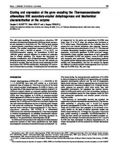
RESULTS Cloning of the gene cluster encoding three subunits of GADH by a direct-expression method. On the assumption that the genes encoding each subunit might be clustered in the genome, we tried to isolate the GADH gene by a direct-expression method as described in Materials and Methods before performing colony hybridization screening with oligonucleotide probes designed from NH2-terminal sequences of GADH subunits or complementation cloning. Usually the activities of
6567
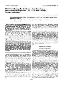
membrane-bound dehydrogenases are estimated as the initial rate of reduction of DCPIP as the electron acceptor by spectrophotometry in the presence of PMS (17). We applied the reduction reaction to the color reaction product on the solid matrix (nitrocellulose filter). To reduce the background color reaction by the host strain on the nitrocellulose membrane, we increased the concentrations of PMS and DCPIP 10 times relative to concentrations used for enzyme assay. The reaction was optimized by using E. cypripedii and E. coli JM109 as a positive and a negative control, respectively. Of 4,000 transformants, one putative positive clone was isolated by a directexpression method and designated clone 301. Clone 301 produced 2KDG in the presence of D-gluconate, indicating that a membrane-bound GADH had been cloned and that it was expressed in E. coli. A recombinant plasmid containing a 10.5-kb DNA insert was purified from the positive clone and designated pGA301. The restriction map of the cloned 10.5-kb EcoRI fragment is shown in Fig. 1. A series of deletion clones of pGA301 were constructed by subcloning the restriction fragments into pUC and pBluescript vectors (Fig. 1). E. coli cells harboring pGA303, pGA308, pGA312, and pGA313 showed dehydrogenase activity by the ferricyanide method, while E. coli cells harboring pGA308 did not convert D-gluconate to 2KDG. These results indicated that the 4.7-kb StuI-SspI DNA fragment covered the whole region of the dehydrogenase subunit genes. Purification of GADH from E. cypripedii ATCC 29267 and amino-terminal amino acid sequencing of subunits. By CMSepharose CL-6B and DEAE-Toyopearl 650 ion-exchange column chromatographies, the GADH of E. cypripedii ATCC 29267 was purified from the membrane fraction. The resulting partially purified enzyme produced a major protein band on native PAGE gels which, upon activity staining, gave a prominent color reaction (data not shown). SDS-PAGE of the enzyme showed three polypeptides of 65, 45, and 20 kDa. The 45-kDa subunit appeared as a red band on unstained SDS gels, suggesting its identity as a cytochrome c subunit. The NH2terminal amino acid sequences of the 65-, 45-, and 20-kDa subunits were determined as NH2-Ala-Asn-Glu-Leu-Lys-LysVal-Asp-Ala-Val-Val-Val-Gly-Phe-Gly, NH2-Asp-Asp-Gln-AlaAsn-Asp-Ala-Leu-Val, and NH2-Ala-Glu-Gln-Ser-Ser-Gly-SerGln-Thr-Ala-Arg-Asp-Tyr-Gln-Pro, respectively. The optimum pH of the enzyme was found to be 5.0, and the optimum temperature was 30°C. Nucleotide sequence of the 4.7-kb fragment containing the genes encoding the three subunits of GADH. The DNA sequence of the 4.7-kb fragment was determined, and the results are shown in Fig. 2. Nucleotide sequencing revealed the presence of three open reading frames (ORFs) corresponding to the dehydrogenase subunit (subunit I), the cytochrome c subunit (subunit II), and the smallest subunit (subunit III). The dehydrogenase subunit is located immediately downstream of the gene coding for the smallest subunit, and the cytochrome c subunit is the next. The three subunit genes were organized in the same transcriptional polarity. The two inverted-repeat sequences of possible transcriptional terminators were found downstream of the cytochrome c subunit gene, which may serve as the rho-independent transcriptional terminator. This indicates that these three genes are in the same operon and are cotranscribed. The ORF corresponding to the smallest subunit might start with either ATG at nucleotides (nt) 258 to 260, ATG at nt 327 to 329, ATG at nt 348 to 350, or ATG at nt 369 to 371 and terminate at TGA at nt 918 to 920. The ATG at nt 258 to 260 seems to be the functional initiator because this ATG was preceded by a possible ribosome-binding sequence, GAGG (nt 247 to 250). The gene consists of 663 bp, encoding
6568
YUM ET AL.
J. BACTERIOL.
FIG. 1. Restriction map of cloned DNA fragment encoding GADH of E. cypripedii. The open arrows indicate the coding sequence for a dehydrogenase (subunit I), a cytochrome c (II), and the smallest subunit (III). Regions covered by plasmids of the pGA series are presented under the restriction map. Enzyme activity was assayed by using ferricyanide as an electron acceptor. Conversion activity was assayed by high-pressure liquid chromatography as described in Materials and Methods.
a polypeptide of 221 amino acids with a calculated molecular weight of 24,471. The NH2-terminal amino acid sequence of the purified smallest subunit was found in this ORF at positions 43 to 56. The extra 42 amino acids at the NH2 terminus of this ORF showed features typical of leader peptides. The molecular mass of the processed subunit deduced from the nucleotide sequence (20 kDa) coincided well with the molecular mass estimated by SDS-PAGE (20 kDa). The coding region of the predicted dehydrogenase gene probably starts with a GTG codon at positions 934 to 936, preceded by a ShineDalgarno (SD) sequence, ATGGA. Another possibility is the ATG codon positioned at nt 925 to 927, but it lacks an SD sequence. The dehydrogenase gene consists of 1,845 bp, encoding a polypeptide of 615 amino acids with a calculated molecular weight of 67,238. The NH2-terminal amino acid sequence of the purified dehydrogenase subunit was found in this ORF at positions 23 to 37. The extra 22 amino acids at the NH2 terminus of this ORF also showed features typical of leader peptides. The molecular mass of the mature subunit (64.9 kDa) was in good agreement with the molecular mass obtained by SDS-PAGE (65 kDa). The ORF corresponding to the cytochrome c subunit was found 11 bp downstream of the ORF coding for the 67-kDa dehydrogenase subunit. A possible ribosome-binding sequence, AGGA, was present 8 nt upstream of the ATG codon. This ORF encodes a 441-aminoacid polypeptide with a molecular weight of 47,094. The NH2terminal amino acid sequence of the cytochrome c subunit was found in this ORF at positions 20 to 28. The extra 19 amino acids at the NH2 terminus of this ORF also showed features typical of leader peptides. The molecular mass of the mature subunit (45 kDa) also coincided well with that estimated by SDS-PAGE (45 kDa). Three possible heme-binding motifs, C-X-X-C-H (20) (nt 2910 to 2924, 3354 to 3368, and 3765 to
3779), the characteristic amino acid sequence of c-type cytochromes, were present within this ORF (Fig. 2). Comparison with other proteins. The flavin adenine dinucleotide (FAD)-dependent enzymes possess the characteristic b1-aA-b2 motif for binding the ADP moiety of FAD (39). This motif is usually located at the amino terminus of the enzyme and contains a so-called glycine box (GXGXXG). The deduced amino acid sequence of the GADH dehydrogenase subunit contained three possible glycine boxes, GFGWAG (nt 1036 to 1053), GTGTGG (nt 1282 to 1299), and GAGGAG (nt 2104 to 2121). A homology search of protein databases revealed that the region containing the first glycine box showed sequence similarity with the FAD-binding motif of cellobiose dehydrogenase (CEDH) from Phanerochaete chrysosporium (12, 24), sorbose dehydrogenase (SDH) from Gluconobacter oxydans (25), choline dehydrogenase (CDH) of Rhizobium meliloti, glucose dehydrogenase of Drosophila melanogaster (38), human monoamine oxidase B (6), and versicolorin B synthase (VBS) from Aspergillus parasiticus (29). The amino acid sequence alignment is shown in Fig. 3A. These data could indicate that the GADH from E. cypripedii is also a flavoprotein like other membrane-bound GADHs (16, 17, 19, 28). In the deduced amino acid sequence of the dehydrogenase subunit, another region with similarity to CEDH, SDH, CDH, and VBS was found (Fig. 3B). However, a Prosite search revealed no functional domain for this region. The predicted amino acid sequence of the cytochrome c subunit showed considerable identity with sequences of Gluconobacter suboxydans cytochrome c-553 (34.1%) (33), Acetobacter pasteurianus alcohol dehydrogenase (ADH) cytochrome c (37.2%) (34), Acetobacter polyoxogenes ADH cytochrome c (39.3%) (35), and Acetobacter aceti ADH cytochrome c (38.9%) (8), which also have signal peptides. The three regions with a C-X-X-C-H sequence are
FIG. 2. Nucleotide and deduced amino acid sequences of the genes encoding the dehydrogenase, the cytochrome c, and the third subunit. The underlined amino acid sequences were determined by an Edman degradation procedure. The vertical arrows indicate the putative signal sequence cleavage sites. Potential ribosomebinding sequences (SD) are marked. Facing arrows show an inverted repeat, which possibly serves as a rho-independent transcriptional terminator. The putative FAD-binding motif is indicated by dashed underlining. The possible heme-binding motifs (C-X-X-C-H) are boxed.
VOL. 179, 1997
GLUCONATE DEHYDROGENASE GENE FROM ERWINIA CYPRIPEDII
6569
6570
YUM ET AL.
J. BACTERIOL.
TABLE 1. Specific GADH activities in E. cypripedii and the recombinant strain of E. colia GADH sp act (U/A600 unit) Strain
E. cypripedii E. coli JM109(pUC18) E. coli JM109(pGA313)
7h
12 h
LB
LB 1 GA
LB
LB 1 GA
0.78 0 0.86
2.36 0 1.85
1.36 0 0.41
2.09 0 2.15
a E. cypripedii ATCC 29267 and E. coli strains were cultivated in LB or LB medium containing 2.5% D-gluconate (GA). Activities were assayed after cultivations for 7 and 12 h by the ferricyanide method described in Materials and Methods.
highly conserved (data not shown). In the membrane-bound ADH, the cytochrome c subunit has been found to be essential for activity on the basis of in situ reconstitution of the subunit in the membranes of an alcohol oxidase activity-deficient strain (15). The pGA308 clone (Fig. 1), in which a part of the Cterminal region of the cytochrome c subunit was deleted, did not convert D-gluconate to 2KDG. This result indicates that the cytochrome c subunit in GADH is also essential for the activity. No protein showing significant similarity to the smallest subunit was found in the protein databases, as was the case with the 20-kDa smallest subunit of A. pasteurianus ADH (9). The function of the third subunit in the 3-subunit membrane-bound dehydrogenases is unknown. Kondo et al. suggested that the 20-kDa smallest subunit of A. pasteurianus ADH had a role in the stability of the dehydrogenase subunit and functioned as a molecular coupler of the dehydrogenase subunit to the cytochrome c subunit on the cytoplasmic membrane (9). Expression of the gene encoding GADH in E. coli. The recombinant E. coli JM109 transformed with pGA303 and E. cypripedii were cultivated in LB or LB medium containing 2.5% D-gluconate. Whole cells were used for assaying GADH activity. As shown in Table 1, the GADH activity was found in the E. coli JM109 cells harboring pGA303 but not in the E. coli JM109 cells harboring vector plasmid pUC18. The activities were found in the cells cultivated both in LB and in LB medium containing D-gluconate, which suggests that the gene encoding GADH is expressed constitutively in both E. cypripedii and E. coli. The higher GADH activity in the presence of D-gluconate was found both in E. cypripedii and in the recombinant strain of E. coli.
FIG. 3. Comparison of the putative FAD-binding region of the GADH dehydrogenase subunit with FAD-dependent enzymes. (A) The amino-terminus region of the dehydrogenase subunit of GADH is aligned with the flavin domain of CEDH from P. chrysosporium (12, 24), SDH from G. oxydans (25), CDH of R. meliloti, glucose dehydrogenase (GDH) of D. melanogaster (38), human monoamine oxidase B (MAO-B) (6), and versicolorin B synthase (VBS) from A. parasiticus (29). (B) The region of the dehydrogenase subunit conserved with CEDH, SDH, CDH, and VBS. Identical amino acids are indicated in boldface type.
Conversion of glucose to 2KDG in E. coli. E. coli JM109 transformed with pGA313 was cultivated in LB medium containing 2.5% glucose, 10 mM PQQ, and ampicillin (100 mg/ml). The supernatant was used for assaying the amounts of Dgluconate z Na and 2KDG z Na converted from D-glucose. The results are shown in Table 2. With E. coli JM109 transformed with pUC18 as a control, the glucose was completely converted to 29.9 mg of D-gluconate/ml in 12 h of cultivation by addition of PQQ to the culture medium. With E. coli JM109(pGA313) in the presence of PQQ, the glucose (25 mg/ml) was almost completely converted to 29.2 mg of 2KDG z Na/ml via D-gluconate during 12 h of cultivation. When culture was carried out with D-gluconate z Na as a carbon source, 30 mg of D-gluconate z Na/ml was converted to 29.2 mg of 2KDG z Na/ml in 12 h. The conversion yields for D-glucose and D-gluconate were 0.86 mol of 2KDG/mol of D-glucose and 0.95 mol of 2KDG/mol of D-gluconate, respectively. D-Glucose (25 mg/ml) was converted to 2KDG
TABLE 2. Production of 2KDG in recombinant strains of E. colia Amt (mg/ml in broth) Strain
Substrate
PQQ Glu
GA z Na
2KDG z Na
YGAb
Y2KDGb
E. coli JM109(pUC18)
Glu Glu
2 1
23.5 0
0 29.9
0 0
0 0.99
0 0
E. coli JM109(pGA313)
Glu Glu
2 1
23.5 0
0 2.4
0 25.9
0 0.08
0 0.86 (0.95c)
E. coli JM109(pUC18)
GA
2
29.8
0
E. coli JM109(pGA313)
GA
2
0
29.2
a
0 0.95
E. coli strains were cultivated in LB medium containing 2.5% D-glucose (Glu) or D-gluconate (GA) for 12 h at 37°C. If needed, PQQ was added to a final concentration of 10 mM. b Molar yield calculated from initial substrate concentration. c Cultivation for 16 h.
VOL. 179, 1997
GLUCONATE DEHYDROGENASE GENE FROM ERWINIA CYPRIPEDII
with a yield of 0.95 mol of 2KDG/mol of glucose in 16 h after inoculation. DISCUSSION We cloned the gene cluster encoding the three subunits of GADH from E. cypripedii by direct expression on a nitrocellulose filter by using PMS and DCPIP as described in Materials and Methods. We also tried to apply the ferricyanide color reaction to the screening of the positive GADH clone on the filter, but it proved to be difficult to distinguish the color difference between the positive and negative controls. This screening method with PMS and DCPIP could be useful for the direct-expression cloning of other membrane-bound dehydrogenases. All GADHs that have been purified and characterized consist of three subunits, a dehydrogenase, a cytochrome c, and a third component of the lowest molecular weight. The molecular masses of the GADH genes of E. cypripedii for the processed subunits deduced from the nucleotide sequences and the subunits estimated from purified protein were 65, 45, and 20 kDa, respectively. These molecular masses are very similar to those of the subunits of GADHs from P. aeruginosa (16, 19), P. fluorescens (17), K. pneumoniae (17), S. marcescens (17), and G. dioxyacetonicus (28). The optimum pH of 5.0 is also in the range of values (pH 4.0 to 5.0) for other GADHs. It has been reported that GADH is highly specific for D-gluconate (17). The substrate specificity of GADH from E. cypripedii is also restricted to D-gluconate (data not shown). These results indicate that the molecular and catalytic properties of membranebound GADHs are similar. To determine whether the substrate, D-gluconate, affects the expression of the GADH gene, the activities of GADH in the growing cells were measured in the presence and absence of D-gluconate in rich medium. In both E. cypripedii and recombinant E. coli, the GADH was constitutively formed and the activity was enhanced more than twofold by addition of Dgluconate to the medium (Table 1). It has been reported that almost the same amounts of the cytochrome c and 15-kDa subunits of ADH from A. pasteurianus were produced regardless of the presence or absence of ethanol (34). The amount of the dehydrogenase subunit of ADH from A. pasteurianus localized in the membrane was smaller in the absence of ethanol due to the incorrect localization (34). In G. suboxydans grown under acidic conditions, an inactive form of ADH was produced and the enzyme had a relatively loose conformation (18). This inactive form was converted to the active form, with a tight conformation, at a neutral culture pH. The relatively lower GADH activity in the absence of D-gluconate might also be due to a conformational change or incorrect localization of GADH. Oxidation of glucose to ketogluconates such as 2KDG, 5keto-D-gluconate (5KDG), and 25DKG in several species of the genera Erwinia, Acetobacter, Gluconobacter, Serratia, and Pseudomonas has been shown to proceed via membrane-bound dehydrogenases (1, 31), and the ketogluconates or their phosphorylated forms are dissimilated through the central metabolic pathway (11, 22, 30, 36). The batch production of 2KDG from D-glucose by S. marcescens (21) and A. pasteurianus (32) has been investigated. In these strains, 2KDG is dissimilated through the central metabolic pathway and the selectivity of glucose oxidation to 2KDG in the ketogluconate producers depends on the culture conditions (23, 32). Since 2KDG, an intermediate in iso-ascorbic acid synthesis, would not be metabolized in this recombinant E. coli strain, it would be accumulated to high levels in the culture medium. As the first step
6571
in our approach, the production of 2KDG from D-glucose by recombinant E. coli in the presence of exogenous PQQ was attempted. As expected, D-glucose (25 mg/ml) was converted to 2KDG with a yield of 0.95 mol of 2KDG/mol of glucose in 16 h after inoculation (Table 2). Considering the fact that the conversion of D-glucose to 2KDG in an E. coli strain unable to metabolize ketogluconate was extremely efficient, the bioconversion process using E. coli cells should be an attractive method. ACKNOWLEDGMENTS We thank E. S. Choi for critical reading of the manuscript and S. J. Ju and H. W. Lee for their excellent assistance in this experiment. REFERENCES 1. Ameyama, M., K. Matsushita, E. Shinagawa, and O. Adachi. 1987. Sugaroxidizing respiratory chain of Gluconobacter suboxydans. Evidence for a branched respiratory chain and characterization of respiratory chain-linked cytochromes. Agric. Biol. Chem. 51:2943–2950. 2. Birnboim, H. C., and J. Doly. 1979. A rapid alkaline extraction procedure for screening recombinant plasmid DNA. Nucleic Acids Res. 7:1513–1523. 3. Biville, F., E. Turlin, and F. Gasser. 1991. Mutants of Escherichia coli producing pyrroloquinoline quinone. J. Gen. Microbiol. 137:1775–1782. 4. Choi, E.-S., E.-H. Lee, and S.-K. Rhee. 1995. Purification of a membranebound sorbitol dehydrogenase from Gluconobacter suboxydans. FEMS Microbiol. Lett. 125:45–50. 5. Goosen, N., H. P. A. Horsman, R. G. M. Huinen, and P. van de Putte. 1989. Acinetobacter calcoaceticus genes involved in biosynthesis of the coenzyme pyrrolo-quinoline-quinone: nucleotide sequence and expression in Escherichia coli K-12. J. Bacteriol. 171:447–455. 6. Grimsby, J., K. Chen, L. J. Wang, N. C. Lan, and J. C. Shih. 1991. Human monoamine oxidase A and B genes exhibit identical exon-intron organization. Proc. Natl. Acad. Sci. USA 88:3637–3641. 7. Hommes, R. W. J., P. W. Postma, O. M. Neijssel, D. W. Tempest, P. Dokter, and J. A. Duine. 1984. Evidence of a quinoprotein glucose dehydrogenase apoenzyme in several strains of Escherichia coli. FEMS Microbiol. Lett. 24:2–3. 8. Inoue, T., M. Sunagawa, A. Mori, C. Imai, M. Fukuda, M. Takagi, and K. Yano. 1992. Nucleotide sequence of the gene encoding the 45-kilodalton subunit of alcohol dehydrogenase from Acetobacter aceti. J. Ferment. Bioeng. 73:419–424. 9. Kondo, K., T. Beppu, and S. Horinouchi. 1995. Cloning, sequencing, and characterization of the gene encoding the smallest subunit of the threecomponent membrane-bound alcohol dehydrogenase from Acetobacter pasteurianus. J. Bacteriol. 177:5048–5055. 10. Laemmli, U. 1970. Cleavage of structural proteins during the assembly of the head of bacteriophage T4. Nature (London) 227:680–685. 11. Levering, P. R., G. Weenk, W. Olijve, L. Dijkhuizen, and W. Harder. 1988. Regulation of gluconate and ketogluconate production in Gluconobacter oxydans ATCC 621-H. Arch. Microbiol. 149:534–539. 12. Li, B., S. R. Nagalla, and V. Renganathan. 1996. Cloning of a cDNA encoding cellobiose dehydrogenase, a hemoflavoenzyme from Phanerochaete chrysosporium. Appl. Environ. Microbiol. 62:1329–1335. 13. Liu, S.-T., L.-Y. Lee, C.-Y. Tai, C.-H. Hung, Y.-S. Chang, J. H. Wolfram, R. Rogers, and A. H. Goldstein. 1992. Cloning of an Erwinia herbicola gene necessary for gluconic acid production and enhanced mineral phosphate solubilization in Escherichia coli HB101: nucleotide sequence and probable involvement in biosynthesis of the coenzyme pyrroloquinoline quinone. J. Bacteriol. 174:5814–5819. 14. Maniatis, T., E. F. Fritsch, and J. Sambrook. 1982. Molecular cloning: a laboratory manual. Cold Spring Harbor Laboratory, Cold Spring Harbor, N.Y. 15. Matsushita, K., Y.-I. Nagatani, E. Shinagawa, O. Adachi, and M. Ameyama. 1991. Reconstitution of the ethanol oxidase respiratory chain in membranes of quinoprotein alcohol dehydrogenase-deficient Gluconobacter suboxydans subsp. a strains. J. Bacteriol. 173:3440–3445. 16. Matsushita, K., E. Shinagawa, O. Adachi, and M. Ameyama. 1979. Membrane-bound D-gluconate dehydrogenase from Pseudomonas aeruginosa. Purification and structure of cytochrome-binding form. J. Biochem. 85:1173– 1181. 17. Matsushita, K., E. Shinagawa, and M. Ameyama. 1982. D-Gluconate dehydrogenase from bacteria, 2-keto-D-gluconate-yielding, membrane-bound. Methods Enzymol. 89:187–193. 18. Matsushita, K., T. Yakushi, Y. Takaki, H. Toyama, and O. Adachi. 1995. Generation mechanism and purification of an inactive form convertible in vivo to the active form of quinoprotein alcohol dehydrogenase in Gluconobacter suboxydans. J. Bacteriol. 177:6552–6559. 19. McIntire, W., T. P. Singer, M. Ameyama, O. Adachi, K. Matsushita, and E.
6572
20. 21. 22.
23. 24.
25.
26. 27. 28.
29.
YUM ET AL.
Shinagawa. 1985. Identification of the covalently bound flavins of D-gluconate dehydrogenases from Pseudomonas aeruginosa and Pseudomonas fluorescens and of 2-keto-D-gluconate dehydrogenase from Gluconobacter melanogenus. Biochem. J. 231:651–654. Meyer, T. E., and M. D. Kamen. 1982. New perspectives on c-type cytochromes. Adv. Protein Chem. 35:105–212. Misenheimer, T. J., R. F. Anderson, A. A. Lagoda, and D. D. Tyler. 1965. Production of 2-ketogluconic acid by Serratia marcescens. Appl. Microbiol. 13:393–396. Pronk, J. T., P. R. Levering, W. Olijve, and J. P. van Dijken. 1989. Role of NADP-dependent and quinoprotein glucose dehydrogenases in gluconic acid production by Gluconobacter oxydans. Enzyme Microb. Technol. 11: 160–164. Qazi, G. N., N. Sharma, and R. Parshad. 1993. Role of dissolved oxygen as a regulator for the direct oxidation of glucose by Erwinia herbicola and Gluconobacter oxydans. J. Ferment. Bioeng. 76:336–339. Raices, M., E. Paifer, J. Cremata, R. Montesino, J. Stahlberg, C. Divne, I. J. Szabo, G. Henriksson, G. Johansson, and G. Pettersson. 1995. Cloning and characterization of a cDNA encoding a cellobiose dehydrogenase from the white rot fungus Phanerochaete chrysosporium. FEBS Lett. 369:233–238. Saito, Y., Y. Ishii, H. Hayashi, Y. Imao, T. Akashi, K. Yoshikawa, Y. Noguchi, S. Soeda, M. Yoshida, M. Niwa, J. Hosoda, and K. Shimomura. 1987. Cloning of genes coding for L-sorbose and L-sorbosone dehydrogenases from Gluconobacter oxydans and microbial production of 2-keto-L-gulonate, a precursor of L-ascorbic acid, in a recombinant G. oxydans strain. Appl. Environ. Microbiol. 63:454–460. Sanger, F., S. Nicklen, and A. R. Coulson. 1977. DNA sequencing with chain-terminating inhibitors. Proc. Natl. Acad. Sci. USA 74:5463–5467. Shinagawa, E., and M. Ameyama. 1982. 2-Keto-D-gluconate dehydrogenase from Gluconobacter melanogenus, membrane-bound. Methods Enzymol. 89: 194–198. Shinagawa, E., K. Matsushita, O. Adachi, and M. Ameyama. 1984. D-Gluconate dehydrogenase, 2-keto-D-gluconate yielding, from Gluconobacter dioxyacetonicus: purification and characterization. Agric. Biol. Chem. 48:1517– 1522. Silva, J. C., R. E. Minto, C. E. Barry III, and K. A. Holland. 1996. Isolation
J. BACTERIOL.
30. 31. 32. 33. 34. 35.
36. 37. 38. 39. 40.
and characterization of the versicolorin B synthase gene from Aspergillus parasiticus. J. Biol. Chem. 271:13600–13608. Sonoyama, T., B. Kageyama, S. Yagi, and K. Mitsushima. 1987. Biochemical aspects of 2-keto-L-gulonate accumulation from 2,5-diketo-D-gluconate by Corynebacterium sp. and its mutants. Agric. Biol. Chem. 51:3039–3047. Sonoyama, T., B. Kageyama, S. Yagi, and K. Mitsushima. 1988. Facultatively anaerobic bacteria showing high productivities of 2,5-diketo-D-gluconate from D-glucose. Agric. Biol. Chem. 52:667–674. Svitel, J., and E. Sturdik. 1995. 2-Ketoglucoic acid production by Acetobacter pasteurianus. Appl. Biochem. Biotechnol. 53:53–63. Takeda, Y., and T. Shimizu. 1991. Cloning and sequencing of the gene encoding cytochrome c-553 (CO) from Gluconobacter suboxydans. J. Ferment. Bioeng. 72:1–6. Takemura, H., K. Kondo, S. Horinouchi, and T. Beppu. 1993. Induction by ethanol of alcohol dehydrogenase activity in Acetobacter pasteurianus. J. Bacteriol. 175:6857–6866. Tamaki, T., M. Fukaya, H. Takemura, K. Tayama, H. Okumura, Y. Kawamura, M. Nishiyama, S. Horinouchi, and T. Beppu. 1991. Cloning and sequencing of the gene cluster encoding two subunits of membrane-bound alcohol dehydrogenase from Acetobacter polyoxogenes. Biochim. Biophys. Acta 1088:292–300. Truesdell, S. J., J. C. Sims, P. A. Boerman, J. L. Seymour, and R. A. Lazarus. 1991. Pathways for metabolism of ketoaldonic acids in an Erwinia sp. J. Bacteriol. 173:6651–6656. Vieira, J., and J. Messing. 1982. The pUC plasmids, an M13mp7-derived system for insertion mutagenesis and sequencing with synthetic universal primers. Gene 19:259–268. Whetten, R., E. Organ, P. Krasney, D. Cox-Foster, and D. Cavener. 1988. Molecular structure and transformation of the glucose dehydrogenase gene in Drosophila melanogaster. Genetics 120:475–484. Wierenga, R. K., P. Terpstra, and W. G. J. Hol. 1986. Prediction of the occurrence of the ADP-binding bab-fold in protein, using an amino acid sequence fingerprint. J. Mol. Biol. 187:101–107. Yanisch-Perron, C., J. Vieira, and J. Messing. 1985. Improved M13 phage cloning vectors and host strains: nucleotide sequences of M13mp18 and pUC19 vectors. Gene 33:103–119.