Immunogenetics (2002) 54:513–519 DOI 10.1007/s00251-002-0492-2
O R I G I N A L PA P E R
Arihiro Iwata · Takashi Iwase · Yoshitaka Ogura Tomihisa Takahashi · Naoyuki Matsumoto Toshiyuki Yoshida · Nobuhiro Kamei Kunihiko Kobayashi · Jiri Mestecky · Itaru Moro
Cloning and expression of the turtle (Trachemys scripta) immunoglobulin joining (J)-chain cDNA Received: 7 June 2002 / Accepted: 15 July 2002 / Published online: 6 September 2002 © Springer-Verlag 2002
Abstract The J-chain protein is a Mr 15,000 polypeptide associated with polymeric IgA and IgM. The complete cDNA sequences of human, mouse, cow, brushtail possum, chicken and frog J chains have been previously reported, but nothing is known about the cDNA and amino acid sequences of reptilian J chain. Here, we determined a turtle J-chain cDNA sequence by RT-PCR and RACE, and examined J-chain mRNA and protein expression by Northern blotting and immunohistochemistry. This turtle J-chain cDNA was 1,934 bp and had an open reading frame of 477 nucleotides, encoding 159 amino acids. The mature J-chain protein is composed of 137 amino acids, Mr ~15,000. The deduced amino acid sequence of the turtle J chain was highly homologous to that of human (60%), mouse (61%), cow (60%), rabbit (60%), chicken (69%), brushtail possum (65%), Rana catesbeiana (47%) and Xenopus laevis (58%). Eight cysteine residues were located at the same positions as in these other species, with the exception of X. laevis. PROSITE database analysis indicated the presence of two N-glycosylation sites in turtle, one of which was novel. Northern blot analysis revealed that turtle J-chain mRNA was expressed in lung, stomach, spleen and intestine. In addition, immunohistochemistry showed J-chain-positive plasTurtle J-chain nucleotide sequence data are available in the DDBJ/EMBL/GenBank nucleotide sequence databases under the accession number AB085611 A. Iwata (✉) · T. Iwase · Y. Ogura · T. Takahashi · N. Matsumoto T. Yoshida · N. Kamei · I. Moro Department of Pathology, Nihon University School of Dentistry, 1-8-13, Kanda-Surugadai, Chiyoda-ku, Tokyo 101-8310, Japan e-mail:
[email protected] Tel.: +81-3-32198124, Fax: +81-3-32198340 K. Kobayashi Department of Pediatrics, Hokkaido University School of Medicine, Kita 15 Jyo-Nishi, Kita-ku, Sapporo 060-8638, Japan J. Mestecky Department of Microbiology, BBRB 757, University of Alabama at Birmingham, Birmingham, AL 35294, USA
ma cells in the intestine and spleen. These results suggest the presence of a mucosal immune system mainly composed of J-chain-containing Ig in the turtle. Keywords Joining chain · Polymeric immunoglobulin · Phylogeny · Turtle · Immunohistochemistry
Introduction Mucosal surfaces are covered by a thin layer of vulnerable epithelium, which is continuously exposed to diverse pathogens such as bacteria and viruses, as well as various harmful molecules. An innate defense mechanism is present in these areas and a mucosal immune system composed of SIgA plays a crucial role in preventing invasion of foreign antigens (Mestecky and McGhee 1987). SIgA consists of dimeric (d) IgA and joining (J) chain, which are produced by plasma cells, and a polymeric immunoglobulin receptor (pIgR) that functions as a receptor for J-chain-containing polymeric immunoglobulins (Igs) (Mestecky and McGhee 1987). The J chain is an acidic polypeptide of Mr 15,000 that has an important role in the polymerization and secretion of polymeric Igs (Brandtzaeg 1985; Brewer et al. 1994; Cattaneo and Neuberger 1987; Johansen et al. 2000; Mestecky et al. 1999; Mestecky and McGhee 1987). However, it has been reported that the J chain is present in IgG- and IgD-containing myeloma cells, as well as in IgA- and IgM-producing cells (Brandtzaeg 1974; Kaji and Parkhouse 1974; Mestecky et al. 1977). The presence of polymeric IgM without J chain in cells and serum has also been reported (Brewer et al. 1994; Kimura et al. 2001; Randall et al. 1990). In addition, polymeric IgM is secreted by a glioma cell line transfected with IgM heavy- and light-chain cDNA, even though this cell line does not produce J chain (Cattaneo and Neuberger 1987). The substitution of serine for cysteine in IgM, which is involved in J-chain binding, results in the formation of polymeric IgM without a J chain (Davis et al. 1989). The presence of J chain also results in the secre-
514
tion of pentameric IgM instead of hexameric IgM and is necessary for a correct assembly of dIgA (Koshland 1989; Randall et al. 1992). Other studies indicated that selective transport of dIgA and pentameric IgM through mucosal epithelial cells is mediated by the pIgR and that the J chain is necessary for pIgR to recognize pIgs (Brandtzaeg 1985). Therefore, the J chain is a key factor for the recognition of pIgs by pIgR localized in glandular epithelial cells. The J-chain genes of human, mouse and chicken consist of four exons and three introns (Matsuuchi et al. 1986; Max and Korsmeyer 1985; Takahashi et al. 2002). However, the expression patterns of J-chain genes are different in these species. The J chain in humans is expressed in pro-B and pre-B cells at an early stage of development before synthesis of Ig (Max and Korsmeyer 1985; Mestecky et al. 1997). On the other hand, murine and chicken J chains are detectable in a late stage of mature B cells expressing cytoplasmic µ chain (Koshland 1985; Takahashi et al. 2000). It has been reported that J-chain mRNA expression ontogenetically occurs 1 week earlier than that of µ chain in the human fetal liver (Iwase et al. 1993). However, these discrepancies have not been clarified. At present, full-length J-chain cDNA has been reported in mammals, birds and amphibia (Adamski and Demmer 1999; Hohman et al. 1997; Kulseth and Rogne 1994; Matsuuchi et al. 1986; Max and Korsmeyer 1985; Takahashi et al. 2000). J-chain cDNA sequencing revealed that mammalian J chains share well-conserved features among mouse, human, cow and brushtail possum (Adamski and Demmer 1999; Kulseth and Rogne 1994; Matsuuchi et al. 1986; Max and Korsmeyer 1985). The amphibian and avian J chains also have a high degree of homology to the cDNA sequences of mammals (Hohman et al. 1997; Takahashi et al. 2000). Although IgM and IgY have been found in reptiles (Kobayashi et al. 1973; Muthukkaruppan et al. 1982; Vaerman et al. 1975; Warr et al. 1995), the presence of J chain has not been documented thus far. Here, we have conducted experiments on the cloning and sequencing of the turtle (Trachemys scripta) J-chain cDNA using reverse transcriptase-polymerase chain reaction (RT-PCR) and rapid amplification of cDNA ends (RACE). In addition, J-chain mRNA and protein expression was examined by Northern blotting and immunohistochemistry, respectively. These results demonstrate that turtle J chain has highly conserved structural features similar to those of mammals, birds and amphibians, and suggest the presence of a mucosal immune system mediated by pIgs in reptiles.
Materials and methods Turtles Ten turtles (Trachemys scripta) were obtained from a dealer (Shimizu kingyo, Tokyo, Japan).
Fig. 1 Primers used are S2 and S4 for RT-PCR; T-GSP-1 and 3 sites adaptor primer for 3′RACE; TS1, TA1, TS2, TA2 and RT primer (5′ phosphorylation) for 5′RACE; T-J-C2 and T-J-C3 for Northern and genomic Southern blotting; T-J-C4 and T-J-C6 for preparation of fusion protein; and Aac-1 and Aac-2 for β-actin
Preparation of RNA Total RNA was extracted by ultracentrifugation methods (Zarlenga and Gamble 1987). Various tissues (stomach, intestine, spleen, lung and heart) from the turtle were dissected into small pieces and each homogenized in a solution containing 4 M guanidine thiocyanate, 25 mM sodium citrate (pH 7.0), 0.5% sarcosyl, and 0.1 M 2-mercaptoethanol. Samples were then layered onto cesium trifluoroacetate (Amersham Pharmacia Biotech, Little Chalfont, Buckinghamshire, UK) and centrifuged at 160,000 g for 16 h at 15 °C. After centrifugation, pellets were extensively washed with 70% ethanol and used as total RNA. Cloning of turtle J-chain cDNA by RT-PCR A random primed first-strand cDNA was synthesized from 1 µg of total RNA from turtle intestine, using 10 units of Rous-associated virus 2 reverse transcriptase (Takara shuzou, Kyoto, Japan) at 42 °C for 1 h. Turtle J-chain primers were used in PCR. cDNA was amplified in a buffer containing 2.5 mM dNTP, 20 pM of both primers and 1 unit of Taq DNA polymerase (Takara shuzou). PCR conditions (35 cycles) were as follows: denature for 1 min at 94 °C, annealing for 2 min at 50 °C and extension for 3 min at 72 °C. PCR products were electrophoresed through 1.8% agarose gels and a fragment that was hybridized with human J-chain cDNA by PCR Southern blotting (data not shown) was subcloned into the pT7 blue T vector (Novagen, Darmstadt, Germany) and then sequenced by Mega BACE 1000 (Molecular Dynamics, Sunnyvale, Calif., USA) using a DYEnamic ET dye terminator cycle sequence kit (Amersham Pharmacia Biotech). DNA sequences obtained were analyzed with Genetyx-Mac version 10.1.1 (Software Development, Tokyo, Japan). In this study, seven kinds of primers were designed to obtain the PCR product of turtle J chain. These were designed through careful examination of sequence conservation patterns based on the published J-chain cDNA sequences of human, mouse, cow, Xenopus laevis and brushtail possum. Various combinations of primer sets were used for PCR and only a combination of S2 and S4 primers successfully functioned to obtain a J-chain product compatible with an expected size. The nucleotide sequences of the S2 and S4 primers are shown in Fig. 1. 3′RACE and 5′RACE To obtain a full-length turtle J-chain cDNA, 3′RACE and 5′RACE were performed using a 3′- and 5′-full RACE core set (Takara shuzou) according to the manufacturer’s instructions. Briefly, the cDNA was synthesized using 1 µg of total RNA, oligo dT-3 sites adaptor primer and reverse transcriptase for 3′RACE. PCR for 3′RACE was performed with J-chain-specific primer (T-GSP-1)
515 and three sites adaptor primer (Fig. 1). PCR conditions (35 cycles) were as follows: denature for 30 s at 94 °C, annealing for 30 s at 55 °C, and extension for 2 min at 72 °C. The PCR product was subcloned into the pT7 blue T vector (Novagen), then sequenced. Furthermore, cDNA was generated with 1 µg of total RNA and RT primer (Fig. 1) for 5′RACE. Intact 5′-end cDNA was obtained with nested PCR using TS1, TA1, TS2 and TA2 primers (Fig. 1), which were designed based on a partial sequence obtained by 3′RACE. The PCR product was subcloned into the pT7 blue T vector (Novagen) and then sequenced. Northern blot analysis Twenty micrograms of total RNA from stomach, intestine, spleen, lung or heart were electrophoresed through 1.5% agarose gels containing 6% formaldehyde and then transferred to nylon membranes. A partial J-chain cDNA fragment (578 bp) was synthesized by PCR using primers T-J-C2 and T-J-C3 (Fig. 1) as a probe. Hybridization was carried out with 32P-labeled probe for 16 h at 42 °C in a solution containing 50% formamide, 0.65 M NaCl, 5×Denhardt’s solution, 5 mM EDTA, 0.1% sodium dodecyl sulfate (SDS), 0.1 M Pipes-NaOH (pH 6.8), and 100 µg/ml salmon sperm DNA. The membrane was washed once with 2×SSC/0.1% SDS, and twice with 0.1×SSC/0.1% SDS at 60 °C for 30 min each, and then exposed to X-ray film for 17 h (Kodak, Rochester, N.Y.). As a control, the turtle β-actin probe was synthesized by RT-PCR, using Aac-1 and Aac-2 primers (Fig. 1). Their designs were based on conserved regions of nucleotide sequences among human (Ng et al. 1985), mouse (Tokunaga et al. 1986) and Halocynthia roretzi (Kusakabe et al. 1991) (data not shown). Genomic Southern blotting Ten micrograms of genomic DNA from turtle liver was digested with BamHI, EcoRI, or XbaI for 12 h at 37 °C, and then electrophoresed through 0.7% agarose gels. Gels were treated with an alkaline solution (1.5 M NaCl, 0.2 M NaOH) for 60 min and denatured genomic DNA was transferred to a nylon membrane. The membrane was hybridized with a 32P-labeled probe for 16 h at 65 °C in a solution containing 5×Denhardt’s solution, 5×SSC, 0.1% SDS and 1 mg/ml salmon sperm DNA. After washing three times with 0.1×SSC and 0.1% SDS for 60 min at 65 °C, the membrane was exposed to X-ray film (Kodak) for 20 h. The same probe used for Northern blotting was utilized for the genomic Southern blotting. Preparation of the turtle J-chain fusion protein An open reading frame was amplified from full-length turtle J-chain cDNA by PCR using primers T-J-C4 and T-J-C6 (Fig. 1), which were designed with EcoRI or SalI sites at the 5′ end, respectively. The PCR product was ligated into EcoRI and SalI sites of the pET32a(+) vector (Novagen) and used to transform E. coli strain BL21 (DE3) (Novagen). Protein expression was induced by addition of 1 mM isopropyl-β-d-thiogalactopyranoside (IPTG) for 4 h at 37 °C. Cells were collected by centrifugation at 7,000 g and suspended in a buffer containing 5 mM imidazole, 0.5 M NaCl, 20 mM Tris-HCl (pH 7.9) and 6 M urea, then sonicated three times at intervals of 30 s on ice. Supernatant was collected by centrifugation at 15,000 g at 4 °C for 30 min, and then applied to His-Bind metal chelation resin (Novagen) according to the manufacturer’s instructions. Purified fusion protein was dialyzed against phosphate buffered saline (PBS) containing 5 M urea and adjusted to a final protein concentration of 1 mg/ml, then assayed by SDS-PAGE. Three rabbits were immunized intradermally with 0.2 mg of fusion protein emulsified in an equal volume of complete Freund’s adjuvant, and then were boosted five times with an emulsion of fusion protein and incomplete Freund’s adjuvant at two-week in-
Fig. 2 Nucleotide sequences of turtle J-chain cDNA. Arrows and solid line indicating each primer site and nucleotide sequence are shown in Fig. 1. The nucleotide sequence data will appear in the DDBJ/EMBL/GenBank nucleotide sequence databases under accession number AB085611 tervals. Two weeks after the last booster, the whole immune serum was isolated and applied to protein A column (Amersham Pharmacia Biotech) and then a polyclonal antibody to the turtle J-chain fusion protein was obtained. After titration, it was used for immunohistochemical staining as an anti-tortoise J-chain IgG. Furthermore, this antibody was absorbed with an excess amount of J-chain fusion protein (absorbed antibody) and used as a control.
516 Fig. 3 Alignment of the amino acid sequences of J chains from human (Max and Korsmeyer 1985), cow (Kulseth and Rogne 1994), mouse (Matsuuchi et al. 1986), rabbit (Hughes et al. 1990), chicken (Takahashi et al. 2000), brushtail possum (Adamski and Demmer 1999), Rana catesbeiana (Mikoryak et al. 1988) and Xenopus laevis (Hohman et al. 1997). Asterisks indicate identical amino acid residues. Dots indicate cysteine residues. The Asn-linked glycosylation sites at position 74 and 116 are boxed. Numbering is according to the turtle J-chain sequence
Immunohistochemical method Tissues from turtle intestine, spleen, stomach and liver were fixed in 96% ethanol at 4 °C for 16 h and then paraffin sections were subjected to immunoperoxidase staining. Briefly, endogenous peroxidase activity was blocked with 0.3% H2O2 in methanol for 30 min. After washing with PBS, sections were incubated with 10% normal goat serum to block non-specific reactions, and subsequently incubated with the anti-turtle J-chain antibody or absorbed antibody for 1 h at room temperature. After washing, sections were incubated with horseradish peroxidase-conjugated F(ab’)2 fragments of goat anti-rabbit immunoglobulins (Biosource International, Camarillo, Calif.) for 1 h at room temperature. The substrate reaction was performed in a solution containing 0.05% 3,3′-diaminobenzidine tetrahydrochloride and 0.005% H2O2 for 10 min at room temperature.
Results The turtle J-chain PCR product, which at 204 bp has a relatively high homology to mammalian, avian and amphibian J chains, was obtained from total RNA by RT-PCR using S2 and S4 primers. To identify full-length turtle J-chain cDNA, sense primers were designed from the sequence of 204 bp for RACE. Both 3′RACE and 5′RACE were performed and 1717 bp and 827 bp products, respectively, were obtained and sequenced. After confirmation of the presence of a poly(A) site and ATG codon, a turtle J-chain cDNA sequence with 1,934 nucleotides was identified (Fig. 2). An open reading frame sequence was reconfirmed by RT-PCR using gene-specific T-J-C2 and T-J-C3 primers and used as a probe for Northern and genomic Southern blotting. As shown in Fig. 2, the open reading frame of turtle J chain consisted of 477 nucleotides, encoding 159 amino acid residues.
Fig. 4 Phylogenetic analysis of J chain from turtle, human, cow, mouse, rabbit, chicken, brushtail possum, Rana catesbeiana and Xenopus laevis. The phylogenetic tree was constructed using the Genetyx-Mac version 10.1.1 (Software Development) of the UPGMA method based on the nucleotide and deduced amino acid sequences
The turtle J-chain cDNA showed a high degree of homology to human (69%), mouse (67%), cow (69%), chicken (76%), brushtail possum (70%), Rana catesbeiana (57%) and Xenopus laevis (65%) J chains. The deduced amino acid sequences of the turtle J chain also showed a high degree of homology to human (60%), mouse (61%), cow (60%), rabbit (60%), chicken (69%), brushtail possum (65%), R. catesbeiana (47%) and X. laevis (58%). In addition, the positions of eight cysteine residues were conserved in these different species
517
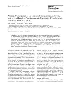
Fig. 6 Northern blot hybridization of turtle J-chain gene expression. Total RNA (20 µg) from various tissues was electrophoresed through 1.5% agarose/6% formaldehyde gels. The membrane was hybridized with the T-J-C2/3 PCR product probe, washed and exposed to X-ray film for 17 h
Fig. 5 Southern blot hybridization of turtle genomic DNA with the turtle J-chain probe. DNA (10 µg) was digested with BamHI (23 kb), EcoRI (2.3 and 3.2 kb), or XbaI (16 kb), and electrophoresed through 0.7% agarose gels. The membrane was hybridized with the T-J-C2/3 PCR product probe, washed and exposed to X-ray film for 20 h
(turtle, human, mouse, cow, rabbit, chicken, brushtail possum, R. catesbeiana) with only X. laevis being an exception (Fig. 3). Genetyx-Mac analysis revealed the presence of two N-glycosylation sites in the turtle J chain. One of these was found at an identical position as in the other eight animals (NISD), but the second (NCSS) has not been described before. Phylogenetic tree analysis performed by the GenetyxMac version 10.1.1 of the UPGMA method based on nucleotide and deduced amino acid sequences indicated that turtle J chain is more closely related to chicken than to the other species examined (Fig. 4). Genomic Southern blotting analysis revealed a 23-kb and 16-kb single band following BamHI or XbaI digestion, respectively. In contrast, EcoRI digestion resulted in the appearance of two bands of 2.3 kb and 3.2 kb (Fig. 5). To examine the expression of J-chain mRNA, RNA extracted from various turtle tissues was examined by Northern blotting. J-chain mRNA was detected in lung, stomach, duodenum, rectum and spleen (Fig. 6). A high level of expression was observed in the small intestine. However, no J-chain expression was observed in heart. Because no anti-turtle J-chain antibody suitable for use in immunohistochemistry was commercially available, we developed an antibody to turtle J-chain fusion protein using the pET32a(+) vector system. Induction by
Fig. 7a, b Immunohistochemical analysis of J-chain protein in turtle. a Spleen; b Small intestine
IPTG resulted in the formation of a fusion protein of Mr ~40,000 (data not shown). This fusion protein, emulsified in adjuvant, was injected into rabbits, and antiturtle J-chain sera were obtained. Immunohistochemical staining revealed the presence of J-chain protein in plas-
518
ma cells of splenic white pulp. A relatively large number of J-chain-positive plasma cells were localized around the white pulp, but only a small number actually in the white pulp. A small number of positive cells were also present in the red pulp. A large number of J-chain-positive plasma cells was detected in the submucosa of the small intestine (Fig. 7a, b). No J-chain-positive cells were detected in spleen and small intestine when absorbed antibody was used as the first antibody instead of an anti-turtle J-chain IgG.
Discussion The turtle J-chain cDNA, as determined in this study, consists of a total of 1,934 bp, containing the signal peptides, a 1,350-bp 3′ untranslated region, and an open reading frame composed of 477 bp, encoding 159 amino acid residues. According to the criteria given by von Heijne (1985), the deduced cleavage site of the signal peptide is glycine or valine at position –1 or –3, respectively. Therefore, mature turtle J chain is composed of 137 amino acids, Mr ~15,000, making it identical to other animal species such as human (Max and Korsmeyer 1985), mouse (Matsuuchi et al. 1986), cow (Kulseth and Rogne 1994) and chicken (Takahashi et al 2000). Based on the deduced amino acid sequence, the positions of the eight cysteine residues in turtle were identical to those established in other species, with the exception of Xenopus laevis (Fig. 3). This suggests that the positions of the cysteine residues in the J chain are well conserved through evolution from reptiles to mammals. A phylogenetic tree based on J-chain amino acid sequences indicated two different but closely linked clusters with common ancestors, one being amphibians and the other the remaining species, including mammals, birds and reptiles. Furthermore, the turtle J chain is more closely related to chicken than to Rana catesbeiana and X. laevis, suggesting that no significant gene mutations accumulated during the evolutionary process from reptile to bird. On the other hand, database analysis revealed two N-glycosylation sites in the turtle J chain. One is at a position identical in all eight animals, but the other has not been previously reported, suggesting a different composition of oligosaccharide chains in turtle. Although the function of the additional N-glycosylation site is not clarified, we hypothesize that it plays some important role in the formation, transport and secretion of turtle pIgs. Mouse pentameric, but not hexameric, IgM contains the J chain (Koshland 1989). It has been reported that IgM does exist in reptiles, but whether the J chain was present was previously unclear (Kobayashi et al. 1973; Muthukkaruppan et al. 1982; Vaerman et al. 1975). Here, we have investigated the presence of J chain in turtle. It becomes clear that J-chain mRNA and protein are present in turtle, but the association of J chain in turtle IgM or IgY is still unclear. Previously, partial cDNA sequences of turtle IgM have been reported
(Turchin and Hsu 1996). By comparison of Cµ3 and Cµ4 between human and turtle, five cysteine residues were shown to be well conserved. The human J chain binds to pentameric IgM via disulfide bonds involving the penultimate cysteine residue in the secretory tailpiece of the µ heavy chain (Mestecky et al. 1999). Because five cysteine residues in turtle IgM were located at the same position as in human, it is speculated that turtle J chain may also bind to the penultimate cysteine residue of IgM. Because the genomic structure of the turtle J-chain gene has not been previously described, genomic Southern blotting was performed to determine the number of J-chain genes in this species. From this, we concluded that because digestion of genomic DNA by BamHI or XbaI each resulted in the appearance of a single band the turtle J-chain gene is a single copy gene, as reported for the amphibian J chain. However, genomic Southern blotting also revealed two bands, approximately 3 kb and 2.3 kb, in turtle DNA digested with EcoRI, demonstrating that the turtle J chain gene has an EcoRI site in the intron, because no EcoRI site was detected in the nucleotide sequence used as a probe. On the other hand, other studies have reported two types of human J-chain genes, a functional gene and pseudogene, which were identified by genomic Southern blotting using DNA digested with EcoRI. This suggests the possibility that there is a similar turtle J-chain pseudogene, as found in human and monkey (Max et al. 1986, 1994). Further study is required to determine the exon/intron organization of the turtle J-chain gene. The J chain is closely associated with SIgA and SIgM, which play an important role in the mucosal immune system (Mestecky and McGhee 1987). It has been reported that tortoise bile and intestinal secretions largely contain 19S Ig (considered to be IgM) and that many cells in the lamina propria of tortoise intestine were stained with an anti-19S antibody (Vaerman et al. 1975). In the present study, expression of J-chain mRNA was observed in lung, stomach, duodenum and rectum, suggesting that J chain in turtle may contribute to the mucosal immune system, as it does in mammals. The spleen is considered to be a lymphopoietic organ in reptiles (Muthukkaruppan et al. 1982) and J-chain mRNA expression was also detected in this organ in the turtle. An immunohistological study revealed that J-chain protein was present in spleen and intestine. Taken together, our results suggest that there may be a mucosal immune system in turtle, and J chain may contribute to the defense of mucosal sites in this species. Acknowledgements We thank Dr. Susumu Tomonaga for helpful discussions and comments on the manuscript. This work was supported in part by grants from the General Joint Research Grant for Nihon University, and from The Promotion and Mutual Aid Corporation for Private Schools of Japan.
519
References Adamski FM, Demmer J (1999) Two stages of increased IgA transfer during lactation in the marsupial, Trichosurus vulpecula (Burushtail possum). J Immunol 162:6009–6015 Brandtzaeg P (1974) Presence of J chain in human immunocytes containing various immunoglobulin classes. Nature 252:418– 420 Brandtzaeg P (1985) Role of J chain and secretory component in receptor-mediated glandular and hepatic transport of immunoglobulins in man. Scand J Immunol 22:111–145 Brewer JW, Randall TD, Parkhause REM, Coley RB (1994) IgM hexamers? Immunol Today 15:165–168 Cattaneo A, Neuberger MS (1987) Polymeric immunoglobulin M is secreted by transfectants of non-lymphoid cells in the absence of immunoglobulin J chain. EMBO J 6:2753–2758 Davis AC, Roux KH, Pursey J, Shulman MJ (1989) Intermolecular disulfide bonding in IgM: effects of replacing cysteine residues in the µ heavy chain. EMBO J 8:2519–2526 Hohman VS, Stewart SE, Willett CE, Steiner LA (1997) Sequence and expression pattern of J chain in the amphibian, Xenopus laevis. Mol Immunol 34:995–1002 Hughes GJ, Frutiger S, Paquet N, Jaton JC (1990) The amino acid sequence of rabbit J chain in secretory immunoglobulin A. Biochem J 271:641–647 Iwase T, Saito I, Takahashi T, Chu L, Usami T, Mestecky J, Moro I (1993) Early expression of human J chain and µ chain gene in the fetal liver. Cell Struct Funct 18:297–302 Johansen FE, Braathen R, Brandtzaeg P (2000) Role of J chain in secretory immunoglobulin formation. Scand J Immunol 52:240– 248 Kaji H, Parkhouse RME (1974) Intracellular J chain in mouse plasmacytomas secreting IgA, IgM and IgG. Nature 249:45–47 Kimura M, Takahashi T, Iwata A, Matsumoto N, Ogura Y, Akagi T, Akima S, Kobayashi K, Moro I (2001) Ontogeny of the murine Ig joining chain gene and protein. Scand J Immunol 54:613–618 Kobayashi K, Vaerman JP, Bazin H, LeBacq-Verheyden AM, Heremans JF (1973) Identification of J-chain in polymeric immunoglobulins from a variety of species by cross-reaction with rabbit antisera to human J-chain. J Immunol 111:1590– 1594 Koshland ME (1985) The coming of age of the immunoglobulin J chain. Annu Rev Immunol 3:425–457 Koshland ME (1989) The immunoglobulin helper: the J chain. In: Honjo T, Alt FW, Rabbitts TH (eds) Immunoglobulin gene XVII. Academic Press, London, pp 345–359 Kulseth MA, Rogne S (1994) Cloning and characterization of the bovine immunoglobulin J chain cDNA and its promoter region. DNA Cell Biol 13:37–42 Kusakabe T, Suzuki J, Saiga H, Jeffery WR, Makabe KW, Satoh N (1991) Temporal and spatial expression of a muscle actin gene during embryogenesis of the ascidian Halocynthia roretzi. Dev Growth Differ 33:227–234 Matsuuchi L, Cann GM, Koshland ME (1986) Immunoglobulin J chain gene from the mouse. Proc Natl Acad Sci USA 83:456–460 Max EE, Korsmeyer SJ (1985) Human J chain gene: structure and expression in B lymphoid cells. J Exp Med 161:832–849 Max EE, McBride OW, Morton CC, Robinson MA (1986) Human J chain gene: chromosomal localization and associated restriction fragment length polymorphisms. Proc Natl Acad Sci USA 83:5592–5596
Max EE, Jahan N, Yi H, McBride WO (1994) A processed J chain pseudogene on human chromosome 8 that is shared by several primate species. Mol Immunol l31:1029–1036 Mestecky J, McGhee JR (1987) Immunoglobulin A (IgA): molecular and cellular interactions involved in IgA biosynthesis and immune response. Adv Immunol 40:153–245 Mestecky J, Winchester RJ, Hoffman T, Kunkel HG (1977) Parallel synthesis of immunoglobulin and J chain in pokeweed mitogen-stimulated normal cells and in lymphoblastoid cell lines. J Exp Med 145:760–765 Mestecky J, Moro I, Moldoveanu Z, Takahashi T, Iwase T, Kubagawa H, Cooper MD (1997) Immunoglobulin J chain: an early differentiation marker of human B cells. Ann NY Acad Sci 815:111–113 Mestecky J, Moro I, Underdown BI (1999) Mucosal immunoglobulins. In: Ogra PL, Mestecky J, Lamm ME, Strober W, Bienenstock J, McGhee JR (eds) Mucosal immunology, 2nd edn. Academic Press, New York, pp 133–152 Mikoryak CA, Margolies MN, Steiner LA (1988) J chain in Rana catesbeiana high molecular weight Ig. J Immunol 140:4279– 4285 Muthukkaruppan VR, Borysenko M, Ridi RE (1982) RES structure and function of the reptilia. In: Cohen N, Sigel MM (eds) The reticuloendothelial system. A comprehensive treatise, vol 3. Phylogeny and ontogeny. Plenum Press, New York, pp 461–508 Ng SY, Gunning P, Eddy R, Ponte P, Leavitt J, Shows T, Kedes L (1985) Evolution of the functional human β actin gene and its multi-pseudogene family: conservation of noncoding regions and chromosomal dispersion of pseudogenes. Mol Cell Biol 5:2720–2732 Randall TD, King LB, Corley RB (1990) The biological effects of IgM hexamer formation. Eur J Immunol 20:1971–1979 Randall TD, Brewer JW, Corley RB (1992) Direct evidence that J chain regulates the polymeric structure of IgM in antibodysecreting B cells. J Biol Chem 267:18002–18007 Takahashi T, Iwase T, Tachibana T, Komiyama K, Kobayashi K, Chen CH, Mestecky J, Moro I (2000) Cloning and expression of the chicken immunoglobulin joining (J)-chain cDNA. Immunogenetics 51:85–91 Takahashi T, Kimura M, Matsumoto N, Iwata A, Ogura Y, Yoshida T, Kamei N, Komiyama K, Mestecky J, Moro I (2002) Cloning of the chicken immunoglobulin joining (J)chain gene and characterization of its promoter region. DNA Cell Biol 21:81–90 Tokunaga K, Taniguchi H, Yoda K, Shimizu M, Sakiyama S (1986) Nucleotide sequence of a full-length cDNA for mouse cytoskeletal β actin mRNA. Nucleic Acids Res 14:2829 Turchin A, Hsu E (1996) The generation of antibody diversity in the turtle. J Immunol 156:3797–3805 Vaerman JP, Picard J, Heremans JF (1975) Structural data on chicken IgA and failure to identify the IgA of the tortoise. Adv Exp Med Biol 64:185–195 Von Heijne G (1985) Signal sequences. The limits of variation. J Mol Biol 184: 99–105 Warr GW, Magor KE, Higgins DA (1995) IgY: clues to the origins of modern antibodies. Immunol Today 16: 392–398 Zarlenga DS, Gamble HR (1987) Simultaneous isolation of preparative amounts of RNA and DNA from Trichinella spiralis by cesium trifluoroacetate isopycnic centrifugation. Anal Biochem 162:569–574