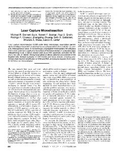
Apr 22, 1996 - Mori for their technical advice and assistance. Care of experimental ... Michael R. Emmert-Buck, Robert F. Bonner, Paul D. Smith,. Rodrigo F.
Apr 22, 1996 - Michael R. Emmert-Buck, Robert F. Bonner, Paul D. Smith,. Rodrigo F. Chuaqui, Zhengping Zhuang, Seth R. Goldstein,. Rhonda A. Weiss ...
site-directed probes.5â7 ABPP probes selectively label active enzymes .... 9400 Amersham Typhoon scanner. ... In the case of OH, only five bands appeared.
Aug 1, 2007 - Heat shock factor protein 4 (HSF4). Z46953.1. 1.841142. 0.006433719. Myb family transcription factor (MYB123). AB029270.1. 1.606206.
Aug 1, 2008 - Arbor, Michigan, and 8Geron, Corporation, Menlo Park, California ...... S. Gryaznov is employed by Geron Corporation and is co-inventor of ...
correlation between focal p53 immunostaining in primary CaP and cancer recurrence after radical prostatectomy. Prostate Cancer and Prostatic Diseases (2003) ...
Nov 29, 2011 - Keywords: Endometrioid endometrial cancer; Laser capture ... Cancer Invest Downloaded from informahealthcare.com by ... Then, it was rinsed with ice-cold RNA nuclease-free water ... cleotide B2, 5.5 µL 20X eukaryotic hybridization con
Graduate School of Medical and Dental Sciences, Niigata University, 2-5274, Gakkocho-dori, Chuo-ku, Niigata ... research fields such as oral embryology [5â10], oral oncology ..... analysis of morphology, histology, immunohistochemistry,.
Jun 2, 2009 - setups; 2) use of a defined kit that incorporates reagents for both LCM RNA ..... Lysis Methodâ13 (see also the Invitrogen user manual for. CellsDirect⢠.... of DNase I (Amp grade 1 U/μL or Turbo 2 U/μL). Mix gently by vortexing .
no realignment by service technicians needed ... Active application support ..... solutions to our customers' needs for
Sep 27, 2012 - filamentous fungi are capable of developing complex, ...... 15 min DNase incubation step (RNase-free DNase Set,. Qiagen, Hilden ..... L, Holford M, Zhang HY, Zhao H, Deng XW, Nelson T: A transcriptome atlas of rice cell ...
treated with Rhodamine-RCA lectin by tail vein injection, and after fluorescence ... Conclusion: Our present study demonstrates that in vivo Rhodamine RCA ...
May 5, 2011 - i fili f i. [Adv Exp Med Biol. 2007] · Review Gene expression profiling of primary · tumor cell populations using laser capture i di ti RNA t. i t lifi ti.
Sep 11, 2017 - 6. Place a small amount of optimal cutting temperature (OCT) medium into .... The use of a brain atlas to guide sectioning and laser capture of ...
Jun 2, 2009 - Fatoumata B. Sow1, Jack M. Gallup1, Randy E. Sacco2, Mark R. ... note: Fatoumata B. Sow and Jack m. ...... pages/reagents/paradise.html).
and that different clones of cells co-exist within a single tumor. Introduction .... lymphatic vessels, muscle or adipose tissue were avoided. Uncoverslipped serial ...
Giovanni Lugli1, Yachana Kataria1, Zachary Richards1, Peter Gann1, Xiaofeng Zhou2, Larisa Nonn1. 1. Department of Pathology, University of Illinois at ...
Jan 4, 2017 - cork tree (Phellodendron amurense) (Zheng et al., 2016). This analysis provided a cell specific lignin profile, but no RNA was isolated.
May 17, 2005 - (7 m) of frozen SSPE brain were prepared on nonplus glass slides (Fisher .... of the eight cells with an identical H chain in clone 3 (IRA-.
Jul 25, 2008 - analyzing gene expression levels in human gastric cancer and colon cancer ... BMC Cancer 2008, 8:210 doi:10.1186/1471-2407-8-210.
stored at 4°C in the presence of desiccant. .... contrast, primers to the carbonic anhydrase, chloro- ... mannitol for 4 h before fixation (toluidine blue O staining).
Methods using manual dissection of brain regions result in an “aver ... enables
investigators to accurately dissect individual groups ... C) Section after microdis.
Mar 15, 2008 - Proteomic Era: Targeting the Genetic Basis of Cancer. Barbara Domazet1 ..... The Cancer Genome Anatomy Project (CGAP) at the. National.
microdissection in combination with laser- pressure catapulting, which allows the fast isolation of single chromosomes for the generation of chromosome-specific ...
Representative PALM Zeiss microdissector images of lifeact-mRuby .... translation initiation factor HA-eIF4E and stained using HA antibodies (green). Panels on ...
Combining laser capture micro-dissection and proteomics reveals an active translation machinery controlling invadosome formation
Ezzoukhry and Henriet et al.
Supplementary Fig. 1: Flowchart of the analytical process (a) Technical flow chart including the metabolic labeling of the proteins to ensure specificity of identification, isolation and collection of fluorescent-labeled structures of interest with an automated laser capture, and finally, protein identification by LC-MS/MS analysis and the enrichment quantification by a label free approach. (b) Detailed workflow of the automated laser capture. A first image is acquired from the Zeiss PALM software. Its positional information is extracted and used by an ImageJ plugin in order to generate a grid of coordinates, covering adjacent field to be explored. For each field, an image is acquired and automatically analyzed by ImageJ to recover the outlines of structures of interest. Structure contours are exported from the ImageJ software as a Zeiss PALM compatible file. The microdissection step is started upon this file’s import. Automatic steps are indicated in squares and manual steps in circles. (b) Assisted invadosome micro-dissection workflow.
Supplementary Fig. 2: Invadosome laser microdissection Representative PALM Zeiss microdissector images of lifeact-mRuby (red)-expressing NIH-3T3-Src cells by transmitted light (a) (scale bar: 30µm). In the higher image (b), the dotted circles surround the rosettes that will be micro-dissected. Blue is Hoechst nuclear staining. The lower image (c) shows the cell after microdissection. Right panels show enlarged views of the boxed regions (scale bar: 10µm).
Supplementary Fig. 3: Functional network of proteins identified in invadosomes and enriched compared to the whole cellular proteome. Annotations were attributed manually for the following functions: “Cancer invasion”, “Invadosomes”, and “Matrix degradation”. Involvement in actin reorganization, cell adhesion, chemotaxis or protein translation was extracted from the Ingenuity® Pathway Analysis Database (Qiagen). Networking was made with the Gephi software. Individual proteins (small circles) are grouped according to depicted molecular functions (large circles) and colorcoded. Proteins that can be attributed to several groups are linked between groups.
Supplementary Fig. 4: Localization and involvement of translation-related proteins (a) Time-course of the number of rosette per nuclei after translation inhibitor treatment. Lifeact-mRubyexpressing NIH-3T3-Src cells were treated with 0.5 µM anisomycin or 35 µM CHX for the indicated time points. Panel on the top shows representative images of the cells for which there is an effect at the earlier time point. The bar graph represents the number of rosettes per nuclei. Error bars represent the SEM (n=20 fields, three independent experiments; ns, not significant, **, P < 0.005; ***, P < 0.001 as compared to the non-treated cells as control). Scale bar: 50 µm. (b) Representative images from time-lapse video microscopy of lifeact-mRuby (red)-expressing NIH3T3-Src cells transfected with eEF2-GFP (green). Scale bar: 10 µm. (c) siRNA screening targeting 19 of the most enriched candidate proteins. Bar graph shows the number of rosettes per nuclei. The black bars represent the controls of the experiment. Control cells were treated with 5 µM GM6001 (metalloproteinase inhibitor) or 5 µM PP2 (Src inhibitor) or with a control siRNA (siCtrl). The grey bars represent the proteins that were further localized. Error bars represent the SEM (n=75 fields, three independent experiments; ns, not significant; *, P