tion to units of Mr 60,300, 44,700, and 39,000 by proteolysis during membrane fractionation. Thus, the analyses presented not only account for the (smaller) ...
Received for publication February 6, 1992 Accepted March 11, 1992
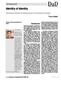
Plant Physiol. (1992) 100, 723-732 0032-0889/92/100/0723/1 0/$01 .00/0
Common Identity of Substrate-Binding Subunit of Vacuolar H+-Translocating Inorganic Pyrophosphatase of Higher Plant Cells1 Philip A. Rea*, Christopher J. Britten, and Vahe Sarafian Plant Science Institute, Department of Biology, University of Pennsylvania, Philadelphia, Pennsylvania 19104-6018 fore, concluded that the substrate-binding subunit of the tonoplast H+-PPase has a common identity in all four organisms.
ABSTRACT There have been conflicting reports in the literature concerning the polypeptide composition of the vacuolar H+-translocating inorganic pyrophosphatase (tonoplast H+-PPase) of plant cells. The major subunit(s) of the enzyme have been attributed to polypeptides of relative molecular weight (Mr) 64,500 (Beta vulgaris), 67,000 (Beta vulgaris), 73,000 (Vigna radiata), and 37,000 to 45,000 (Zea mays). Here, we reconcile these differences to show, through the combined application of independent purification, affinitylabeling, sequencing, and immunological procedures, that the major polypeptide associated with the H+-PPase from all of these organisms, and Arabidopsis thaliana, corresponds to the same moiety. The principal polypeptide components of the H+-PPase purified from Beta and Vigna by independent procedures have similar apparent subunit masses when subjected to sodium dodecyl sulfate-polyacrylamide gel electrophoresis (SDS-PAGE) under identical conditions (M,(Be,a) = 64,500; Mr(vigna) = 66,000) and exhibit identical kinetics of irreversible inhibition and ligand-modified labeling by ['4C]-N-ethylmaleimide. Similarly, the M, 64,500 and 67,000 polypeptides isolated from Beta by independent methods (cf. C.J. Britten, J.C. Turner, P.A. Rea [1989] FEBS Lett 256: 200-206 versus V. Sarafian and R.J. Poole [1989] Plant Physiol 91: 34-38) are indistinguishable: the two polypeptides comigrate when electrophoresed under the same conditions and yield tryptic fragments with identical overlapping sequences. Because both the Nterminal sequence of the Mr 66,000 subunit of the H'-PPase isolated from Vigna and the direct sequence data from Beta align precisely with the deduced amino acid sequence of cDNAs encoding the H -PPase of Arabidopsis, all three enzymes are inferred to be highly conserved structurally. Accordingly, immunoblots of membranes prepared from Arabidopsis, Beta, Vigna, and Zea, probed with antibody affinity purified against the magnesium inorganic pyrophosphate-binding, Mr 66,000 polypeptide of Vigna, reveal a single immunoreactive band at Mr 64,500 to 67,000 in all four preparations. The Mr 66,000 polypeptide of Zea membranes is, however, prone to proteolysis during membrane fractionation and selective aggregation during sample denaturation for SDSPAGE. The anomalous Mr 37,000 to 45,000 subunit pattern previously ascribed to the H -PPase from Zea (A. Chanson and P.E. Pilet [1989] Plant Physiol 90: 934-938) is attributed to loss of the Mr 66,000 subunit and the appearance of polypeptide fragments of M, 44,700 and 39,000 through the combined effects of sample aggregation before SDS-PAGE and proteolysis, respectively. It is, there-
A striking feature of the vacuo-lysosomal compartment of higher plant cells is the presence of two primary H+ pumps on the bounding (vacuolar) membrane: an H+-ATPase (EC 3.6.1.3) (13) and a PPi-energized H+-PPase2 (EC 3.6.1.1) (5, 19). Both enzymes catalyze inward electrogenic H+-translocation (from cytosol to vacuole lumen) to establish an insideacid, inside-positive ATH+ for the energization of secondary H+-coupled solute transport (26), but the H+-PPase has the unusual characteristic of exclusively utilizing PPi as energy source (20, 21). H+-translocating PPases have been identified in the energy-coupling membranes of mitochondria, chloroplasts, and phototrophic bacteria (3), but the vacuolar (tonoplast) H+PPase is the first example of a PPi-energized H+-translocase in a 'nonenergy' coupling membrane (18). In view of the crucial role played by the PPi bond in biological energy transduction, the unique status of PPi as the limiting case of a high-energy phosphate, and the increasing recognition of PPi as a key metabolite in plants (21, 27, 28), the existence of an energy-conserving H+-PPase on the vacuolar membrane of plant cells is of potentially profound bioenergetic
significance. The tonoplast H+-ATPase is now reasonably well characterized. The major subunits of the complex have been identified (14), the gross partitioning of function between subunits has been elucidated (10, 14, 16, 17), and three of the subunits have been cloned and sequenced (13). By comparison, our understanding of the protein chemistry of the tonoplast H+PPase is still rudimentary. Although the enzyme is ubiquitous in the vacuolar membranes of plant cells and capable of 2 Abbreviations: PPase, inorganic pyrophosphatase, A7A,,+, transtonoplast H+-electrochemical potential difference in millivolts (inside versus outside); AVP, cDNA encoding Arabidopsis Vacuolar (H+translocating) Pyrophosphatase; FPLC, fast protein liquid chromatography; LPC, L-a-lysophosphatidylcholine; NEM, N-ethylmaleimide (N-ethyl[2,3-'4C]maleimide); PVP-40, PVP with average mol wt 40,000; Tris-GDEM buffer, 20 mm Tris-acetate (pH 7.5), 20% (v/v) glycerol, 1 mM DTT, 1 mm Tris-EGTA, 2 mM MgCl2; TTBS, Tween in Tris-buffered saline (0.07% [w/v] Tween-20, 0.9% [w/v] NaCl, 10 mM Tris-Cl [pH 7.3]).
l This work was supported by the National Science Foundation (grant No. DCB-9005330), the Department of Energy (grant No. DEFG02-91ER20055), the University Research Foundation, University of Pennsylvania, and a grant from the Biological Sciences Research Group, University of Pennsylvania. C.J.B. was partly supported by the Science and Engineering Research Council (United Kingdom).
723
724
REA ET AL.
making a substantial contribution to the transtonoplast AAH+ (21), a major impediment to the structural analysis of the tonoplast H+-PPase is the confusion in the literature concerning its precise molecular identity. Britten et al. (4) and Sarafian and Poole (24) attributed polypeptides of Mr 64,500 and 67,000, respectively, to the enzyme from red beet (Beta vulgaris) storage root, Maeshima and Yoshida (11) attributed a polypeptide of Mr 73,000 to the enzyme from etiolated hypocotyls of mung bean (Vigna radiata), and Chanson and Pilet (6) attributed polypeptides of Mr 37,000 to 45,000 to the enzyme from corn (Zea mays) roots. Thus, our objective in this investigation was to determine whether the reported differences in size and/or identity for the major subunits(s) of the H+-PPase from different sources have a structural basis or simply reflect differences in methodology between laboratories. To this end, we have examined the H+-PPase of Arabidopsis, Beta, Vigna, and Zea through the combined application of purification, affinity-labeling, sequencing, and immunological approaches. The results of our studies demonstrate that the Mr 64,500 to 67,000, substrate-binding (MgPPi-binding) subunit of the H+-PPase, first identified and purified from Beta (4), has a common identity and shows a remarkable degree of structural conservation among a wide range of plant species, including the previously most discrepant, Z. mays.
MATERIALS AND METHODS
Plant Physiol. Vol. lOO, 1992
mortar and pestle in extraction buffer (1.1 M glycerol, 5 mm Tris-EDTA, 5 mm DTT, 1% [w/v] PVP-40, 1 mm PMSF, 70 mM Tris-Mes [pH 8.0]) and centrifuged at 6,000g for 20 min. The pellet from the first centrifugation was discarded, and the microsomes in the supernatant were pelleted at 100,OOOg for 30 min. The microsomal pellet was resuspended in 1 to 2 mL of suspension medium (1.1 M glycerol, 1 mm Tris-EGTA, 5 mM DTT, 30 mm Tris-Mes [pH 8.0]) to a protein concentration of 2 to 4 mg/mL. Two procedures were used for the preparation of membranes from roots of 4-d-old, dark-grown corn (Z. mays) seedlings. In the first procedure, tonoplast vesicles were isolated as described by Chanson and Pilet (6). In the second procedure, microsomes were prepared by a rapid fractionation procedure, after it was recognized that the H+-PPase
from this source is prone to extensive proteolysis (see 'Results and Discussion'). Approximately 50 g of roots were homogenized at high speed in a Waring blender for 20 s in 100 mL of homogenization medium (250 mm sucrose, 2 mm TrisEDTA, 1 mm DTT, 0.1% [w/v] BSA, 1 mm PMSF, 50 mm TrisMes [pH 8.0]), filtered through cheesecloth, and immediately centrifuged at 6000g for 10 min. The supernatant from the low-speed centrifugation was centrifuged again at 100,OOOg for 20 min, and the resulting microsomal pellet was resuspended in 1 to 2 mL of suspension medium. All membrane preparations were frozen in liquid nitrogen immediately after preparation and stored at -850C for sub-
Preparation of Membranes
sequent use.
Tonoplast vesicles were isolated from red beet (Beta vulgaris L.) storage root by differential and sucrose density gradient centrifugation as described elsewhere (22). Tonoplast vesicles were prepared from etiolated hypocotyls of mung bean (Vigna radiata) by a combination of the procedures of Maeshima and Yoshida (11) and Rea and Poole (19). Hypocotyls were harvested from 4-d-old seedlings of Vigna, washed in ice-cold distilled water, and homogenized in a Waring blender at a tissue fresh weight to medium ratio of 1:1.3 (w/ v) in homogenization medium (1.1 M glycerol, 5 mm TrisEGTA, 1.5% [w/v] PVP-40, 1% [w/v] ascorbic acid, 1 mm PMSF, 50 mm Tris-Mes [pH 7.6]). The homogenate was successively filtered through two and then four layers of cheesecloth and centrifuged at 3600g for 10 min. The pellet was discarded, and the supernatant was centrifuged at 100,OOOg for 35 min. The microsomal pellet from the highspeed centrifugation was resuspended in suspension medium (1.1 M glycerol, 1 mm Tris-EGTA, 2 mm DTT, 5 mm Tris-Mes [pH 7.6]), dispersed with a Dounce homogenizer, and layered onto sucrose step gradients constituted of 10% (w/w) and 23% (w/w) sucrose in suspension medium. The gradients were centrifuged at 100,OOOg for 2 h in a swing-out rotor, after which time the tonoplast vesicles at the 10/23% sucrose interface were removed. The membranes were diluted 10- to 20-fold with suspension medium, sedimented at 100,OOOg for 35 min, and resuspended in 1 to 2 mL of the same medium to a protein concentration of 1 to 3 mg/mL. Microsomes were prepared from 10-d-old, dark-grown seedlings of Arabidopsis thaliana by differential centrifugation. Seedlings (12 g fresh weight) were homogenized with a
Purification of H+-PPase The H+-PPase of Beta tonoplast vesicles was purified by two methods. In the first method, Triton X-100-solubilized tonoplast vesicles (20) were subjected sequentially to gel filtration on Sephacryl S400 and anion-exchange FPLC on Mono-Q as described by Britten et al. (4). In the second
method, KI-treated, ATPase-depleted tonoplast vesicles (16) were solubilized with Triton X-100 and chromatographed on
DEAE-Sephacel in Triton X-100-containing buffer (24). The peak fractions from anion-exchange chromatography were precipitated with 30% saturated ammonium sulfate and finally gel filtered on Sephacryl S400 in running buffer containing 3-[(cholamidopropyl)dimethylammonio]-1-propanesulfonic acid (24). The H+-PPase of Vigna tonoplast vesicles was purified by differential detergent solubilization (11) followed by anionexchange chromatography (4, 11). Tonoplast vesicles were diluted 15- to 20-fold in Tris-GDEM buffer (20% [v/v] glycerol, 1 mm DTT, 1 mn-m Tris-EGTA, 2 mM MgCl2, 20 mi Trisacetate [pH 7.5]) and centrifuged at 100,OOOg for 30 min. The pellet was resuspended in Tris-GDEM buffer to a final protein concentration of 1 mg/mL and brought to 50 mm with 1 M KCl and 2 mg/mg of protein with 5% (w/v) sodium deoxycholate. The suspension was mixed by inversion, incubated on ice for 10 min, and centrifuged at 150,000g for 30 min at 40C. The supernatant from the deoxycholate treatment was discarded, and the membrane pellet was solubilized by stirring in Tris-GDEM buffer containing 0.4% (w/v) LPC for 10 min at room temperature. After the material was centrifuged
H+-INORGANIC PYROPHOSPHATASE SUBUNIT IDENTITY
at 150,000g for 50 min, the supernatant was removed, frozen in liquid nitrogen, and stored at -850C. The LPC-solubilized membranes were further fractionated by chromatography on DEAE-Sephacel. LPC supematant (12 mL) was diluted 1:1 (v/v) with running buffer (40 mm KCl, 0.1% [w/v] Triton X-100, 0.05 mg/mL of type IV-S phospholipid, 10% [v/vl glycerol, 4 mIM MgCl2, 0.5 mm DTT, 5 mM Tris-HCl [pH 6.0]) and applied to an 18.5- x 1.6-cm diameter column packed with DEAE-Sephacel. The sample was eluted with a linear 40 to 320 mm KCl gradient in running buffer at a flow rate of 8 to 12 mL/h. Fractions (1-2 mL) were collected, and aliquots were assayed for PPase activity. PPase and ATPase Assays
PPase and ATPase activity were determined by measuring the rate of liberation of Pi from PPi or ATP, respectively, at 370C. The reaction media contained 30 mm Tris-Mes (pH 8.0), 50 mnm KCl, 5 FM gramicidin-D, 1.3 mm or 3.0 mm MgSO4, and 0.3 mm Tris-PPi or 3.0 mm Tris-ATP, respectively. Reaction was initiated by the addition of membrane protein, and Pi was measured by the method of Ames (2) after precipitation of LPC, Triton X-100, and/or added phospholipid with ice-cold 5% (w/v) TCA, 2% (w/v) perchloric acid (20). To maximize the activity of the solubilized membranes and fractions from chromatography, the reaction media were supplemented with 1.33 mg/mL of sonicated type IV-S phospholipid (20). PPase activity was calculated as half the rate of Pi liberation from PPi (= ,mol of PPi consumed/ unit time).
PPi- and ATP-Dependent HW-Translocation PPi- and ATP-dependent intravesicular acidification were assayed fluorimetrically at 250C with the monoamine dye acridine orange at excitation and emission wavelengths of 495 and 540 nm, respectively, and a slitwidth of 5 nm for both excitation and emission (19, 22). The assay media contained 5 mm Tris-Mes (pH 8.0), 50 mm KCl, 0.3 mm Tris-PPi (or 3 mm Tris-ATP), and 2.5 AM acridine orange. H+-Translocation was initiated by the addition of 1.3 mm (or 3.0 mM) MgSO4 to the assay media.
Inhibition and Labeling by NEM The standard mixture for reaction with NEM contained 30 mm Tris-Mes (pH 8.0), ['2C]- or [14C]NEM (0-100 AM), and the indicated concentrations of ligands (PPi and/or Mg2") (4). Membrane protein was added, and the mixture was incubated for 5 min at 0°C. Reaction was terminated by the addition of 500 AM DTT, and aliquots of the mixture were assayed for PPase activity.
Affinity labeling was performed by pretreating the membranes with 50 AM ['2C]NEM in the presence of 0.3 mm TrisPPi + 1.3 mM MgSO4 ('MgPPi'). NEM, DTT, and ligands were removed by two successive 100-fold dilutions of the membranes in suspension medium (minus DTT), centrifugation at 80,000g, and resuspension of the membrane pellet in suspension medium (minus DTT). The membranes were then treated with 50 FM [1'4C]NEM (6 mCi/mmol) in reaction
725
medium containing 0.3 mm Tris-PPi ('free PPi') or MgPPi at 0°C for 5 min before the addition of 500 AM DTT. Ligands and unreacted ["4C]NEM were removed by dilution of the membranes into suspension medium, sedimentation, and resuspension in 1 to 2 mL of suspension medium.
SDS-PAGE and Fluorography One-dimensional SDS-PAGE was performed as described by Laemmli (9) on concave exponential gradient gels (14). Detergent and lipid were removed from the membranes and chromatographic fractions before the addition of denaturation buffer by extraction with acetone:ethanol (1:1; -200C) (14). All samples intended for SDS-PAGE were routinely denatured in denaturation buffer (10 mm Tris-Cl [pH 8.0], 5% [w/v] SDS, 2% [w/v] 2-mercaptoethanol) for 20 min at 500C before loading. For the investigations of the effects of denaturation temperature on recovery of the catalytic subunit (Fig. 6; see 'Results and Discussion'), some samples were denatured at 1000C. The gels were stained with silver stain (Bio-Rad) or Coomassie blue or double stained (14). '4Clabeled polypeptides were detected fluorographically after destaining the Coomassie blue-stained gels in 7% (v/v) acetic acid. The gels were infiltrated with Amplify (Amersham International PLC, Amersham, England), dried, and exposed to preflashed Kodak X-Omat x-ray film at -850C.
Microsequence Analyses Purified PPase intended for microsequence analysis was delipidated with acetone-ethanol (14) and exhaustively dialyzed against 2 mm Tris-HCl (pH 8.0) in 2-mL collodion tubes (mol wt cut-off 25,000) before lyophilization and denaturation for electrophoresis. The denatured samples were electrophoresed on gradient gels as described above. Highpurity (Fisher Electrophoresis Grade) chemicals were used throughout, the stock solutions of acrylamide/bisacrylamide were deionized with mixed bed ion-exchange resin (Amberlite MB-3), and the gels were polymerized for at least 18 h before sample loading (12). To minimize oxidative modification of the samples during electrophoresis, 0.1 mm thioglycollate was included in the cathodic buffer reservoir (12). After electrophoresis, PPase protein was electrotransferred to 0.45-Am nitrocellulose filters at 60 V for 1.5 h at 40C in a Mini Trans-Blot transfer cell (Bio-Rad). Because SDS was found to diminish the retention of sample on the filters, the electrotransfers were performed in transfer buffer minus SDS (20% [v/v] methanol, 153 mm glycine, 20 mm Tris). Immediately after transfer, the transferred protein was reversibly stained with Ponceau-S (1) by immersion of the filters for 60 s in an aqueous solution of 0.1% (w/v) Ponceau-S and 1 % (v/v) acetic acid. Excess stain was removed from the blots by gentle agitation for 1 to 2 min in 1% (v/v) acetic acid followed by a 60-s rinse in distilled water. The protein band corresponding to the substratebinding subunit of the H+-PPase (see 'Results and Discussion') was excised immediately after destaining. The excised filter strips were stored wet at -850C and shipped on dry ice to the Harvard Microchemistry Facility for in situ tryptic digestion and gas-phase sequence analysis by the method of Aebersold et al. (1).
726
Plant Physiol. Vol. 100, 1992
REA ET AL.
Preparation of Antibody for Immunoscreens The rabbit polyclonal antibody used in these studies had been raised against total peak PPase chromatographic fractions purified from Vigna (11). To ensure monospecificity, the antibody was fractionated further by affinity purification against the M, 66,000 MgPPi-binding subunit of the H+PPase from Vigna (see 'Results and Discussion"). Vigna tonoplast protein (1 mg) was subjected to preparative SDS-PAGE on a single-well, 1.5-mm, 7 to 12% (w/v) concave exponential gradient gel (14) and electrotransferred to nitrocellulose (7). The nitrocellulose filters were rinsed for 10 min in TTBS and blocked for 1 h in TTBS containing 5% (w/v) powdered skimmed milk. After three 5-min rinses in TTBS, the filters were incubated overnight in antibody (1:300 dilution). Two narrow vertical strips were cut from each end of the filters and incubated in TTBS containing horseradish peroxidaseconjugated anti-rabbit immunoglobulin G (Boehringer Mannheim) for 1 h. Immunoreactive bands were visualized with 0.03% (w/v) H202, 0.6 mg/mL of diaminobenzidine, and 0.03% (w/v) NiCl2 (7), after which the strips were aligned with the remainder of the blot, and a narrow horizontal band corresponding to the Mr 66,000 subunit of the H+-PPase was excised. The excised band was rinsed in 0.15 M NaCl for 20 min and given three brief rinses with Tris-buffered saline. Bound antibody was eluted with 0.2 mm glycine and 1 mM EGTA (pH 2.8). The eluate was immediately neutralized with Tris base and made lx with Tris-buffered saline and 0.02% (w/v) with sodium azide.
Immunoblots The component polypeptides of the acetone-/ethanolwashed membrane preparations were electrotransferred from 0.75-mm, 7 to 14% (w/v) concave exponential gradient gels to 0.45-,um nitrocellulose filters (7). After the blots were reversibly stained with Ponceau-S (1), they were processed as described above for the affinity purification of antibody. Immunodetection was with H202/diaminobenzidine/NiCl2 (7) after reaction with 1:100 diluted, affinity-purified antibody.
RESULTS AND DISCUSSION Relative Abundance of H+-PPase
The most homogeneous preparations of the tonoplast H+PPase reported are those from storage root of B. vulgaris (4) and etiolated hypocotyls of V. radiata (11). The H+-PPase from Beta has been purified by 85-fold from Triton X-100solubilized tonoplast vesicles by gel filtration on Sephacryl S400 and anion-exchange FPLC on Mono-Q to yield enzyme constituted of one major polypeptide of Mr 64,500 (4). Differential detergent solubilization and QAE-Toyopearl chromatography of the H+-PPase from Vigna tonoplast vesicles, on the other hand, results in an 8- to 10-fold enrichment of activity and purification of one major polypeptide of Mr 73,000 (11). Our aim at the outset of these investigations was to determine whether the principal polypeptides associated with these two preparations were functionally and structurally equivalent. In partial fulfillment of this objective, the relative activities of the H+-PPase in tonoplast vesicles isolated from Beta and Vigna were enumerated to gain an indication of the relative degrees to which the respective enzymes need be enriched to yield preparations of comparable purity. The results of the estimates of relative catalytic activity are summarized in Figure 1 and Table I. Tonoplast vesicles isolated from Vigna contain 4- to 6-fold greater H+-PPase activity than the corresponding membrane fraction from Beta, whether activity is measured as substrate hydrolysis (Table I) or PPi-dependent H+-translocation (Fig. 1, Table I). The tonoplast H+-ATPase activities of Vigna and Beta are, by contrast, similar. Hence, if it is assumed that the differences in PPase activity between Beta and Vigna are largely attributable to differences in the relative abundances of the enzyme in the two membrane preparations, i.e. the enzymes from both organisms have similar turnover numbers, it would be predicted that the H+-PPase from Vigna need be purified by only 14- to 21-fold to yield a preparation of comparable purity to the 85-fold purified enzyme from Beta (4). The purification data support this contention.
Purification of H -PPase from Vigna and Beta
Protein Estimations Protein was estima.ed by a modification of the method of Lowry (15).
Chemicals Sephacryl S400, DEAE-Sephacel, and FPLC supplies were from Pharmacia LKB Biotechnology Inc. (Piscataway, NJ). Type IV-S phospholipid (partially purified soybean L-a-phosphatidylcholine), LPC (type I, approximately 99% from egg white), and Ponceau-S were purchased from Sigma Chemical Co. ['4C]NEM (6 mCi/mmol) and horseradish peroxidaseconjugated sheep anti-rabbit immunoglobulin G were obtained from Amersham Corp. and Boehringer Mannheim Corp., respectively. The collodion tubes (2-mL capacity, mol wt cut-off 25,000) and nitrocellulose filters were obtained from Schleicher and Schuell (EMSCO, Philadelphia, PA). All of the general reagents were from Fisher Scientific (Pittsburgh, PA) or Sigma.
Differential detergent solubilization and DEAE-Sephacel chromatography of Vigna tonoplast vesicles yields enzyme of similar specific activity (Table II) and purity to the enzyme purified from Beta by gel filtration and FPLC (Fig. 2). However, the degrees of enrichment required to obtain similar levels of homogeneity differ markedly between the two membrane sources. In agreement with the activity assays on isolated tonoplast vesicles, the H+-PPase of Vigna need be purified by only 15-fold to yield enzyme of comparable quality to the 85-fold enriched preparation from Beta. Because the purifications reported here for the H+-PPase from Vigna coincide both quantitatively (Table I) and qualitatively (Fig. 2) with those achieved by Maeshima and Yoshida (11) and the purified enzymes from Beta and Vigna have similar apparent subunit masses (Mr(Beta)= 64,500, Mr(vigna) = 66,000; Fig. 2), we conclude that the Mr 73,000 polypeptide identified by Maeshima and Yoshida (11) and the Mr 66,000 polypeptide identified by us are equivalent.
H+-INORGANIC PYROPHOSPHATASE SUBUNIT IDENTITY
A 100r
B Mg2+
100
V 000000
00 00 00 800 00 0o 0 0 0 8 0o 0 0 0 0 0 0 0 0 0 0 0 0
0° vigna
80 K
Beta PPase
60
-
0. % 0.0
'I
a 40 1
0 0 0 C 0
1.1
o o
40
60 H
0
0 0 0 0 0 0 0 0 0 0 0 0
00
0 0 0 0 0 0 0 0
Van * Ps *
0G0
U
40
0 0 0 o 0 0 0 0 0 0 0
0 0 0 0 0
-
ATPase
00 00 0 0 0 0 0 0 0 0 0 0 0 0 0 0 0 0 0 0 0 0 0 0 o 0 o 0. 0o o o o o o 0o o o
GDt
Vigna PPase 08
80
20
0 0 0 0 0 o 0 0 0 0 0
0
0000000 0
0
8
CD~~~~~~~~~ 00
aC. a 0o00 00
V go
*o80
GD1
Mg2+ oo00
00000000 80s
727
o
-
0 o
o o0 o0000000000 Ps 0000 Ain o
o
o~~~~~~~
o~~~~~~~~~
Ovigna PPaseicdin° 20
[-
°°oooooooi° Gramicidin-D I 0
2
4
6
8
10
12
____j
14
16
Time (min)
0
2
4
6
8
10
12
14
16
Time (min)
Figure 1. Comparison of rates of PPi-dependent and ATP-dependent HW-translocation by tonoplast vesicles isolated from etiolated hypocotyls of V. radiata and storage root of B. vulgaris. A, Vigna PPase versus Beta Ppase. B, Vigna ATPase versus Vigna PPase. Formation of ApH, indicated by fluorescence quenching of acridine orange (2.5 MM), was initiated by the addition of 1.3 mm (or 3.0 mM) MgSO4 to buffers containing 0.3 mm Tris-PPi (or 3.0 mm Tris-ATP), 50 mm KCI, and 10 mm Tris-Mes (pH 8.0). Uncoupler (gramicidin-D) was added at a concentration of 5 Mm. The same amount of membrane protein (28 Mg) was used in each assay.
Inhibition and Covalent Modification by NEM The results of affinity-labeling experiments confirm and extend the conclusions derived from the purification data by showing that the Mr 64,500 and 66,000 polypeptides of Beta and Vigna tonoplast vesicles, respectively, both directly participate in substrate (MgPPi) binding. The H+-PPase activities of Beta and Vigna tonoplast vesicles are subject to ligand-modified irreversible inhibition by NEM (Fig. 3). Inhibition is pseudo-first order and approximates the relationship %C
=
100 (1
-
exp)(k°t[NEM])
where %C is percentage inhibition (percentage of control), ko is the first-order rate constant, t is the time, and [NEM] is the concentration of NEM. Quantitative protection is conferred by MgPPi (k'Beta = 0.22 x 10³ M-1 min-'; k°vigna = 0.25 x 10³ M-1 min-'), whereas free PPi increases the potency of NEM by a factor of 2 (kIBeta= 4.09 X 10³ M-1 min-'; k°Vigna= 6.51 X 10³ M-1 min-') versus control samples incubated with NEM in the absence of ligands (W°Beta = 2.30 x 10³ M-1 min-'; k°Vigna = 3.12 x 10³ M-1 min-'). Inhibition by NEM is independent of membrane protein concentration (Fig. 3, inset) and neither MgPPi nor free PPi affect the kinetics of inhibition of the tonoplast H+-PPase (4). The kinetics and specificity of inhi-
bition by NEM, therefore, provide a means of identifying the MgPPi-binding subunit of the H+-PPase on the basis of its susceptibility to MgPPi-protectable, free PPi-potentiated labeling by ['4C]NEM. SDS-PAGE and fluorography of tonoplast vesicles labeled with 50 /IM ['4C]NEM after pretreatment with 50 Mm [12C]NEM in the presence of MgPPi, to block nonprotectable, Table I. Comparison of PPase and ATPase Activities of Tonoplast Vesicles Isolated from Etiolated Hypocotyls of V. radiata and Storage Root of B. vulgaris PPase activity was assayed in a reaction system containing 30 mm Tris-Mes (pH 8.0), 50 mm KCI, 5 zm gramicidin-D, 1.3 mM MgSO4, and 0.3 mm Tris-PPi. ATPase activity was measured in the same medium containing 3 mM MgSO4 and 3 mm Tris-ATP instead of MgPPi. PPi- and ATP-dependent HW-translocation were measured as described in the legend to Figure 1, and the rate of H+-translocation (%F/min) was estimated graphically (22). ATPase activity (umol/mg.-min) PPase activity (,4mol/mg. min)
PPi-dependent HW-translocation (%F/min) ATP-dependent HW-translocation (%F/min)
Vigna
Beta
0.26 1.08
0.33 0.20
80 23
20 50
728
REA ET AL.
Table II. Purification of Vigna Tonoplast H+-PPase
Tonoplast vesicles (1-2 mg/mL), equilibrated with Tris-GDEM buffer, were treated with 0.4% (w/v) sodium deoxycholate and 50 mM KCI for 10 min at 4'C before centrifugation at 150,000g for 30 min. The supernatant (DOC supernatant) was decanted and the pellet resuspended in 0.4% (w/v) LPC and stirred gently for 10 min at 21 'C. After centrifugation at 150,000g, for 50 min, the supernatant (LPC supernatant) was combined 1:1 (v/v) with running buffer and applied to a DEAE-Sephacel column equilibrated with running buffer. The sample was eluted with a linear 40 to 320 mm KCI gradient. PPase activity was determined on 10- to 30-jAL aliquots of the fractions before (-PL) and after (+PL) the addition of sonicated phospholipid (1.33 mg/mL) to the reaction media. Native membranes were assayed in the presence of 5 AM gramicidin-D to ensure H+/cation equilibration. Peak fraction, column fraction containing the highest PPase activity. Activity
Purification
Step -PL
+PL Step (+PL) Overall (+PL)
pmol/mg- min Tonoplast DOC supernatant LPC supernatant
0.85 0.38 1.34
fold
-
1.00
1.00
0.66 3.36
3.96
3.96
DEAE-Sephacel: peak fraction 1.56 12.50
3.72
14.72
nonessential NEM-reactive groups and to protect protectable groups, generates two prominent '4C-labeled polypeptides of Mr 64,500 and 23,000 in Beta and one labeled polypeptide of Mr 66,000 in Vigna (Fig. 4). In both preparations, the Mr 64,500 to 66,000 polypeptides, alone, are subject to labeling under conditions that cause maximal inhibition of the H+PPase: labeling is abolished by MgPPi and potentiated by free PPi. The purification and affinity-labeling data are, therefore, in strict agreement. Purified preparations of the H+-PPase from Beta and Vigna are primarily constituted of polypeptides of Mr 64,500 and 66,000, respectively, and the same polypeptides are susceptible to MgPPi-protectable, free PPi-potentiated labeling by [14C]NEM in isolated tonoplast vesicles. From the close correspondence between inhibition of substrate hydrolysis and covalent modification by [14C]NEM, the Mr 64,500 and 66,000 polypeptides of Beta and Vigna are deduced to be catalytic in function, a conclusion consistent with the likelihood that MgPPi (29) and/or Mg2PPi (A.A. Baykov, N.P. Bakuleva, and P.A. Rea, unpublished data) are the true substrates) for the enzyme. The Mr 23,000, 14C-labeled polypeptide of Beta is unlikely to be a subunit of the H+-PPase: it is resolved from PPase activity (and the Mr 64,500 subunit) when solubilized membranes are fractionated by FPLC on Mono-Q (4), is absent from Vigna tonoplast vesicles, which contain at least 4-fold more H+-PPase activity (Table I), and is maximally labeled by ['4C]NEM when inhibition is minimal, i.e. when MgPPi is included in the NEM reaction medium (Figs. 3 and 4).
Sequence Data The ostensive size differences between the Mr 64,500 and 67,000 polypeptides of the H+-PPase purified from Beta by the methods of Britten et al. (4) and Sarafian and Poole (24),
Plant Physiol. Vol. 100, 1992
respectively, appear not to have a structural basis. The major polypeptides from the two preparations comigrate when electrophoresed under the same conditions (data not shown) and possess overlapping sequences (Fig. 5). In situ tryptic digestion of the purified Mr 67,000 (Beta-i [24]) and Mr 64,500 (Beta-2 [4]) polypeptides after electrotransfer to nitrocellulose (1) yields peptide fragments with identical sequences. Of a total of six tryptic fragments subjected to gas-phase sequence analysis (three from Beta-l and three from Beta-2), two fragments from each preparation overlap precisely with two of the fragments from the other preparation (Fig. 5). Moreover, all six microsequences, including those unique to Beta-i and Beta-2, are present in the open reading frame of cDNAs encoding the substrate-binding subunit of the H+-PPase from Arabidopsis (23). Clone pAVP-3, which was isolated from a cDNA library constructed in the expression vector XZAP by immunoscreens with antibody affinity purified against the Mr 66,000, MgPPi-binding subunit of the enzyme from Vigna (see 'Materials and Methods'), encodes a polypeptide showing extensive sequence identities to the Mr 64,500 to 67,000 polypeptide of Beta. The direct sequence from Beta and the deduced amino acid sequence of the polypeptide encoded by pAVP-3 show near-complete identity over a span of 66 amino acid residues except for two conservative (Val -- Ile, Gln -- His) substitutions and one
A
1 -
_
.. O
mi-
3_l_
.%VW mat _;.. *qmp
I..&
a
Figure 2. SDS-PAGE of H+-PPase purified from Vigna and Beta. A, Enzyme purified from Vigna. Lane 1, Tonoplast (20 jg); lane 2, 150,000g supernatant (20 ,ug) from solubilization with sodium deoxycholate; lane 3, 1 50,000g supernatant (12 jag) from solubilization with lysophosphatidylcholine; lane 4, peak PPase fraction (8 jag) from DEAE-Sephacel chromatography of supernatant from solubilization with lysophosphatidylcholine. B, Enzyme purified from Beta. Lane 1, Tonoplast (20,jg); lane 2, pooled peak PPase fractions from FPLC on Mono-Q (8 jag). The samples were run on 7 to 14% concave exponential gradient gels after delipidation with acetoneethanol. The gels were double stained with silver stain and Coomassie blue. Arrowheads, Mr in thousands.
H+-INORGANIC PYROPHOSPHATASE SUBUNIT IDENTITY
A
B
90-0°
1oo'
729
loo0
o
o
o
Iv\
a~ ~ ~
o--
~
~
~
--o
0
~
w~~~~~~ I.
A 100w
A
oo
I.b
1\1
a
0\
0
w A
++\ -t
I
.
IC.l
..
0
10
O
1I A' 0
'
.
'
2D
0.30
.
40
'
'
40
20
SO
-
'
'
60
80
^
s
100
-
s
120
in
0
20
40
.
-I
60
l-
.
80
100
(NEMI, tM
[NEM1, tM Figure 3. Kinetics of inhibition of H+-PPase from Vigna and Beta by NEM. Effects of no ligands (0), free PPi (0.3 mm Tris-PPi, A), and MgPPi (0.3 mm Tris-PPi + 1.3 mM MgSO4, 0) on inhibition of H+-PPase of Beta (A) and Vigna (B) tonoplast vesicles by NEM. Inset, Effect of membrane protein concentration on kinetics of inhibition of H+-PPase of Beta tonoplast vesicles by NEM at membrane protein concentrations of 1 (x), 2 (M), and 4 mg/mL (+). Membranes were reacted with NEM in reaction media containing 30 mm Tris-Mes (pH 8.0) and the indicated concentrations of ligands (PPi and/or Mg2+) for 5 min at 0°C. Reaction was terminated by the addition of 500 AM DTT, and aliquots of the reaction media were assayed for PPase activity.
nonconservative
(Ser
-k
Gly)
substitution.
Comparison
of the
deduced N-terminal sequence of pAVP-3 and the N terminus of the substrate-binding subunit of the H+-PPase from Vigna (11), on the other hand, reveals 19 identities and 5 conservative substitutions within a span of 30 amino acid residues. These results not only demonstrate that pAVP-3 encodes the substrate-binding subunit of the H+-PPase but also show that the sequence of the polypeptide concemed is highly conserved among Arabidopsis, Beta, and Vigna.
Immunological Cross-Reactivity
Although the data presented do not preclude the participation of polypeptides in addition to the MgPPi-binding subunit in H+-PPase function, the Mr 37,000 to 45,000 polypeptides identified by Chanson and Pilet (6) for the enzyme from Zea are unlikely candidates for the catalytic, or even major, subunits of the translocase. Immunoblots of membranes from Arabidopsis, Beta, Vigna, and Zea show that all four preparations contain an Mr 64,500 to 67,000 polypeptide (Mr(Arabidopsis) = 66,800; Mr(Beta) = 64,500; Mr(Vigna) = Mr(Zea) = 66,000) that is immunoreactive with antibody raised and purified against the Mr 66,000 subunit of Vigna (Fig. 6). Although the results of immunoblots taken alone can be equivocal in that the specificity of polyclonal antibody is determined by the purity of the original immunogen, such a criticism is not applicable to these findings. The antibody used was affinity purified against the Mr 66,000 subunit of
Vigna, this same subunit is known to directly participate in substrate binding (Fig. 4), and immunoscreens of the Arabidopsis expression library using the same antibody yield cDNA clones with extensive sequence identities to this subunit and the corresponding subunit from Beta (Fig. 5). Two factors are probably responsible for the results obtained by Chanson and Pilet (6): protein aggregation during the preparation of samples for SDS-PAGE and proteolysis during membrane fractionation. Critical for detection of the Mr 66,000 subunit of Zea on SDS gels is the sample denaturation method used. Samples incubated in denaturation buffer at 500C before SDS-PAGE show an intense immunoreaction at Mr 66,000 (Fig. 6C). The boiling procedure used by Chanson and Pilet (6), by contrast, results in total loss of the immunoreactive Mr 66,000 band (Fig. 6C). Moreover, the same result is obtained with the partially purified H+-PPase from Vigna (Fig. 6C) and Beta (data not shown). It is suggested, therefore, that the hydrophobic MgPPi-binding subunit, common to the H+-PPases from different sources, is liable to aggregation at high temperatures and this prevents this polypeptide from entering the separating phase of SDS gels. The Mr 37,000 to 45,000 polypeptides identified by Chanson and Pilet (6) appear to arise by proteolysis of the Mr 66,000 polypeptide (Fig. 6B). Preparation of microsomes in media containing protease inhibitor (PMSF) by a rapid fractionation procedure (see 'Materials and Methods') results in a single, Mr 66,000 immunoreactive band on SDS gels (Fig.
Plant Physiol. Vol. 100, 1992
REA ET AL.
730
-.4
lawwom, :., f.
-:
e
_
f14, -,. %:
48mwdffi..
..TliOw, '49". 'ANNW.
Figure 4. SDS-PAGE and fluorography of ["4C]NEM-treated tonoplast vesicles from Beta and Vigna. A (Beta): Lanes 1 and 2, Coomassie blue-stained gel; lanes 3 and 4, fluorogram. Beta tonoplast vesicles (50 Ag) were labeled with 50 tM [14C]NEM in the presence of MgPPi (lanes 1 and 3) or free PPi (lanes 2 and 4). B (Vigna): Lanes 1 and 2, Coomassie blue-stained gel; lanes 3 and 4, Vigna tonoplast vesicles labeled with [14C]NEM in the presence of MgPPi (lanes 1 and 3) or free PPi (lanes 2 and 4). All samples were pretreated with 50 AM [12C]NEM + MgPPi (= 1.3 mM MgSO4 + 0.3 mm Tris-PPi) before incubation with 50 Mm [14C]NEM + free PPi (= 0.3 mm TrisPPi) or MgPPi (= 1.3 mM MgSO4 + 0.3 mm Tris-PPi). The samples were prepared for SDS-PAGE and electrophoresed as described in the legend to Figure 2. Arrowheads, M, in thousands.
6C). Omission of protease inhibitors from the fractionation media and preparation of tonoplast vesicles by the more protracted protocol of Chanson and Pilet (6), on the other hand, generates membranes containing immunoreactive bands at Mr 60,300, 44,700, and 39,000, in addition to 66,000 (Fig. 6A). Because the affinity-purified antibody is monospecific for the Mr 64,500 to 66,800 polypeptides of Arabidopsis, Beta, and Vigna, it is concluded that the Mr 39,000 to 60,300 polypeptides of Zea tonoplast vesicles are immunologically cross-reactive with each other and the Mr 66,000 polypeptide because they are derived from the latter by proteolysis. The simplest explanation of the results is that the H+-PPase from Zea, like its counterparts in Arabidopsis, Beta, and Vigna, is constituted of one major Mr 64,500 to 66,800 polypeptide. CONCLUSIONS The investigations described demonstrate that the Mr 64,500 to 67,000 substrate-binding subunit of the tonoplast H+-PPase from Arabidopsis, Beta, Vigna, and Zea has a common identity. The Mr 64,500 and 66,000 polypeptides of Beta and Vigna, respectively, strictly copurify with H+-PPase activity, have relative abundances commensurate with the activity of the H+-PPase, and undergo MgPPi-protectable, free PPi-potentiated labeling by [14C]NEM. The previously identified Mr 64,500 and 67,000 subunits of the H+-PPase purified from Beta by two different methods (4, 24) also correspond to the same moiety: they have identical apparent masses on
SDS gels and sequences that align with each other and the deduced sequence of Arabidopsis H+-PPase cDNA clone pAVP-3 (23). The deduced N terminus of pAVP, in turn, aligns with the direct sequence data derived from the N terminus of the corresponding subunit of the enzyme from Vigna (11). Although previous investigations of the H+-PPase from Zea were interpreted to indicate a novel multisubunit composition for the enzyme from C4 plants (6), our findings seem to exclude this possibility. The MA 37,000 to 45,000 subunit composition ascribed to the enzyme from Zea by Chanson and Pilet (6) is explicable in terms of selective elimination of the Mr 66,000 polypeptide through temperature-dependent aggregation during sample denaturation and its fragmentation to units of Mr 60,300, 44,700, and 39,000 by proteolysis during membrane fractionation. Thus, the analyses presented not only account for the (smaller) differences in apparent subunit size between the tonoplast H+-PPases from Arabidopsis, Beta, and Vigna but also the seemingly exceptional subunit composition of the enzyme from Zea. The MgPPi-binding subunit of the tonoplast H+-PPase appears to be the sole polypeptide constituting the functional
Arabidopsis Vigna
Arabi dops is Beta-2 (NT25-2)
1-MVAPA LLPZL WTEIL VPXCA VIGIA FSLFQWY : :11:1 1111 :1: 11 11 1 1111 1-GAA ILPDL GTEIL IPV-A VITIA FALFQWL
255-AADVG ADLVGK IE 11111 111111 :1 AADVG ADLVGK VE
Beta-2 (NT22)
530-TDALD AAGNT TAAIGK 11111 11111 111111 TDALD AAGNT TAAIGK 1iii 11111 11111 DACD AAGNT TAAIG
Arabi dops is
567-AGINT VDVLT PK
Arabi dops is
Beta-i
(NT25)
1:1 Beta-l (NT31)
Arabidopsi s Beta-i (NT53)
11
722-AAVIG DTIGDP LKDTS GPSLN ILIK I 11111 111111 11111 11111 AAVIG DTIGDP LKDTS GPSLN IL-K
III Beta-2 (NT4l)
11111
ASIQT VDVLT PK
11111
VIG DTIGD? L?D
Figure 5. Alignment of direct sequence data of H+-PPase isolated from Beta and Vigna and deduced amino acid sequence of Arabidopsis cDNA clone pAVP-3. The sequences of the tryptic fragments derived from the Mr 67,000 (Beta-1) and Mr 64,500 (Beta-2) polypeptides of the H+-PPase purified from Beta by the methods of Sarafian and Poole (24) and Britten et al. (4), respectively, were obtained after SDS-PAGE and electrotransfer to nitrocellulose. In situ tryptic digestion and gas-phase analysis of the peptide fragments after separation by narrow-bore reverse-phase HPLC were performed as described by Aebersold et al. (1). The N-terminal sequence data for the enzyme from Vigna were taken from Maeshima and Yoshida (11). cDNA clone pAVP-3, encoding the H+PPase from Arabidopsis, was isolated by immunoscreens of a XZAP expression library with antibody affinity purified against the Mr 66,000 subunit of the enzyme from Vigna (23). NT, nitrocellulose transfer.
H+-INORGANIC PYROPHOSPHATASE SUBUNIT IDENTITY
A
C
B
12 3 4
731
1
12 3 4
3 4 _-V
2
-116
-116 -
-
97
97
_~~66
_
*I __ 4:
-66
60-3 ..-
o
.410
_ _w
-44.7 --
39
-45
AM_
Alm
-45 -29
.....*S w
-29
40._
Figure 6. A and B, Immunoblots of membranes prepared from Arabidopsis, Beta, Vigna, and Zea and probed with antibody affinity purified against the Mr 66,000 subunit of the H+-PPase from Vigna. A, Double-stained gel loaded with 20 Ag membrane protein. B, Immunoblot of membranes (6 jig) probed with affinity-purified antibody. Lane 1, Arabidopsis microsomes; lane 2, Beta tonoplast vesicles; lane 3, Vigna tonoplast vesicles; lane 4, Zea tonoplast vesicles. The apparent Mr values of the immunoreactive bands were 66,800 (Arabidopsis), 64,500 (Beta), 66,000 (Vigna) and 66,000, 60,300, 44,700, and 39,000 (Zea). C, Effect of temperature of sample denaturation on detection of M, 66,000 subunit of H+-PPase from Zea and Vigna. Lanes 1 and 2, immunoblots of 6-,ug aliquots of Zea microsomes probed with antibody after sample denaturation for 20 min at 50'C (lane 1) and 100'C (lane 2). Lanes 3 and 4, Silver-stained gel of partially purified H4-PPase from Vigna (8 jqg) after denaturation for 20 min at 50'C (lane 3) and 1000C (lane 4). The gels were run and the blots were prepared and probed as described in "Materials and Methods." Arrowheads, Mr in thousands.
complex. This component alone copurifies with PPase activity during detergent solubilization and chromatography and is the only polypeptide of tonoplast vesicles to undergo MgPPiprotectable, free PPi-potentiated labeling by ["4C]NEM. By extension, selective purification of the MgPPi-binding subunit of the tonoplast H+-PPase from Vigna and its incorporation into artificial liposomes results in the reconstitution of both MgPPi hydrolysis and MgPPi-dependent H+-translocation (C.J. Britten, P.A. Rea, in preparation). In conjunction with the amino acid sequence data deduced from pAVP-3, which show that the MgPPi-binding polypeptide of the tonoplast H+-PPase of Arabidopsis is extremely hydrophobic and contains at least 13 transmembrane spans, in addition to hydrophilic domains containing potential MgPPi-binding motifs (23), these findings strongly suggest that subunits in addition to the polypeptide encoded by AVP-3 need not be invoked to account for the capacity of the tonoplast H+PPase for PPi-energized transtonoplast HW-translocation. The open reading frame of the cDNA insert of pAVP-3 encodes a 770-amino acid polypeptide with a predicted Mr of 80,800 (molecular mass, 81 kD) (23). The corresponding immunoreactive polypeptide of Arabidopsis microsomes, on the other hand, has an apparent MA of 66,800 on SDS gels (Fig. 6). Two important corollaries follow. First, in common with a number of hydrophobic membrane proteins, the Mr
of the MgPPi-binding subunit estimated by SDS-PAGE only has a relative meaning. Binding of nonsaturating amounts of SDS, resulting in the exposure of charged amino acid residues or irregularities in the shape of the SDS-protein complex, can cause large shifts in apparent Mr (23). Second, the anomalous migration of the H+-PPase on SDS gels might explain why estimates of the functional mass of the enzyme have been difficult to reconcile with its electrophoretic characteristics. Although the PPase elutes as a dimer during gel filtration (4, 24) and may function as a homotrimer during H+-translocation (25), recent estimates of the target size ('functional mass') of the tonoplast H+-PPase by radiation inactivation yield values between 91,000 and 96,000 D for (uncoupled) substrate hydrolysis (25). Hence, when account is taken of the tendency for radiation inactivation to give overestimates of size through the secondary, nonnihilatory effects of exposure to high-energy-ionizing radiation (8), the minimum functional or monomeric mass of the enzyme (91-96 kD) and the predicted mass of the polypeptide encoded by the Arabidopsis H+-PPase clone (81 kD) are in remarkably close agreement. ACKNOWLEDGMENTS We wish to thank Dr. W. Lane, Harvard Microchemistry Facility, Cambridge, MA, for performing the microsequence analyses, Dr. Joe Ecker, Plant Science Institute, Department of Biology, University of
732
REA ET AL.
Pennsylvania, for constructing and generously providing us with an aliquot of the Arabidopsis XZAP library, Dr. M. Maeshima, Institute of Low Temperature Science, Hokkaido University, Japan, for an aliquot of antibody directed against the H+-PPase of V. radiata, and Dr. D. Brauer, Eastern Regional Center, U.S. Department of Agriculture, Philadelphia, PA, for providing the Zea tonoplast vesicles used in preliminary experiments.
1.
2. 3.
4. 5.
6.
7. 8. 9. 10. 11.
12. 13.
LITERATURE CITED Aebersold RH, Leavitt J, Saavedra RA, Hood LE, Kent SBH (1988) Intemal amino acid sequence analysis of proteins separated by one- and two-dimensional gel electrophoresis after in situ protease digestion on nitrocellulose. Proc Natl Acad Sci USA 84: 6970-6974 Ames BN (1966) Assay of inorganic phosphate, total phosphate and phosphatases. Methods Enzymol 8: 115-118 Baltscheffsky M, Nyren P (1987) PPi in the energy conversion system of Rhodospirillum rubrum. In A Torriani, ed, Phosphate Metabolism and Cellular Regulation in Microorganisms. American Society for Microbiology, Washington, DC, pp 260-263 Britten CJ, Turner JC, Rea PA (1989) Identification and purification of substrate-binding subunit of higher plant H+-translocating inorganic pyrophosphatase. FEBS Lett 256: 200-206 Chanson A, Fichmann J, Spear D, Taiz L (1985) Pyrophosphate-driven proton transport by microsomal membranes of corn coleoptiles. Plant Physiol 79: 159-164 Chanson A, Pilet PE (1989) Target molecular size and sodium dodecyl sulfate-polyacrylamide gel electrophoresis analysis of the ATP- and pyrophosphate-dependent proton pumps from maize root tonoplast. Plant Physiol 90: 934-938 Harlowe E, Lane D (1988) Antibodies: A Laboratory Manual. Cold Spring Harbor Laboratory, Cold Spring Harbor, NY Kempner ES, Miller JH (1983) Radiation inactivation of glutamate dehydrogenase hexamer: lack of energy transfer between subunits. Science 222: 586-589 Laemmli UK (1970) Cleavage of structural proteins during the assembly of the head of bacteriophage T4. Nature 227: 680-683 Lai S, Randall S, Sze H (1988) Peripheral and integral subunits of the tonoplast H+-ATPase from oat roots. J Biol Chem 263: 16731-16737 Maeshima M, Yoshida Y (1989) Purification and properties of vacuolar membrane proton-translocating inorganic pyrophosphatase from mung bean. J Biol Chem 264: 20068-20073 Matsudaira PT (1989) A Practical Guide to Protein and Peptide Purification for Microsequencing. Academic Press, New York Nelson N, Taiz L (1989) The evolution of H+-ATPases. Trends Biochem Sci 14: 113-116
Plant Physiol. Vol. 100, 1992
14. Parry RV, Turner JC, Rea PA (1989) High purity preparations
of higher plant vacuolar H+-ATPase reveal additional subunits: revised subunit composition. J Biol Chem 264:
20025-20032 15. Peterson GL (1977) A simplification of the protein estimation method of Lowry et al. which is generally more applicable. Anal Biochem 83: 346-356 16. Rea PA, Griffith CJ, Manolson MF, Sanders D (1987) Irreversible inhibition of H+-ATPase of higher plant tonoplast by chaotropic anions: evidence for peripheral location of nucleotide-binding subunits. Biochim Biophys Acta 904: 1-12 17. Rea PA, Griffith CJ, Sanders D (1987) Purification of DCCDbinding proteolipid of higher plant tonoplast H+-ATPase. J Biol Chem 262: 14745-14752 18. Rea PA, Kim Y, Sarafian V, Poole RJ, Britten CJ (1992) Vacuolar H+-translocating inorganic pyrophophatase: biochemistry and molecular biology. In DT Clarkson, DT Cooke, eds, Transport and Receptor Proteins of Plant Membranes. Plenum, New York, London, pp 25-38 19. Rea PA, Poole RJ (1985) Proton-translocating inorganic pyrophosphatase in red beet (Beta vulgari L.) tonoplast vesicles. Plant Physiol 77: 46-52 20. Rea PA, Poole RJ (1986) Chromatographic resolution of H+translocating pyrophophatase from HW-translocating ATPase of higher plant tonoplast. Plant Physiol 81: 126-129 21. Rea PA, Sanders D (1987) Tonoplast energization: two pumps, one membrane. Physiol Plant 71: 131-141 22. Rea PA, Turner JC (1990) Tonoplast adenosine triphosphatase and inorganic pyrophosphatase. Methods Plant Biochem 3: 385-405 23. Sarafian V, Kim Y, Poole RJ, Rea PA (1992) Molecular cloning and sequence of cDNA encoding the pyrophosphate-energized vacuolar membrane proton pump (H+-PPase) of Arabi-
dopsis thaliana. Proc Natl Acad Sci USA 89: 1775-1779 24. Sarafian V, Poole RJ (1989) Purification of an H+-translocating inorganic pyrophosphatase from vacuolar membranes of red beet. Plant Physiol 91: 34-38 25. Sarafian V, Potier M, Poole RJ (1992) Radiation inactivation study of vacuolar H+- ATPase and H+-pyrophosphatase. Biochem J 283: 493-497 26. Sze H (1985) H+-translocating ATPases: advances using membrane vesicles. Annu Rev Plant Physiol 36: 175-206 27. Takeshige K, Tazawa M (1989) Intracellular pyrophosphate distribution in Chara corallina. J Biol Chem 264: 3262-3266 28. Weiner H, Stitt M, Heldt HW (1987) Subcellular compartmentation of pyrophosphate and alkaline pyrophosphatase in leaves. Biochim Biophys Acta 893: 13-21 29. White PJ, Marshall J, Smith JAC (1990) Substrate kinetics of the tonoplast H+-translocating inorganic pyrophosphatase and its activation by free Mg2+. Plant Physiol 93: 1063-1070