segmental analysis and force platform methods can be considered to provide the most accurate results for. COM vertical excursion during human hopping in a.
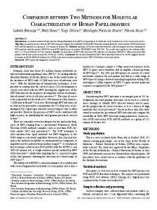
Journal of Applied Biomechanics, 2008, 24, 271-279 © 2008 Human Kinetics, Inc.
Comparison Between Kinematic and Kinetic Methods for Computing the Vertical Displacement of the Center of Mass During Human Hopping at Different Frequencies Alberto Ranavolo,1 Romildo Don,1 Angelo Cacchio,1 Mariano Serrao,1,2 Marco Paoloni,1 Massimiliano Mangone,1 and Valter Santilli1 1
“La Sapienza” University and 2IRCCS Neuromed
Kinematic and kinetic methods (sacral marker, reconstructed pelvis, segmental analysis, and force platform methods) have been used to calculate the vertical excursion of the center of mass (COM) during movement. In this study we compared the measurement of vertical COM displacement yielded by different methods during able-bodied subjects’ hopping at different frequencies (varying between 1.2 and 3.2 Hz). ANOVA revealed a significant interaction between hopping frequency and method (p < 0.001), showing that increasing hopping frequency reduced the differences between methods. A post hoc analysis revealed a significant difference between all methods at the lowest hopping frequency and between the force platform and both the sacral marker and reconstructed pelvis methods at the intermediate hopping frequencies, with differences ranging from 16 to 67 millimeters (all p < 0.05). Results are discussed in view of each methods’ limits. We conclude that the segmental analysis and force platform methods can be considered to provide the most accurate results for COM vertical excursion during human hopping in a large range of hopping frequency. Keywords: biomechanics, stiffness, movement analysis
The displacement of the center of mass (COM) has been mostly used to evaluate walking and running in terms of external work (Blickhan & Full, 1993; Cavagna
Ranavolo, Romildo, Cacchio, Paoloni, Mangone, and Santilli are with the Department of Physical Medicine and Rehabilitation, “La Sapienza” University, Rome, Italy; and Serrao is with “La Sapienza” University–Polo Pontino–ICOT, Latina, Italy, and IRCCS Neuromed, Pozzilli (IS), Italy.
et al., 1976, 2002), biomechanical performance (Kerrigan et al., 1995), and changes of mechanical energy (Iida & Yamamuro, 1987) in healthy subjects. Moreover, COM displacement has been used to determine whole-body stiffness during walking, running, and hopping (Farley & Morgenroth, 1999; Farley et al., 1991, 1998; Ferris & Farley, 1997), to assess balance during perturbation tests (Pai et al., 1998), and to calculate the moment of the ground reaction force during sit to stand (Papa & Cappozzo, 2000). As the positions of the COM that cannot be measured directly, calculations based on kinematic and kinetic methods are required. Kinematic methods require the use of an optoelectronic system to track the trajectory of reflective markers, and differ according to the process used to approximate the COM vertical coordinate. The sacral marker method uses the cutaneous projection of the sacral prominence (Kerrigan et al., 1995), the reconstructed pelvis method uses the center of the pelvis (Saini et al., 1998; Whittle, 1997), and the segmental analysis method uses the weighted average of the individual body segments’ centers of mass (Iida & Yamamuro, 1987; Zatsiorsky & Seluyanov, 1983). Kinetic methods require the use of a force platform to record ground reaction force. The force platform method applies Newton’s second law to the human body, allowing one to calculate the COM vertical excursion by double integration of the vertical component of the ground reaction force (Blickhan, 1989; Cavagna, 1985). When aiming at evaluating COM displacement during movement, it is important to be aware that the investigated motor task may diversely affect the accuracy of the different methods, depending, for example, on the amount of body segment displacement. Hence, when comparing the different methods, the conclusions drawn for a specific motor task cannot be automatically applied to activities with different features. 271
272 Ranavolo et al.
To our knowledge, the various methods used to calculate vertical COM displacement have not previously been compared during hopping, nor has the influence of the hopping frequency on such methods been investigated. Hopping is a motor task commonly used to evaluate biomechanical properties (Farley & Morgenroth, 1999; Farley et al., 1991, 1998; Ferris & Farley, 1997; Granata et al., 2002) and to assess motor performance (Rousanoglou & Boudolos, 2006; Vanneste et al., 2001), and can be divided into a contact and a flight phase. The contact phase is further subdivided in a landing phase, characterized by the downward vertical deceleration of the body mass and, in a take-off phase, characterized by the upward vertical acceleration of the body mass (Blickhan, 1989; Farley et al., 1991; Fritz & Peikenkamp, 2001; McMahon & Cheng, 1990; Rousanoglou & Boudolos, 2006). The aim of this study was to compare the calculation of vertical COM displacement during the landing phase of two-legged hopping in place as yielded by sacral marker, reconstructed pelvis, segmental analysis and force platform methods and to investigate the influence of hopping frequency on these methods.
rigid-link model of the body, consisting of a six-segment model for the lower limbs, one segment for the pelvis, and one segment for the upper body (including trunk, upper limbs, and head). We also acquired data from a standing trial for each subject by adding two heel makers to determine the offset angles. Joint centers of rotation and angular excursions were determined by combining anthropometric and kinematic data (Davis et al., 1991). Vertical COM displacement was calculated between the 6th and the 15th hops during each landing phase. The beginning and termination of the landing phase were identified by the onset and the peak, respectively, of the vertical component of the ground reaction force (Figure 1; Ferris & Farley, 1997). Trials were considered acceptable if the hopping frequency was within 2% of the designated metronome frequency (Farley et al., 1998). The COM vertical displacement between the start and end of the landing phase was calculated by four methods. For the sacral marker method (Gard et al.,
Methods Ten healthy male subjects (body mass 78.3 ± 2.98 [SD] kg) aged 24–32 years participated in this study. The research was approved by the local ethics committee and all the subjects gave their informed written consent. The subjects were instructed to hop barefoot with their trunk extended and hands on their hips, at a frequency designated by a digital auditory metronome, on the extremely stiff surface of a force platform (platform stiffness = 12,000 kN/m, as specified by the manufacturer). They performed a training session to practice matching the beat of the metronome and keeping their balance while hopping. The subjects were tested at 1.2, 1.6, 2.0, 2.4, 2.8, and 3.2 Hz; each test consisted of a series of 20 hops and the order of the hopping frequencies was randomized. Computerized movement analysis was performed using the ELITE system (BTS, Milan, Italy; Ferrigno & Pedotti, 1985) with eight infrared video cameras for the acquisition of kinematic data and a Kistler force platform (Kistler Instruments, Winterthur, Switzerland) for the acquisition of kinetic data. Kinematic and kinetic data were acquired simultaneously and digitized with respective sampling rates of 100 and 500 Hz. All kinematic and kinetic data were filtered using a fourth-order, zero lag, low-pass Butterworth filter with a cut-off frequency of 6 Hz. Anthropometric data were collected for each subject (height, weight, lower limb length, pelvic width, pelvic height, knee and ankle width, foot length) and 18 retroreflective spherical markers were placed using a validated procedure (Davis et al., 1991), modified by excluding the four lower limb bar markers and adding left and right posterior-superior iliac spine markers. The protocol allowed the creation of an eight-segment,
272
Figure 1 — Vertical reaction force, sacrum position, and velocity during hopping.
Kinematic and Kinetic Methods for Computing Vertical Displacement 273
2004; Saini et al., 1998), COM vertical displacement (∆ySMM) was calculated according to the displacement of the sacral marker. For the reconstructed pelvis method, COM vertical displacement (∆yRPM) was calculated according to the displacement of the pelvic center, defined as the point anterior to the sacral marker at a distance of about 5 cm along a line starting from the sacral marker and perpendicular to the plane defined by the sacral and the posterior-superior iliac spine markers (Saini et al., 1998). For the segmental analysis method (Saini et al., 1998), the COM vertical displacement (∆ySAM) was calculated according to the weighted average of the individual body segments’ center of mass (Equation 1):
(1)
where mlower represents the sum of the lower limb segment masses; mpelvis represents the mass of the pelvic segment; mupper represents the mass of the upper body segment (including head, arms, and trunk); and ylower(t), ypelvis(t), and yupper(t) represent the instantaneous COM of the lower, pelvic, and upper segments on the vertical axes, respectively. The masses of the pelvic and lower limb segments and their relative COM were calculated using equations derived from Zatsiorsky and Seluyanov (1983). The mass of the upper body segment was calculated by subtracting mlower and mpelvis from the total body mass. To calculate the yupper(t), we adopted the following procedure. First, during standing, the ybody was assumed to correspond to the ypelvis (Saini et al., 1998). Thus, the position of the upper body COM during standing was calculated using Equation 1, obtaining
Second, the vertical position of the upper body COM compared with the pelvic COM was expressed as a percentage (y%) of the upper body segment vertical length lupper, as calculated by Davis’s protocol:
Third, during hopping trials, yupper(t) was calculated by means of the following formula: where lupper(t) represents the instantaneous projection of the upper body segment length on the vertical axis. For the force platform method, COM vertical displacement (∆yFPM) was calculated as the difference between the values of the position function dy(t) at the initial and final instant of the landing phase. The position function dy(t) was calculated by double integration of the ground reaction force, F(t), in relation to the time (Blickhan, 1989; Cavagna, 1985):
where v0 and r0 are the integration constants relative to the velocity and position of the COM. The constant v0 was assumed equal to zero because it is the velocity value at the end of the landing phase (Gutierrez-Farewik et al., 2006; Saini et al., 1998; Thirunarayan et al., 1996) and r0 is an unknown constant that disappears in the displacement calculation.
273
274 Ranavolo et al.
Statistical Analysis The average vertical COM excursions were calculated for each subject and hopping frequency using the sacral marker, reconstructed pelvis, segmental analysis, and force platform methods. The data yielded by these methods were analyzed using a two-way repeated-measures ANOVA. We also performed a one-way repeated-measures ANOVA at each hopping frequency to determine whether there were any significant differences between the methods. Post hoc analyses were performed using a paired t test with Bonferroni’s corrections when signifi-
cant differences were observed in the ANOVA. P-values less than 0.05 were considered statistically significant.
Results The results are shown in Tables 1–3 and plotted in Figure 2. Briefly, we detected a significant effect of both the method and hopping frequency (all p < 0.05) on the calculation of COM vertical displacement. Moreover, a highly significant interaction effect was observed (Table 1). Likewise, we observed significant differences between
Figure 2 — COM vertical excursion computed with kinematic and kinetic methods at different frequencies.
Table 1 Results of the Two-Way ANOVA Investigating the Effects of Varying Method and Hopping Frequency on the Calculations of COM Vertical Excursion Factor Method Frequency Method × Frequency
F
p value*
32.621 54.841 10.904
0.00863 0.00023 0.00002
*Boldface values indicate statistical significance.
Table 2 Calculation of COM Vertical Displacement With Different Methods at Variable Hopping Frequencies Hopping frequency (Hz) 1.2 1.6 2.0 2.4 2.8 3.2
SMM (m)
SAM (m)
RPM (m)
FPM (m)
F *
p value*
0.292 ± 0.013 0.175 ± 0.020 0.118 ± 0.015 0.092 ± 0.009 0.073 ± 0.010 0.054 ± 0.012
0.269 ± 0.015 0.155 ± 0.025 0.098 ± 0.025 0.078 ± 0.019 0.057 ± 0.007 0.040 ± 0.011
0.311 ± 0.014 0.189 ± 0.027 0.127 ± 0.019 0.099 ± 0.018 0.077 ± 0.002 0.056 ± 0.014
0.244 ± 0.021 0.154 ± 0.014 0.089 ± 0.007 0.072 ± 0.005 0.051 ± 0.007 0.041 ± 0.008
196.550 13.608 27.912 38.261 5.223 12.935
0.00061 0.02977 0.01081 0.00685 0.1039 0.03192
Note. Values are mean ± SD; SMM, sacral marker method; RPM, reconstructed pelvis method; SAM, segmental analysis method; FPM, force platform method. *One-way ANOVA. Boldface values indicate statistical significance. 274
Kinematic and Kinetic Methods for Computing Vertical Displacement 275
Table 3 Post hoc Paired t Test With Bonferroni Corrections Methods SMM–SAM SMM–RPM SMM–FPM SAM–FPM SAM–RPM RPM–FPM
1.2 0.0018 0.0056 ∆ySMM > ∆ySAM; Figure 2) and the kinetic method—the force platform method—yields a vertical COM excursion that is less than that calculated using all kinematic methods at 1.2 Hz, and less than that calculated using the reconstructed pelvis and sacral marker methods at 1.6, 2.0, and 2.4 Hz. These differences can be attributed to the peculiar approximation of each method. Mainly, the motion of the COM within the body occurring during hopping, due to changes in the body segment geometry, can affect the calculation of COM excursion by the different methods. In the following paragraphs, we discuss the effects of varying hopping frequencies on the calculation of the COM vertical excursion for each method. As far as concerns the sacral marker method, it uses the position of the sacral marker to approximate the COM position. This assumption is based on the fact that, in the standing position, the COM is located in the midline of the body at a position just anterior to the second sacral vertebra of the spine (Gard et al., 2004). Indeed, the
assumption introduces an error because when standing the actual position of the COM is not on the back surface, but anterior to the vertebral bones (Gard et al., 2004); hence, pelvic tilt influences the assessment of the vertical position of the COM even when not associated with the movement of other body segments. However, the main error caused by this approximation is that the COM cannot be considered as stationary within the body during the landing phase of hopping as there is a significant displacement of the body segments due to joint motion. These motions determine a vertical shift of the COM within the body (Saini et al., 1998), owing to compression of the lower body (due to ankle dorsiflexion and knee and hip flexion) and upper body (due to moderate flexion of the trunk over the lower limbs). The amount of the COM motion within body is greater at lower hopping frequencies, due to the increase of joints motion (Figure 3). In addition, anterior pelvic tilt increases as the frequency decreases, thereby affecting the calculation of COM displacement even more markedly because the superficial position of the sacral marker determines an overestimation of the vertical coordinate of the COM at the end of the landing phase, and consequently an underestimation of COM excursion with respect to the center of the pelvis (Figure 2). The sacral marker method should be considered as a reliable means of calculating COM excursion during hopping only at high frequencies (>2 Hz), when the body configuration is similar to that of the standing position throughout the hopping trial (to ensure the validity of the assumption regarding COM stationarity) and the pelvic tilt is minimal (to ensure the validity of the sacral marker level). When, instead, a considerable degree of joint rotation and pelvic motion occurs (hopping frequencies ≤2 Hz), other methods that account for these factors are more reliable (Figures 3–4). Like the sacral marker method, the reconstructed pelvis method is also based on the assumption of COM stationarity within the body. In this method, the COM position is approximated to the center of the pelvis, as identified by the position of the sacral and posteriorsuperior iliac spine markers (Saini et al., 1998; Whittle, 1997). Hence, the considerations regarding changes in the position of the COM within the body due to joint angular excursion for the reconstructed pelvis method are the 275
Figure 3 — Kinematic sagittal behavior of pelvis, hip, knee, and ankle at different frequencies.
Figure 4 — COM vertical position as determined by the different methods at the start and end of the landing phase. 276
Kinematic and Kinetic Methods for Computing Vertical Displacement 277
same as those for the sacral marker method. However, the error due to the pelvic tilt is avoided in the reconstructed pelvis method since the COM is assumed to be located in a position that minimizes the effects of pelvic tilt (Saini et al., 1998; Figures 3–4). During hopping, a large degree of lower joint excursion, as it occurs at low frequencies, should be considered as a factor that affects the calculation of COM excursion in the reconstructed pelvis method because the assumption of stationarity loses validity (Figures 3–4). Thus, although the reconstructed pelvis method eliminates the error due to pelvic tilt present in the sacral marker method, the limitations regarding COM stationarity within the body are almost the same as those in the sacral marker method. The reconstructed pelvis method may, like the sacral marker method, can be considered as a reliable method for calculating COM displacement during hopping only when minimal leg joints rotations occur, such as at frequencies >2 Hz (Figures 3–4). The segmental analysis method is the only kinematic method not based on the assumption of COM stationarity within the body. The COM position is determined by using the weighted average of the COM positions of the individual body segments, as calculated from the combination of anthropometric data, regression equations predictive of body segment masses, and kinematic data (Iida & Yamamuro, 1987; Saini et al., 1998; Zatsiorsky & Seluyanov, 1983). In this respect, the segmental analysis method is the most sophisticated of the kinematic methods. The assumptions of the segmental analysis method are related to the rigidity of the body segments (Cappozzo et al., 2005; Whittle, 1997), the invariability of their density, and the negligible intersubject variability of the proportions between segment masses, which are obtained from specimens (Saini et al., 1998). In the other models, it is the complexity of the biomechanical model that affects the accuracy of the calculation most, whereas the error attributable to the segmental analysis method assumptions is not influenced by the motor task being investigated. In this study, we used a model whose main approximation regarded the modeling of the trunk, head, and upper limbs as a single segment. However, the hopping technique was designed in such a way as to ensure that the trunk was extended and the hands fixed on the hips, thereby minimizing changes in the upper body geometry in the vertical direction irrespective of the hopping frequency; by contrast, this method is sensitive to compression of the lower body due to the flexion of the hip, knee, and ankle joints and to the anterior pelvic tilt, thus allowing to accurately describe the COM vertical behavior (Figure 3). The segmental analysis method therefore appears to be the most accurate kinematic method for determining COM vertical excursion during the hopping landing phase, and subsequent considerations will be referred to the data obtained by this method. In the current study, we found that there were differences between the COM vertical excursions yielded
by the different kinematic methods at the lowest hopping frequency (Figure 2), whereas no significant differences were found at the higher hopping frequencies. At low hopping frequencies, the body posture at the start and end of the landing phase may influence the calculation of COM vertical excursion. Indeed, the ensemble of joints positions will affect the COM position within the body, owing to its effects on the centers of mass of the different body segments. For example, increasing trunk and knee flexion will move the upper and lower body centers of mass, respectively, downward, with opposite effects on the COM position within the body (Figures 3–4). The segmental analysis calculations yield lower values for COM displacement than the sacral marker and reconstructed pelvis calculations. This can be explained by the greater sensitivity of the segmental analysis method to COM motion within the body. As the sacral marker and reconstructed pelvis methods overestimate COM excursion when compared with the segmental analysis method, the main factor that affects measurements may be the upward motion of the COM within the body during the landing phase. This motion is due above all to knee flexion and ankle dorsiflexion, which together move the lower body center of mass downward, thus “pushing” the COM up toward the pelvis. In this regard, the counteractive effect of trunk flexion, which moves the upper body center of mass downward, thus “pulling” the COM down toward the pelvis, reduces the error of the sacral marker and reconstructed pelvis methods by attenuating the upward motion of the COM within the body. Hence, the error of the sacral marker method due to the anterior pelvic tilt contributes to compensate for the error due to the motion of the lower limb segments because the sacral marker method records greater upward displacement of the COM than the reconstructed pelvis method during this movement (Figures 3–4). When the motion at leg joints (i.e., the motion responsible for the errors in the sacral marker and reconstructed pelvis methods) progressively decreases as the frequency increases, the differences between these methods and the segmental analysis method lose statistical significance, though a trend toward an overestimation of the COM excursion persists (Figure 2). By contrast, the progressive lowering of the anterior pelvic tilt at higher frequencies reduces the trend toward an overestimation of the reconstructed pelvis method if compared with the sacral marker method because the attenuating effect of this motion on the sacral marker method error is eliminated. The force platform method yields lower COM excursion if compared with all the kinematic methods at 1.2 Hz, whereas at higher hopping frequencies the force platform method did not show significant differences when compared with the segmental analysis method. Hence, the force platform method provides COM excursion data similar to that obtained by the segmental analysis method at hopping frequencies higher than 1.2 Hz. As far as technical aspects are concerned, systematic and random instrumental errors can affect the accuracy
277
278 Ranavolo et al.
of the different methods. With regard to the kinematic methods, the main source of error concerns the accuracy of the optoelectronic system in reconstructing the threedimensional marker trajectories. In our study, the accuracy with which marker positions were reconstructed, according to the instrumentation properties and the volume of acquisition, was lower than 0.3 mm (Chiari et al., 2005). Soft tissue artifacts, that causes marker movements with respect to the underlying bone (Harris & Wertsh, 1994; Saini et al., 1998; Leardini et al., 2005) cause negligible errors with respect to the magnitude of segment movements on the sagittal plane; thus, they cannot affect our results (Leardini et al., 2005). Another potential source of error is the anatomical landmark misplacement, which can affect determination of position and motion of the centers of mass of the different body segments (Harris & Wertsh, 1994; Saini et al., 1998; Della Croce et al., 2005). We tried to minimize the error by accurately following the procedures indicated by Davis et al. (1991). All these errors are amplified when a large number of markers are used (Harris & Wertsh, 1994; Saini et al., 1998), as occurs in the segmental analysis method, thus reducing the accuracy of the method. With regard to the force platform method, the main source of error is the mathematical process required for the calculation. The double-integration process may determine a smoothing of the signal spectrum that may partly explain the lower COM excursion yielded by the force platform method with respect to the kinematic methods (Saini et al., 1998). Finally, in regard to the potential error due to the sampling process of both kinematic and kinetic signals (Chiari et al., 2005), according to the Nyquist theorem it can be considered negligible due to the low hopping frequencies. In conclusion, the segmental analysis and force platform methods can be considered to provide the most accurate results for COM vertical excursion during human hopping in a large range of hopping frequency.
References Blickhan, R., & Full, R.J. (1993). Mechanical work in terrestrial locomotion. In A.A. Biewener (Ed.), Biomechanics: Structures and Systems. New York: Oxford University Press. Blickhan, R. (1989). The spring-mass model for running and hopping. Journal of Biomechanics, 22, 1217–1227. Cappozzo, A., Leardini, A., Della Croce, U., & Chiari, L. (2005). Human movement analysis using stereophotogrammetry. Part 1: theoretical background. Gait & Posture, 21, 186–196. Cavagna, G.A., Thys, H., & Zamboni, A. (1976). The sources of external work in level walking and running. The Journal of Physiology, 262, 639–657. Cavagna, G.A., Willems, P.A., Legramandi, M.A., & Heglund, N.C. (2002). Pendular energy transduction within the step in human walking. The Journal of Experimental Biology, 205, 3413–3422. Cavagna, G.A. (1985). Force platforms as ergometers. Journal of Applied Physiology, 39, 174–179.
278
Chiari, L., Della Croce, U., Leardini, A., & Cappozzo, A. (2005). Human movement analysis using stereophotogrammetry. Part 2: instrumental errors. Gait & Posture, 21, 197–211. Davis, R.B., Ounpuu, S., Tyburski, D., & Gage, J.R. (1991). A gait analysis data collection and reduction technique. Human Movement Science, 10, 575–587. Della Croce, U., Leardini, A., Chiari, L., & Cappozzo, A. (2005). Human movement analysis using stereophotogrammetry. Part 4. Assessment of anatomical landmark mislocation and its effects on joint kinematics. Gait & Posture, 21, 226–237. Farley, C.T., & Morgenroth, D.C. (1999). Leg stiffness primarily depends on ankle stiffness during human hopping. Journal of Biomechanics, 32, 267–273. Farley, C.T., Blickhan, R., Saito, J., & Taylor, C.R. (1991). Hopping frequency in humans: A test of how springs set stride frequency in bouncing gaits. Journal of Applied Physiology, 71, 2127–2132. Farley, C.T., Houdijk, H.H.P., van Strien, C., & Louie, M. (1998). Mechanism of leg stiffness adjustment for hopping on surfaces of different stiffness. Journal of Applied Physiology, 85, 1044–1055. Ferrigno, G., & Pedotti, A. (1985). ELITE. A Digital Dedicated Hardware System for Movement Analysis Via Real Time Signal Processing. IEEE Transactions on Bio-Medical Engineering, 32, 943–950. Ferris, D.P., & Farley, C.T. (1997). Interaction of leg stiffness and surface stiffness during human hopping. Journal of Applied Physiology, 82, 15–22. Fritz, M., & Peikenkamp, K. (2001). Simulating the impact during human jumping by means of a 4-degrees-offreedom model with time-dependent properties. Journal of Motor Behavior, 33, 286–294. Gard, S.A., Miff, S.C., & Kuo, A.D. (2004). Comparison of kinematic and kinetic methods for computing the vertical motion of the body center of mass during walking. Human Movement Science, 22, 597–610. Granata, K.P., Padua, D.A., & Wilson, S.E. (2002). Gender differences in active musculoskeletal stiffness. Part II. Quantification of leg stiffness during functional hopping tasks. Journal of Electromyography and Kinesiology, 12, 127–135. Gutierrez-Farewik, E.M., Bartonek, A., & Saraste, H. (2006). Comparison and evaluation of two ommon methods to measure center of mass displacement in three dimension during gait. Human Movement Science, 25, 238–256. Harris, G.F., & Wertsh, X.X. (1994). Procedures for Gait Analysis. Archives of Physical Medicine and Rehabilitation, 75, 216–225. Iida, H., & Yamamuro, T. (1987). Kinetic analysis of the center of gravity of the human body in normal and pathological gaits. Journal of Biomechanics, 20, 987–995. Kerrigan, D.C., Viramontes, B.E., Corcoran, P.J., & LaRaia, P.J. (1995). Measured versus predicted vertical displacement of the sacrum during gait as tool to measure biomechanical gait performance. American Journal of Physical Medicine & Rehabilitation, 74, 1–8. Leardini, A., Chiari, L., Della Croce, U., & Cappozzo, A. (2005). Human movement analysis using stereophotogrammetry. Part 3: Soft tissue artifact assessment and compensation. Gait & Posture, 21, 212–225. McMahon, T.A., & Cheng, G.C. (1990). The mechanics of running: How does stiffness couple with speed? Journal of Biomechanics, 23(Suppl. 1), 65–78.
Kinematic and Kinetic Methods for Computing Vertical Displacement 279
Pai, Y.C., Rogers, M.W., Patton, J., Cain, T.D., & Hanke, T.A. (1998). Static versus dynamic predictions of protective stepping following waist-pull perturbations in young and older adults. Journal of Biomechanics, 31, 1111–1118. Papa, E., & Cappozzo, A. (2000). Sit-to-stand motor strategies investigated in able-bodied young and elderly subjects. Journal of Biomechanics, 33, 1113–1122. Rousanoglou, E.N., & Boudolos, K.D. (2006). Rhytmic performance during a whole body movement: Dynamic analysis of force-time curves. Human Movement Science, 25, 393–408. Saini, M., Kerrigan, D.C., Thirunarayan, M.A., & Duff-Raffaele, M. (1998). The vertical displacement of the center of mass during walking: a comparison of four measurement methods. Journal of Biomechanical Engineering, 120, 133–139. Thirunarayan, M.A., Kerrigan, D.C., Rabuffetti, M., Della Croce, U., & Saini, M. (1996). Comparison of three methods for estimating vertical displacement of center
of mass during level walking in patients. Gait & Posture, 4, 306–314. Vanneste, S., Pouthas, V., & Wearden, J.H. (2001). Temporal control of rhythmic performance: A comparison between young and old adults. Experimental Aging Research, 27, 83–102. Whittle, M.W. (1997). Three-dimensional motion of the center of gravity of the body during walking. Human Movement Science, 16, 347–355. Zatsiorsky, V.M., & Seluyanov, V.N. (1983). The Mass and Inertia Characteristics of the Main Segments of the Human Body. In H. Matsui & K. Kobayashi (Eds.), Biomechanics VIII-B. Champaign, IL: Human Kinetics.
279