Magn Reson Med Sci, Vol. 13, No. 4, pp. 221–229, 2014
doi:10.2463/mrms.2013-0068
© 2014 Japanese Society for Magnetic Resonance in Medicine
MAJOR PAPER
Comparison between Two Separate Injections and a Single Injection of Double-dose Contrast Medium for Contrast-enhanced MR Imaging of Metastatic Brain Tumors Tomoko OCHI1,2*, Toshiaki TAOKA1, Ryosuke MATSUDA3, Masahiko SAKAMOTO1, Toshiaki AKASHI1, Tetsuro TAMAMOTO4, Tadashi SUGIMOTO3, Hiroshi SAKAGUCHI2, Masatoshi HASEGAWA4, Hiroyuki NAKASE3, and Kimihiko KICHIKAWA1 1
Department of Radiology, Nara Medical University Department of Radiology, Nara Prefectural Mimuro Hospital 1–14 –16 Mimuro, Sangouchou, Ikoma, Nara 636–0802, Japan 3 Department of Neurosurgery, Nara Medical University 4 Department of Radiation Oncology, Nara Medical University (Received July 18, 2013; Accepted April 2, 2014; published online August 27, 2014) 2
Purpose: As stereotactic radiotherapy (SRT) becomes widespread, precise information including number, location, and margin of lesions is required when magnetic resonance (MR) imaging of brain metastasis is performed. We compare methods using 2 separate injections and a single injection for the administration of a double dose of contrast medium for contrastenhanced MR imaging. Materials and Methods: We divided 40 patients with brain metastasis into 2 groups of 20 patients. Group A received 2 separate injections (0.2 + 0.2 mL/kg) of contrast medium (gadoteridol); Group B received a single injection of the same total dose (0.4 mL/kg). Group A underwent spin echo (SE) T 1 -weighted imaging (T 1 WI) and magnetization prepared rapid acquisition with gradient echo sequence (MPRAGE) after each injection, and Group B underwent the same MR studies at the same timing as Group A. We evaluated the number, signal-to-noise ratio (SNR), diameter, margin delineation, and volume of lesions and compared them between early and delayed studies by the 2 methods. Results: The number of detected lesions was largest in delayed studies of MPRAGE in both groups. The SNR of the lesions was statistically lower in early studies of Group A than other studies. Delayed studies of Group B showed statistically better margin delineation than other studies on both SE-T 1 WI and MPRAGE studies. Diameter and enhanced volume were statistically significantly larger on delayed phase than early phase in both groups. Conclusion: Use of a single injection of double-dose contrast medium and longer delay time may improve margin delineation of lesions for the study of brain metastasis. Enhanced volume was larger on delayed phase, and it may influence selection of therapeutic strategy. Keywords: contrast-enhanced MRI, double-dose contrast study, metastatic brain tumor, number of injections trast material are known to improve visualization of brain metastasis on magnetic resonance (MR) imaging. 1,2 For patients with suspected brain metastasis, gadoteridol is approved for use in a triple dose (0.3 mmol/kg body weight [BW] cumulative dose) in the United States and England and in a double
Introduction High dose, concentration, and relaxitivity of con*Corresponding author, Phone: +81-745-32-0505, Fax: +81745-32-0517, E-mail:
[email protected] 221
222
dose (0.2 mmol/kg BW cumulative dose) in Japan. When no lesion is detected or when enhancement is insufficient after initial administration of a single dose (0.1 mmol/kg BW), Japanese regulations allow additional administration of 0.1 mmol/kg BW within 30 min. 3 Although current regulations allow only the separate injection, contrast MR study with a single double dose injection of contrast material (0.2 mmol/kg BW) is expected to improve enhancement and shorten imaging time. However, the effect of the number of injections and timing of image acquisition has not been fully explored. We evaluated the number of injections and image acquisition using 2 methods for administering a double dose of contrast material (gadoteridol), comparing methods using 2 separate injections and a single injection with respect to lesion enhancement, detection, and delineation for contrast-enhanced study for brain metastasis.
Materials and Methods Japanese regulations do not permit the administration of double-dose gadoteridol in a single injection, so we obtained approval of our institutional review board to alter the injection pattern of the contrast medium. We obtained written informed consent from all patients after explaining that possible side effects might include nausea, vomiting, obstructed liver function, hives, anaphylactic shock, and convulsion as well as usual contrast enhanced MRI. We also explained to patients that the single injection of a double dose of gadoteridol is approved in the United States. The corresponding author (T.O.) had complete access to the results of the study, and all authors had control of the data and statistical results included in this article. This prospective study was performed from January 2011 to February 2012. Subjects were 40 patients with a known primary malignancy and brain metastases detected by previous imaging including plain or contrast-enhanced computed tomography (CT) or MR imaging. Primary neoplasms included lung cancer, 34 cases; breast cancer, 4 cases; anal fistula cancer, one case; and colon cancer, one case. Subjects were randomly divided into 2 groups of 20 each–Group A (10 men, 10 women; mean age, 69 years, range, 48 to 84 years) and Group B (13 men, 7 women; mean age, 63 years; range, 48 to 86 years). Patients in Group A received 2 separate injections of contrast material (0.2 + 0.2 mL/kg), and patients in Group B received a single injection (0.4 mL/kg). Imaging was performed on a 1.5-tesla clinical MR system (Magnetom Avanto, Siemens, Munich,
T. Ochi et al.
Germany). The protocol included acquisition of non-contrast T 1 - and T 2 -weighted images followed by contrast-enhanced study. We administered gadoteridol as the contrast medium (Gd-10-[2-hydroxypropyl]-1,4,7,10-tetraazacyclododecane-1,4,7-triacetic acid; ProHance µ , Eisai, Tokyo, Japan) at 0.1 mmol/kg BW for a single dose and 0.2 mmol/kg BW for a double dose. The contrast medium was administered intravenously through a peripheral vein as a bolus at a rate of 0.5 to 1.0 mL/s followed by a flush with 20 mL of saline using a power injector. For the contrast-enhanced images, we applied 2 imaging sequences–spin-echo T 1 -weighted sequence (SE-T 1 WI) (repetition time [TR], 480 ms; echo time [TE], 8.1 ms; flip angle [FA], 80°; field of view [FOV], 23; matrix, 256 © 256; 5-mm thickness; image acquisition time, 2A45AA) and magnetization prepared rapid acquisition with gradient echo sequence (MPRAGE) (TR, 10 ms; TE, 3.5 ms; FA, 10°; FOV, 23; matrix, 256 © 256; 2-mm thickness; image acquisition time, 3A47AA). We applied MPRAGE to obtain thin-slice images to prevent overlooking small lesions and to adapt the planning software for stereotactic radiotherapy (SRT), and we applied SE-T 1 WI, which has a higher signalto-noise ratio (SNR) than the gradient echo method, to reduce such oversight. The imaging protocol for Group A was: non-contrast T 1 /T 2 , injection of a single dose of contrast medium (30AA), SE-T 1 WI (acquisition time 2A45AA, scan initiation time after injection (SIT) 0A30AA), MPRAGE (acquisition time 3A47AA, SIT 3A15AA), additional injection of a single dose of contrast medium, SE-T 1 WI (acquisition time 2A45AA, SIT 7A45AA), and MPRAGE (acquisition time 3A47AA, SIT 10A45AA). The imaging protocol for Group B was: non-contrast T 1 /T 2 , injection of a double dose of contrast medium, SE-T 1 WI (acquisition time 2A45AA, SIT 0A30AA), MPRAGE (acquisition time 3A47AA, SIT 3A15AA), pause (30AA), SE-T 1 WI (acquisition time 2A45AA, SIT 7A45AA), and MPRAGE (acquisition time 3A47AA, SIT 10A45AA). Thus, the timing for corresponding postcontrast imaging was identical between the 2 groups (Fig. 1). Regarding the dose and timing of contrast medium administration, early phase images in Group A were acquired as single-dose contrast images including SE-T 1 WI (SE single) and MPRAGE (MPRAGE single), and delayed phase images in Group A were acquired as double-dose contrast images including SE-T 1 WI (SE separate double) and MPRAGE (MPRAGE separate double). In contrast, early phase images in Group B were acquired as double-dose contrast images including SE-T 1 WI (SE double early) and MPRAGE (MPRAGE double early), and delayed Magnetic Resonance in Medical Sciences
Single Injection of Double Dose for CE MRI
223
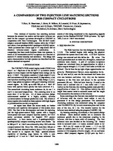
Fig. 1. Protocol for contrast study. The patients in Group A received 2 separate injections. Each injection was a single dose. Patients underwent spin echo (SE) T1-weighted imaging (T1WI) and magnetization prepared rapid acquisition with gradient echo sequence (MPRAGE) after each injection. The patients in Group B received a single injection of a double dose of the contrast medium and underwent the same magnetic resonance (MR) studies with the same timing as Group A.
phase images in Group B were acquired as doubledose contrast images including SE-T 1 WI (SE double delayed) and MPRAGE (MPRAGE double delayed). Two readers blinded to the injection parameters (T.O., T.T.) evaluated images by consensus. MPRAGE images were evaluated in their source images. The readers assessed 5 variables–number of lesions, SNR of lesions, diameters of enhanced lesions, delineation of margins of enhanced lesions, and volume of enhanced lesions. We evaluated the number of detected lesions in the early and delayed phases from Groups A and B, including SE single, MPRAGE single, SE separate double, MPRAGE separate double, SE double early, MPRAGE double early, SE double delayed, and MPRAGE double delayed. We measured signal intensities of lesions to give the SNR for the images listed above and compared them between early and delayed phase in Groups A and B. The SNR was calculated for each post-contrast image based on the measured signal intensity (SI) values. We placed a region of interest (ROI) in a lesion and its background to cover the lesion as completely as possible. We measured diameters of lesions on MPRAGE and compared them between the early and delayed phases. We scored the delineation of lesion margins from 0 to 5 (0, no enhancement; 1, ill-defined lesion margin; 3, half circumference; 5, definition of the whole circumference [Fig. 2]; scores of 2 and 4 were intermediate between 1 and 3 and 3 and 5.) and compared the distribution of scores between the early and delayed phases of Vol. 13 No. 4, 2014
Groups A and B. We calculated enhanced lesion volume with iPLAN µ image radiation therapy planning system software (BRAINLAB AG, Feldkirchen, Germany) based on MPRAGE images and compared volumes between the early and delayed phases. We classified lesions by enhanced lesion volume based on the delayed phase as follows: small lesions, 10 mL; and large lesions, >10 mL. We used a t-test to evaluate quantitative results for the mean number of enhanced lesions per case, and their SNR, mean diameter, and volume and used Mann-Whitney U test to evaluate quantitative results of delineation of enhanced lesions. P < 0.01 was considered statistically significant.
Results Number of lesions: The number of detected lesions was largest in the delayed phase of MPRAGE in both groups (Table 1). The number of lesions detected on early and delayed phases did not differ significantly. The mean numbers of detected lesions per case on the delayed phase of MPRAGE were 4.5 in Group A and 3.9 in Group B. The numbers of lesions detected in the 2 groups did not differ significantly. Signal-to-noise ratios of lesions: In SE-T 1 WI, the SNR of lesions was statistically lower in the early phase of Group A (SE single) than in other studies. The SNRs did not differ significantly among the delayed phase of Group A (SE separate double), early phase of Group B (SE double early), and de-
224
T. Ochi et al.
Fig. 2. Scoring of margin delineation. Delineation was graded 1 for ill-defined margin (a), 2 when it fell between 3 and 1 (b), 3 for half circumference (c), 4 when it fell between 5 and 3 (d), and 5 for delineation of the whole circumference (e).
Table 1.
Table 2.
Number of lesions Number of lesions Early phase
Separated injection SE T1WI (Group A) MPRAGE SE T1WI Single injection (Group B) MPRAGE
80 84 67 75
(89.9%) (94.4%) (87.0%) (97.4%)
Delayed phase 88 89 72 77
(98.9%) ( 100%) (93.5%) ( 100%)
MPRAGE, magnetization prepared rapid acquisition with gradient echo sequence; SE, spin echo; T1WI, T1-weighted imaging The number of detected lesions was largest on delayed studies of MPRAGE in both groups.
layed phase of Group B (SE double delayed). We obtained the same results using MPRAGE (Fig. 3). Diameter of enhanced lesions: The diameter of lesions was larger during the delayed phase than the early phase for both Groups A and B (Table 2). Delineation of margins of enhanced lesions: The score of margin delineation was the lowest of all on SE-T 1 WI of SE single (early phase of Group A) and the second lowest score on SE double early (early phase of Group B). The score was larger for SE separate double (delayed phase of Group A) than SE double early and highest for SE double delayed (delayed phase of Group B). Differences were sig-
Mean diameter of lesions Mean diameter of lesions (mm) Early phase Delayed phase P value
Separated injection 7.09 («SD) (Group A) Single injection 8.01 («SD) (Group B)
7.81 («SD)