Journal of Geriatric Cardiology (2016) 13: 1522 ©2016 JGC All rights reserved; www.jgc301.com
Research Article
Open Access
Comparison between type-2 and type-1 myocardial infarction: clinical features, treatment strategies and outcomes Angel López-Cuenca1, Miriam Gómez-Molina2, Pedro J Flores-Blanco2, Marianela Sánchez-Martínez2, Andrea García-Narbon3, Ignacio De Las Heras-Gómez3, María J Sánchez-Galian2, Esther Guerrero-Pérez2, Mariano Valdés2,4, Sergio Manzano-Fernández2,4 1
Department of Internal Medicine, Hospital de la Vega Lorenzo Guirao, Cieza, Spain Division of Cardiology, University Hospital Virgen de la Arrixaca, School of Medicine, IMIB, Murcia, Spain 3 Department of Biochemistry, University Hospital Virgen de la Arrixaca, School of Medicine, IMIB, Murcia, Spain 4 Department of Internal Medicine, University of Murcia, Murcia, Spain 2
Abstract Objective To assess the differences in incidence, clinical features, current treatment strategies and outcome in patients with type-2 vs. type-1 acute myocardial infarction (AMI). Methods We included 824 consecutive patients with a diagnosis of type-1 or type-2 AMI. During index hospitalization, clinical features and treatment strategies were collected in detail. At 1-year follow-up, mortality, stroke, non-fatal myocardial infarction and major bleeding were recorded. Results Type-1 AMI was present in 707 (86%) of the cases while 117 (14%) were classified as type-2. Patients with type-2 AMI were more frequently female and had higher co-morbidities such as diabetes, previous non-ST segment elevation acute coronary syndromes, impaired renal function, anaemia, atrial fibrillation and malignancy. However, preserved left ventricular ejection fraction and normal coronary arteries were more frequently seen, an invasive treatment was less common, and anti-platelet medications, statins and beta-blockers were less prescribed in patients with type-2 AMI. At 1-year follow-up, type-2 AMI was associated with a higher crude mortality risk (HR: 1.75, 95% CI: 1.142.68; P = 0.001), but this association did not remain significant after multivariable adjustment (P = 0.785). Furthermore, we did not find type-2 AMI to be associated with other clinical outcomes. Conclusions In this real-life population, compared with type-1, type-2 AMI were predominantly women and had more co-morbidities. Invasive treatment strategies and cardioprotective medications were less used in type-2, while the 1-year clinical outcomes were similar. J Geriatr Cardiol 2016; 13: 1522. doi:10.11909/j.issn.1671-5411.2016.01.014 Keywords: Coronary vessels; Follow-up studies; Myocardial infarction
1 Introduction The definitions of the five different clinical types of acute myocardial infarction (AMI) have recently been updated:[1] type-1 AMI is caused by an acute atherothrombotic coronary event; type-2 AMI is a more heterogeneous entity, where a condition other than coronary artery disease (CAD) contributes to an acute imbalance between oxygen supply (e.g., hypoxemia, anemia, hypotension) and demand (e.g., tachycardia, hypertension). In critically ill patients, or in Correspondence to: Angel López-Cuenca, MD, PhD, Department of Internal Medicine, Hospital de la Vega Lorenzo Guirao, Cieza, Spain, Carretera de Abarán, s/n, 30530 Cieza, Murcia, Spain. E-mail:
[email protected] Telephone: +34-968-77555
Fax: +34-968-455632
Received: September 15, 2015
Revised: November 20, 2015
Accepted: November 27, 2015
Published online: January 15, 2016
patients undergoing major (non-cardiac) surgery, elevated values of cardiac biomarkers may appear, due to the direct toxic effects of endogenous or exogenous high circulating catecholamine levels. Also coronary vasospasm and/or endothelial dysfunction have the potential to cause type-2 AMI. Evidence-based treatment recommendations for type-1 AMI are clearly established, however for type-2 AMI these recommendations are lacking. Moreover, treatment strategies in clinical practice in these patients are frequently limited because of a higher co-morbidity of this population. A recent study showed that patients with type-2 AMI are more frequently managed non-invasively and received less frequently cardio-protective drugs.[2,3] Also, there are controversial data about the prognosis of these patients. While some authors have shown this population is strongly associated with a high mortality rate,[4,5] other studies have demonstrated that mortality compared with those patients with
http://www.jgc301.com;
[email protected] | Journal of Geriatric Cardiology
López-Cuenca A, et al. Comparison between type 1 vs. 2 MI
16
type-1 AMI is similar after multivariate adjustment, probably reflecting the poor clinical profile of this group compared with type-1 AMI patients.[3] Thus, the aim of the present study was to compare the patient clinical profiles, treatment strategies, mortality and other clinical outcomes such as recurrent MI, stroke or major bleeding (MB) complications between patients with type-2 and type-1 AMI.
2 Methods The present study is a retrospective analysis of a tertiary university hospital registry. Between January 1, 2012 and September 30, 2013, 824 consecutive patients admitted to the cardiology division with a diagnosis of type-1 or type-2 AMI were included. Patients were classified as having type-1 or type-2 AMI according to the third universal MI definition,[1] and for each case a consensus reached by three cardiologists was needed. Two of these three cardiologists assigned the cause of type-2 AMI. For patients with more than one potential cause, these two doctors selected the initial or fundamental cause. A third cardiologist was consulted, if there was a difference of opinion, to get a consensus. During the index hospitalization, data on demographic and clinical characteristics, medication as well as laboratory, ECG, echocardiography, angiography parameters and clinical complications were collected in detail. The study complied with the Declaration of Helsinki and was approved by the Institutional Review Board of the University Hospital Virgen de la Arrixaca-University of Murcia. Patients were followed-up from admission date to occurrence of death or until day 365 using a standardized protocol that included outpatient clinic attendance, telephone contact and review of the medical notes. Six patients were lost to follow-up. The end-point of the study was the occurrence of all-cause mortality, non-fatal MI, stroke and MB complications. Information on deaths was ascertained from available medical records and death certificates. MI was defined as detection of rise in cardiac biomarkers of necrosis with at least one measurement above the 99th percentile upper reference limit, together with evidence of myocardial ischemia with at least one of the following: electrocardiographic changes indicative of new ischemia (new ST-T changes or new left bundle branch block), new pathological Q waves in at least two contiguous leads, imaging evidence of new loss of viable myocardium or new wall motion abnormality.[1,6,7] Stroke was defined as any clinical manifestation of acute cerebral ischemia or hemorrhage that was ascertained by objective diagnostic/imaging testing.[8] MB was defined according to the Bleeding Academic Research Consortium Definition criteria as bleeding types 3–5.[9]
Categorical variables are presented as frequency values and compared by χ2-tests. Continuous variables are presented as mean ± SD or as medians and IQRs. Differences in continuous variables were evaluated using independent samples t-tests and Mann–Whitney tests, as appropriate. Hazard ratios (HRs) were assessed from Cox regression models. The independent effect of AMI type on clinical outcomes was calculated using a Cox multivariate regression analysis. The covariates were chosen based on clinical considerations and confounders known from risk-stratification models. Linearity assumption was tested using Martingale residuals. Log-cumulative hazard plots, time-dependent covariates, and Schoenfeld residuals were used to evaluate adherence of the proportional hazard assumptions of the Cox model. All P-values (2 tailed) < 0.05 were accepted as statistically significant. Given that this is a retrospective cohort study, it was necessary to achieve comparability of both groups (type-1 and type-2 AMI) with regard to potential confounding variables. This was accomplished using propensity score matching. Variables used to compute the propensity score were those which showed differences between both AMI types and those related with the clinical endpoints. If two variables were related (for example, serum creatinine and estimated glomerular filtration rate), we only selected one of them according to criteria of reproducibility, objectivity and less missing value. Finally, medications at discharge were not used to compute the propensity score because a significant reduction in sample size generated by excluding patients with in-hospital death. We used generalized boosted models attempting a 1: 1 ratio, with no interactions included. Balance between both groups was assessed by unweighted standardized mean differences, variance ratios, histograms and jitter plots of propensity score distribution and visual inspection of QQ plots. HRs calculated in matched population with multivariate analysis were adjusted by those variables not properly balanced after propensity score matching. Statistical analysis was performed using statistical software SPSS 15.0 for Windows.
3 Results 3.1 Clinical characteristics of the study population The study population consisted of 824 patients. Of them, 707 (86%) had type-1 AMI and 117 (14%) had type-2 AMI. The most common causes of type-2 AMI were tachyarrhythmias (36.7%), aortic stenosis (14.5%) and heart failure (13.7%). Tables 1-4 show patients characteristics before and after propensity score matching. Compared with patients with type-1 AMI, those with type-2 were older, more frequently women and had higher co-morbidities such as hypertension,
Journal of Geriatric Cardiology |
[email protected]; http://www.jgc301.com
López-Cuenca A, et al. Comparison between type 1 vs. 2 MI
17
Table 1. Study population clinical characteristics as a function of acute myocardial infarction type. Variables Age, yrs
Whole population
P
Type 1 (n = 707) Type 2 (n = 117) 68 ± 13
72 ± 12
< 0.001
Male
539 (76%)
61 (52%)
< 0.001
Diabetes mellitus
336 (48%)
52 (44%)
0.536
Hypertension
522 (74%)
103 (88%)
0.001
Hyperlipidemia
530 (75%)
89 (76%)
0.798
Current smoking
232 (33%)
23 (20%)
< 0.001
Previous STEMI
101 (14%)
19 (16%)
0.587
Previous NSTE-ACS
160 (22%)
40 (34%)
0.007
Previous PCI
196 (28%)
40 (34%)
0.152
Previous CABG
31 (4%)
12 (10%)
0.008
Chronic heart failure
42 (6%)
21 (18%)
< 0.001
Previous stroke
81 (12%)
20 (17%)
0.085
Peripheral artery disease
57 (8%)
11 (9%)
0.626
Atrial fibrillation/flutter
< 0.001
103 (15%)
51 (44%)
Malignancy
48 (7%)
15 (13%)
0.023
COPD
71 (10%)
17 (15%)
0.145
Data are expressed as mean ± SD or n (%). CABG: coronary artery bypass; COPD: chronic obstructive pulmonary disease; NSTE-ACS: non-ST-segment acute coronary syndrome; STEMI: ST-segment elevation myocardial infarction; PCI: percutaneous coronary intervention.
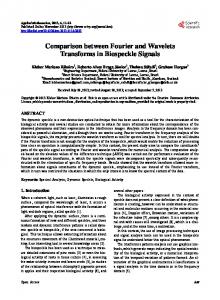
heart failure, impaired renal function, anaemia, atrial fibrillation and malignancy (Table 1). After matching, type-1 and type-2 AMI patients were similar with regards to almost all baseline covariates introduced in the propensity matching analysis (Table 4). Our propensity score matching reduced standardized differences for almost all observed covariates below 20% in absolute value, demonstrating substantial improvement in covariate balance across the AMI type groups (Figure 1). 3.2 Symptoms, signs and complementary studies As shown in Table 2, the main symptom of presentation differed from type-2 to type-1 AMI. While dyspnoea was more common in type-2 AMI, chest pain was more frequent in patients with type-1 AMI. At hospital admission, patients with type-2 AMI had higher heart rate than patients with type-1 AMI. Moreover, pulmonary crackles, legs oedema and cardiomegaly on chest X-ray were more frequently in type-2 AMI patients. In patients with type-2 AMI, the admission ECG showed ST-segment depression and transient ST-segment elevation more often than in patients with type-1 AMI (Table 2). However, persistent ST-segment elevation and pathological Q waves were less frequent in patients with type-2 AMI. Laboratory analyses showed that patients with type-2 AMI had lower estimated glomerular filtration
Figure 1. Absolute standardized differences before and after propensity score matching comparing covariate values for type-1 and type-2 acute myocardial infarction. AF: atrial fibrillation; COPD: chronic obstructive pulmonary disease; eGFR: estimated glomerular filtration rate; LVEF: left ventricular ejection fraction; NSTE-ACS: non-ST-segment acute coronary syndrome; PAD: peripheral arterial disease; SBP: systolic blood pressure.
rate, haemoglobin and higher sensitivity troponin T concentrations. In addition, these patients also had more often significant aortic stenosis and mitral regurgitation, but similar left ventricular ejection fraction on echocardiogram (Table 2). 3.3 Management Reperfusion strategies and invasive treatment was less common in patients with type-2 AMI than those with type-1 AMI (Table 3). Moreover, among patients who underwent coronary angiography, normal coronary arteries or non-obstructive CAD (< 50% stenosis) were more common in type-2 than in type-1 AMI (67% vs. 9%, P < 0.001). During hospitalization period, patients with type-2 AMI underwent invasive coronary angiography were less frequently and less often revascularized than patients with type-1 AMI (Table 3). As expected, the use of thrombolytic agents and glycoprotein IIB/IIIA inhibitors were less frequent in type-2 AMI as compared with type-1 AMI. At hospital discharge, cardio-protective medications such as β-blockers, ACE inhibitors and statins were less often prescribed to type-2 AMI patients. Antiplatelet drugs were also less often prescribed, while anticoagulants and diuretics were more often prescribed to patients with type-2 AMI. By contrast, use of anti-aldosterone antagonists and angiotensin II receptor blockers did not differ between the groups (Table 3). 3.4 Prognosis In both whole population and propensity matched cohort,
http://www.jgc301.com;
[email protected] | Journal of Geriatric Cardiology
López-Cuenca A, et al. Comparison between type 1 vs. 2 MI
18 Table 2. Symptoms, signs and complementary studies findings as a function of acute myocardial infarction type.
Table 3. Study population management as a function of acute myocardial infarction type.
Whole population Variables
Whole population P
Type 1
Type 2
(n = 707)
(n = 117)
618 (87%)
87 (74%)
< 0.001
Dyspnea
38 (6%)
22 (19%)
< 0.001
Other symptoms
51 (7%)
8 (7%)
0.987
Cardiac arrest
19 (3%)
2 (2%)
0.755
SBP, mmHg
134 ± 29
135 ± 31
0.693
DBP, mmHg
73 ± 16
72 ± 17
0.532
Symptoms and signs Chest pain
P
Type 1
Type 2
(n = 707)
(n = 117)
Coronary angiography
622 (88%)
46 (39%)
PCI
486 (69%)
11 (9%)
< 0.001
Drug eluting stent
390 (55%)
7 (6%)
< 0.001
CABG
28 (4%)
0 (0%)
0.024
Thrombolytic
28 (4%)
0 (0%)
0.024
111 (16%)
0 (0%)
< 0.001
In-hospital procedures
Heart rate, beats/min
80 ± 36
102 ± 36
< 0.001
Pulmonary crackles
152 (22%)
40 (34%)
0.003
S3
22 (3%)
6 (5%)
0.266
Legs edema
33 (5%)
13 (11%)
0.005
Chest X-ray Cardiomegaly
148 (22%)
41 (36%)
0.001
Pulmonary congestion
116 (17%)
25 (22%)
0.193
49 (7%)
32 (27%)
< 0.001
Admission ECG findings Atrial fibrillation/flutter
Variables
Glycoprotein IIB/IIIA inhibitors
< 0.001
Medications at discharge* β-blocker
614 (93%)
86 (78%) < 0.001
ACEI
438 (66%)
53 (48%) < 0.001
Angiotensin receptor blockers
160 (24%)
35 (32%)
0.095
Antialdosterone antagonist
131 (20%)
22 (20%)
0.998
Diuretic
230 (35%)
70 (63%) < 0.001
Statins
648 (96%)
92 (83%) < 0.001
Aspirin
647 (97%)
72 (65%) < 0.001
35 (5%)
10 (9%)
0.214
Other antiplatelet
621 (94%)
46 (41%) < 0.001
Q waves
156 (22%)
12 (10%)
0.006
Oral anticoagulant
89 (13%)
44 (40%) < 0.001
ST-segment elevation
225 (32%)
1 (0.9%)
< 0.001
Data are expressed as n (%). ACEI: angiotesin converter enzyme inhibitor;
19 (3%)
9 (8%)
0.011
CABG: coronary artery bypass; PCI: percutaneous coronary intervention.
ST-segment depression
152 (22%)
35 (30%)
0.044
*Referred to patients alive at discharge (type 1, n = 666; type 2, n = 111)
Symmetric negative T waves
100 (14%)
9 (8%)
0.056
Glucose, mg/dL
168 ± 87
158 ± 93
0.230
Serum creatinine, mg/dL
1.1 ± 0.5
1.2 ± 0.6
0.034
eGFR, mL/min per 1.732 m2
80 ± 36
63 ± 28
< 0.001
Hemoglobin, g/dL
13.8 ± 1.9
12.5 ± 2.1
< 0.001
Leucocytes, 103/µL
10.4 ± 4.7
9.5 ± 4.4
0.042
hs-troponin T, ng/L
70 [26283]
Left bundle branch block
Transient ST-segment elevation
Laboratory parameters
36 [22131] < 0.001
Echocardiogram findings LVEF, %
54 ± 13
56 ± 15
0.172
Aortic stenosis
66 (5%)
27 (24%)
Aortic insufficiency
23 (3%)
8 (7%)
0.071
Mitral regurgitation
67 (10%)
23 (20%)
0.001
Tricuspid regurgitation
21 (3%)
7 (6%)
0.106
Pericardial effusion
20 (3%)
2 (2%)
0.757
Moderate/severe valvulopathy < 0.001
Data are expressed as mean ± SD, median [interquartile range] or n (%). DBP: diastolic blood pressure; eGFR: estimated glomerular filtration rate; LVEF: left ventricular ejection fraction; SBP: systolic blood pressure.
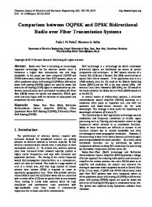
there is a lower incidence of in-hospital MI in patients with type-2 versus those with type-1 AMI (whole population: 0% vs. 4%, P = 0.038 and matched population: 0% vs. 6%, P = 0.029). However, the incidence of all other in-hospital complications was comparable in both groups (Table 5). Patients with type-2 AMI had significantly higher 1-year mortality compared to patients with type-1 AMI (23.3% vs.
14.4%, P = 0.02) (Figure 2). By contrast, both type-2 AMI and type-1 AMI had similar rate of non-fatal MI (9.8% vs. 10.3%, P = 0.87), stroke (3% vs. 0.9%, P = 0.35) and MB complications (5.7% vs. 7.8%, P = 0.39) (Figure 2). In univariate Cox regression analysis (Table 6), type-2 AMI was associated with a higher mortality risk (HR: 1.75, 95% CI: 1.14–2.68; P = 0.001), but this association did not remain significant after multivariable adjustment (P = 0.785). Furthermore, we did not find type-2 AMI to be associated with other clinical outcomes neither using univariate nor multivariate Cox regression analyses (Table 6). As shown in Table 6, there is no difference in events rate in analysis after propensity score matching.
4 Discussion In the present study, we described clinical characteristics, management and prognosis of a consecutive cohort of patients with type-2 AMI in comparison with type-1. Despite the important differences in baseline characteristics, clinical presentation and treatment strategy between the two groups, the 1-year adjusted mortality was similar. Moreover, we showed that the incidence of in-hospital complications and 1-year ischemic or hemorrhagic events was similar in type-1
Journal of Geriatric Cardiology |
[email protected]; http://www.jgc301.com
López-Cuenca A, et al. Comparison between type 1 vs. 2 MI
19
Table 4. Characteristics of patients as a function of acute myocardial infarction type after propensity score matching. Matched population
Variables Age, yrs
Absolute standardized
Type 1 (n = 98)
Type 2 (n = 98)
differences
74 ± 1
71 ± 12
19.8
Variance ratio
P
1.11
0.138
Sex, male
55 (56%)
54 (55)
2.1
1.00
0.886
Diabetes mellitus
57 (58%)
44 (45)
26.5
1.02
0.063
Hypertension
83 (85%)
88 (90)
16.8
0.71
0.284
Current smoking
43 (44%)
40 (41)
6.2
0.98
0.665 0.306
Previous NSTE-ACS
42 (43%)
35 (36)
14.8
0.94
Chronic heart failure
15 (15%)
17 (17)
5.4
1.11
0.699
Previous stroke
16 (16%)
18 (18)
5.24
1.09
0.706
Peripheral artery disease
13 (13%)
10 (10)
10.1
0.79
0.506
Atrial fibrillation/flutter
36 (37%)
39 (40)
6.2
1.03
0.659
Malignancy
11 (11%)
13 (13)
5.9
1.15
0.663
COPD
11 (11%)
13 (13)
5.9
1.15
0.663
Cardiac arrest
3 (3%)
2 (2)
7.2
0.67
1.000
Killip ≥ 2
68 (69%)
63 (64)
10.6
1.08
0.448
SBP, mmHg
130 ± 29
138 ± 32
25.6
1.18
0.062
Heart rate, beats/min
90 ± 29
95 ± 33
16.4
1.36
0.338 0.868
2
62 ± 24
62 ± 24
5.4
1.03
Hemoglobin, g/dL
12.6 ± 2.0
12.6 ± 2.1
2.1
1.13
0.999
hs-troponin T, ng/L
49 [24191]
34 [20126]
4.9
1.01
0.267
LVEF, %
eGFR, mL/min per 1.732 m
54% ± 15%
55% ± 15%
9.1
0.94
0.566
Aortic stenosis (moderate/severe)
29 (30%)
22 (22%)
17.0
0.84
0.254
Mitral regurgitation (moderate/severe)
19 (19%)
19 (19%)
0
1
1.000
Data are expressed as mean ± SD, median [interqueartile range] or n (%). CABG: coronary artery bypass; COPD: chronic obstructive pulmonary disease; eGFR: estimated glomerular filtration rate; LVEF: left ventricular ejection fraction; NSTE-ACS: non-ST-segment acute coronary syndrome; SBP: systolic blood pressure.
Table 5. In hospital complications as a function of acute myocardial infarction type. Whole population
Complications Type 1 (n = 707) Use of inotropic agents Intra-aortic balloon pump
62 (9%)
Matched population P
Type 1 (n = 98)
Type 2 (n = 98)
P
8 (7%)
0.488
9 (9%)
5 (5%)
0.267
0
0.248
1 (1%)
0
1.000
Non-invasive mechanical ventilation
47 (7%)
6 (5%)
0.535
11 (11%)
5 (5%)
0.118
Invasive mechanical ventilation
48 (7%)
4 (3%)
0.165
6 (6%)
4 (4%)
0.516
1 (0.9%)
0.709
0
0
-
Haemodialysis and/or hemofiltration Ventricular thrombus Vascular access complications
8 (1%)
Type 2 (n = 117)
4 (0.6%) 14 (2%) 8 (1%)
1 (0.9%)
0.399
0
1 (1%)
1.000
0
0.609
2 (2%)
0
0.497
Ventricular arrhythmias
39 (6%)
4 (4%)
0.410
7 (7%)
3 (3%)
0.145
Atrial fibrillation
29 (4%)
5 (4%)
0.807
4 (4%)
3 (3%)
1.000
High degree atrioventricular block
28 (4%)
4 (3%)
1.000
4 (4%)
4 (4%)
1.000
Death
41 (6%)
6 (5%)
0.772
8 (8%)
4 (4%)
0.233
Myocardial infarction
25 (4%)
0.038
6 (6%)
0
0.029
0
Stent thrombosis
6 (0.8%)
0
0.602
0
0
-
Stroke
7 (1%)
0
0.623
1 (1%)
0
1.000
3 (3%)
1.000
5 (5%)
2 (2%)
0.445
Major bleeding
18 (3%)
Data are expressed as n (%). http://www.jgc301.com;
[email protected] | Journal of Geriatric Cardiology
López-Cuenca A, et al. Comparison between type 1 vs. 2 MI
20
Figure 2. One-year clinical outcomes as a function of AMI type. AMI: acute myocardial infarction.
and type-2 AMI. Also, in our propensity score matched study, we did not find differences with respect to the incidence of 1-year events in both groups. The third Universal Definition of MI consensus document defines type-2 AMI in instances in which a supply/ demand imbalance leads to myocardial injury with necrosis that is not caused by acute coronary syndrome, including arrhythmias, aortic dissection, severe aortic valve disease, hypertrophic cardiomyopathy, shock, respiratory failure, severe anemia, hypertension with or without left ventricular hypertrophy, coronary spasm, coronary embolism or vasculitis, and coronary endothelial dysfunction without CAD.[1] In our study, the most common cause of all was tachycardia (36.7%), which represents a similar percentage to that re-
ported in the literature,[10] and the ratio type-2/type-1 AMI was 16.5%. Also, the other main causes were similar (heart failure, aortic stenosis, anaemia/bleeding), with a range between 5% and 15%, although in our cohort coronary vasospasm was determined in a higher percentage of patients.[10] Previously[11] reported global incidence of type-2 AMI varies from 1.6% to 29.6%.[1113] This wide range reflects the lack of clear and objective diagnostic criteria, where it is difficult to differentiate type-1 from type-2 AMI and also type-2 AMI from myocardial injury of multi-factorial genesis.[14] Saaby, et al.[13] have proposed specific criteria for type-2 AMI, in order to avoid the implicit subjectivity in the clinical diagnosis. However, their application is difficult because of multifactorial nature of the pathophysiologic mechanism of ischemia in these patients. Considering clinical characteristics of our patients with type-2 AMI compared to type-1, we noted that they did not significantly differ from those showed in other reports.[2,10,15] Thus, patients with type-2 AMI were more often women, older and had a higher prevalence of cardiovascular risk factors or co-morbidities, such as hypertension, heart failure, impaired renal function, anaemia, aortic stenosis, atrial fibrillation and malignancy. Prevalence of peripheral artery disease and chronic obstructive pulmonary disease was similar between both groups, although other authors with larger samples have indeed reported differences.[2,15] However, little information exists about the clinical presentation of these patients,[15] which more frequently presented with dyspnea at admission, with higher heart rate and with more physical examination and radiographic signs of heart failure.
Table 6. Cox regression risk analysis of type-2 acute myocardial infarction for prediction of 1-year clinical events. Events
HR
95% CI
P
HR
95% CI
P
Death Unadjusted HR
1.75
1.142.68
0.001
0.84
0.461.53
0.569
Adjusted HR
0.88
0.501.53
0.785*
0.88
0.481.63
0.692**
Non-fatal myocardial infarction Unadjusted HR
0.76
0.411.41
0.376
1.20
0.522.78
0.667
Adjusted HR
2.12
0.905.28
0.196*
1.38
0.593.22
0.463**
Unadjusted HR
2.64
0.3519.61
0.348
0.25
0.32.21
0.210
Adjusted HR
0.15
0.021.50
0.106*
0.24
0.12.18
0.203**
Stroke
Major bleeding Unadjusted HR
0.61
0.281.27
0.176
0.83
0.292.28
0.710
Adjusted HR
1.17
0.413.38
0.768*
0.89
0.322.51
0.825**
*HRs calculated in total population with multivariate analysis adjusted by age, diabetes mellitus, previous NSTE-ACS, chronic heart failure, atrial fibrillation, previous stroke, peripheral artery disease, malignancy, Killip class, heart rate, SBP, eGFR, hemoglobin, hs-troponin T and LVEF; **HRs calculated in matched population with multivariate analysis adjusted by diabetes mellitus and SBP. eGFR: estimated glomerular filtration rate; HR: harzad ratio; LVEF: left ventricular ejection fraction; NSTE-ACS: non-ST-segment acute coronary syndrome; SBP: systolic blood pressure; STEMI: ST-segment elevation myocardial infarction. Journal of Geriatric Cardiology |
[email protected]; http://www.jgc301.com
López-Cuenca A, et al. Comparison between type 1 vs. 2 MI
The differences in ECG were also noteworthy. Type-2 AMI patients presented more frequently with ST-segment depression and rarely with persistent elevation. This is consistent with data from other previous studies.[2,10] Non-STsegment elevation MI and ST-segment elevation MI terms may be used with caution in patients with type-2 AMI because they may confuse the healthcare community, who has associated these terms with plaque rupture and all its attendant therapies. Globally in our study, as in previous works,[2] patients with type-2 AMI were less likely to undergo coronary angiography or percutaneous coronary angioplasty or to take dual antiplatelet therapy than patients with type-1. The reason for this discrepancy is probably multifactorial. First, these patients have more co-morbidities and their physicians may tend to use more conservative strategies and avoid aggressive treatments. Second, the impact of antithrombotic and/or antiplatelet therapies, as well as the role of reperfusion in patients without plaque rupture are uncertain and might be detrimental or contraindicated in many cases, e.g., in a patient with type-2 AMI in the setting of severe anemia due to an acute gastrointestinal hemorrhage. And finally, the older patients with type-2 AMI may have been less likely to agree to undergo invasive procedures or to take multiple medications.[16,17] On the other hand, patients with type-2 AMI less often received secondary preventive treatment such as β-blockers, statins or angiotensin-converting-enzyme inhibitors and more commonly receive specific treatment for concomitant diseases, as anticoagulants for atrial fibrillation or diuretics for heart failure. All these discrepancies in management of both groups of patients are common in studies published before and are due to the absence of guidelines addressing the acute or long-term treatment of this entity.[11] So, there is an urgent need for evidence-based diagnostic and therapeutic strategies, primarily randomized, controlled clinical trials. Finally, we have analyzed in detail the prognosis of these patients. We did not find differences regarding in-hospital complications and 1-year incidence of ischemic (non-fatal MI, stent thrombosis or stroke) or MB events. To our knowledge, only it has been published a study that included in-hospital complications,[15] where most of these complications were more common of patients with type-1 compared to patients with type-2 AMI. However, the absence of multivariate analysis in this study makes it difficult to identify predictors for and risk-stratification of type-2. Therefore we analyzed the prognosis of these patients showing the importance of each of these predictors. Moreover, in univariate analysis, 1-year mortality was higher in type-2 AMI patients but after adjustment for confounding factors this difference did not achieve statistical significance. Previous studies
21
have shown contradictory results regarding long-term mortality. In the Swedish study of Baron et al.,[10] the crude 1-year mortality was higher in type-2 AMI than type-1 but after adjustment background characteristics, treatments and clustering by treating hospitals, the difference was attenuated and did not reach statistical significance, reflecting that the higher crude mortality in type-2 AMI may be caused by factors other than the type of AMI itself. However, Saaby, et al.[2] reported that type-2 AMI was a significant predictor of an adverse outcome using multivariable regression analysis. This controversy may be probably explained by the heterogeneity of the patients included in these studies due to the subjectivity of the diagnostic criteria for type-2 AMI and by the different diagnostic methods used (only the Danish study, like us, used a high sensitivity troponin assay for all patients).[18] Also, unlike previous studies, we used propensity score matching to control for several potential confounding variables unevenly distributed between groups. The “negative” results in our study need to be interpreted in the context of whether these might be type II errors. So, further studies are needed to clarify the diagnosis, treatment and prognosis of patients with type-2 AMI. There are some limitations in the present study that need to be considered. It is small and reflective of the experience of one hospital in Spain. Only patients admitted to our unit, which is equipped to perform coronary angiography and coronary revascularizations, were included; the applicability of the present results should therefore be viewed with caution in centers with other types of populations and medical facilities, and should be considered as hypothesis generating. However, single-center studies offer the advantage of evaluating homogeneous populations and care processes, unlike multicenter studies, which often differ in the availability of their logistical resources and management habits. The small sample size is a critical limitation that makes it difficult to draw firm conclusions. A study with a larger sample size and more registered events would provide more power. Nonetheless, the demographics and outcomes of our study subjects are comparable to other type-2 AMI. Complete cardiac examinations were not performed in all patients. Thus, diagnostic procedures and supplementary blood sampling were done at the discretion of the treating physicians. The lower rate of coronary angiography in type-2 AMI may, in part, reflect verification bias of an unexpected finding of culprit lesion, which can lead to reclassification to type-1 AMI. As the patients with type-2 AMI were older and had more comorbidities, they might more likely have been treated in clinical departments other than cardiac care units and, therefore, not registered in our registry. Thus, the true incidence of type-2 AMI might be underestimated in the present study.
http://www.jgc301.com;
[email protected] | Journal of Geriatric Cardiology
López-Cuenca A, et al. Comparison between type 1 vs. 2 MI
22
sent study. Finally, when analyzing a single baseline variable, propensity score matching in one of the most robust ways of approaching observational data in order to reduce confounding and assess possible causality. In this study, acceptable balance between type-1 and type-2 AMI groups was achieved. However, regardless of rigorous statistical efforts, residual confounding almost certainly exists. In conclusion, in this real-life population, type-2 AMI were predominantly women and had more comorbidities compared with type-1. Although invasive treatment strategies and cardio-protective medications were less used in type-2 AMI, the 1-year clinical outcomes were similar.
8
9
References
10
1
11
2
3 4
5
6
7
Thygesen K, Alpert JS, Jaffe AS, et al. Third universal definition of myocardial infarction. J Am Coll Cardiol 2012; 60: 1581–1598. Saaby L, Poulsen TS, Diederichsen ACP, et al. Mortality rate in type 2 myocardial infarction: observations from an unselected hospital cohort. Am J Med 2014; 127: 295–302. Sandoval Y, Smith SW, Apple FS. Type 2 myocardial infarction: the next frontier. Am J Med 2014; 127: e19. Bonaca MP, Wiviott SD, Braunwald E, et al. American College of Cardiology/American Heart Association/European Society of Cardiology/World Heart Federation universal definition of myocardial infarction classification system and the risk of cardiovascular death: observations from the TRITONTIMI 38 trial (Trial to Assess Improvement in Therapeutic Outcomes by Optimizing Platelet Inhibition With Pra- sugrelThrombolysis in Myocardial Infarction 38). Circulation 2012; 125: 577–583. El-Haddad H, Robinson E, Swett K, et al. Prognostic implications of type 2 myocardial infarctions. World J Cardiovasc Dis 2012; 2: 237–241. Mendis S, Thygesen K, Kuulasmaa K, et al. World Health Organization definition of myocardial infarction: 2008-09 revision. Int J Epidemiol 2011; 40: 139–146. Thygesen K, Alpert JS, White HD, Joint ESC/ACCF/AHA/ WHF task force for the redefinition of myocardial infarction. Universal definition of myocardial infarction. Eur Heart J 2007; 28: 2525–2538.
12
13
14
15
16
17
18
Journal of Geriatric Cardiology |
[email protected]; http://www.jgc301.com
Adams HP, del Zoppo G, Alberts MJ, et al. Guidelines for the early management of adults with ischemic stroke: a guideline from the American Heart Association/American Stroke Association Stroke Council, Clinical Cardiology Council, Cardiovascular Radiology and Intervention Council, and the Atherosclerotic Peripheral Vascular Disease and Quality of Care Outcomes in Research Interdisciplinary Working Groups: the American Academy of Neurology affirms the value of this guideline as an educational tool for neurologists. Stroke J Cereb Circ 2007; 38: 1655–1711. Mehran R, Rao SV, Bhatt DL, et al. Standardized bleeding definitions for cardiovascular clinical trials: a consensus report from the Bleeding Academic Research Consortium. Circulation 2011; 123: 2736–2747. Baron T, Hambraeus K, Sundström J, et al. Type 2 myocardial infarction in clinical practice. Heart 2015; 101: 101–106. Sandoval Y, Smith SW, Thordsen SE, Apple FS. Supply/demand type 2 myocardial infarction: should we be paying more attention? J Am Coll Cardiol 2014; 63: 2079–2087. Melberg T, Burman R, Dickstein K. The impact of the 2007 ESC-ACC-AHA-WHF Universal definition on the incidence and classification of acute myocardial infarction: a retrospective cohort study. Int J Cardiol 2010; 139: 228–233. Saaby L, Poulsen TS, Hosbond S, et al. Classification of myocardial infarction: frequency and features of type 2 myocardial infarction. Am J Med 2013; 126: 789–797. Alpert JS, Thygesen KA, White HD, et al. Diagnostic and therapeutic implications of type 2 myocardial infarction: review and commentary. Am J Med 2014; 127: 105–108. Stein GY, Herscovici G, Korenfeld R, et al. Type-II myocardial infarction--patient characteristics, management and outcomes. PloS One 2014; 9: e84285. Dziewierz A, Siudak Z, Rakowski T, et al. In-hospital management and mortality in elderly patients with non-ST-segment elevation acute coronary syndromes treated in centers without on-site invasive facilities. Cardiol J 2008; 15: 451–457. Krumholz HM, Radford MJ, Wang Y, et al. National use and effectiveness of beta-blockers for the treatment of elderly patients after acute myocardial infarction: National Cooperative Cardiovascular Project. JAMA 1998; 280: 623–629. Collinson PO. Type 2 myocardial infarction. Heart 2015; 101: 89–90.