a mobile phase of 74:25 I (v/v/v) methanol:water:acetic acid. Aspirin was stored m ..... [4] PhilliPS, TA. et al. (1993) J. Leukoc. ... [22] O'Neill, G.P. et al. (1994) Mol.
FEBS Letters 371 (1995) 315-320
FEBS 16002
Comparison of recombinant cyc1ooxygenase-2 to native isoforms: aspirin labeling of the active site Lawrence P. Wennogle*, Hongbin Liang, Joseph C. Quintavalla, Benjamin R. Bowen, James Wasvary, Donna B. Miller, Albin Allentoff, William Boyer, Michele Kelly, Paul Marshall Research Department, CIBA Pharmaceuticals Divl,lion, 556 Moms Avenue, Summa. NJ 07901, USA
Received 21 July 1995
Abstract The search for isoform-specific enzyme inhibitors has been the focus of much recent research effort. Towards this goal, human recombinant cyclooxygenase-2 (EC 1.14.99.1, prostaglandin H synthase) was expressed in insect cells and purified to >98% purity. Recombinant enzyme was characterized both by physical methods and activity measurements and shown to be fully active with kinetic properties similar to native COX-2 and COX-I. After detergent extraction, the enzyme had hydrodynamic properties indistinguishable from native bovine COX-I and corresponded to the enzyme dimer as measured with size-exclusion chromatography. Peptide mapping via Lys-C protease identified a site of N-Iinked glycosylation and the aspirin covalent modification site. In the presence of heme, aspirin-specifically acetylated Ser-516. The enzyme will be suitable for biophysical studies and may lead to isoform-specific enzyme inhibitors.
Key words. Aspirin; Cyclooxygenase; Labeling; Expression; Purification
I. Introduction The recent discovery [1,2] and cloning [3-8] of an inducible isoform of cyclooxygenase (prostaglandin G/H synthase) has initiated a reinvestigation of inflammatory prostanoid biosynthesis. We and several others [8-10] have put forward a model III which the constitutive form of cyclooxygenase (COX-I) plays a key role in cellular housekeeping functions while the inducible form (COX-2), when up regulated by mediators, such as IL-IfJ or TNF-a, produces pro-inflammatory amounts of cyclooxygenase products during an inflammatory response. Most non-sterOidal anti-inflammatory drugs (NSAIDs) are known to inhibit both isoforms of cyclooxygenase with only modest selectivity for one isozyme. The model presented previously, however, posits that drugs which are selective for COX-2 might give the anti-inflammatory benefits of NSAIDs while avoiding their deleterious side-effects. These side-effects, which include gastric ulceration and renal insufficiency, can reasonably be ascribed to inappropriate inhibitIOn of COX-I. Therefore, the design of COX-2-specific inhibitors has attracted considerable interest among enzymologists and molecular pharmacologists. Exploitation of the enzymatic and structural differences between the COX-I and COX-2 isoenzymes constitutes one strategy to develop COX-2-specific inhibitors [11-13]. Structural informatIOn about COX-I [14] indicates that the amino acid *Corresponding author. Fax. (1) (90S) 277-4739.
residues which are involved in binding heme and fatty acid substrates are highly conserved in the COX-2 sequence. The enzymologic properties of COX-I are well-studied, as are the properties of certain COX-I inhibitors. For COX-2, no structural information has been revealed and the enzymologic properties have been investigated only minimally. Whereas COX-I is known to be covalently modified by aspirin at Ser-530 [14-17], the site of covalent modification of COX-2 has not yet been definitively demonstrated (see also Lecomte et al. [18]). Aspirin inhibited prostaglandin synthesis III whole cells or in microsomal assays using native or recombinant enzymes [10,14,18-20]. However, COX-2 was shown to be different than COX-I in these systems as it caused an increased production of an alternate product 15-HETE [18,20-23]. In this study, we employed recombinant human COX-2 produced in the baculovirus/insect cell expression system and purified by a two-step process. The resulting enzyme has been characterized for substrate preferences, heme stoichiometry and kinetic properties. Using this well-characterized enzyme and peptide mapping strategies, aspirin was demonstrated to label Ser-516 specifically and effiCiently. The site is analogous to residue Ser-530 of COX-I. 2. Materials and methods 2 I. Chenllcals Chemicals obtamed were ram semmal vesicle COX-I and sheep placental COX-2 from Cayman (Ann Arbor, MI), Lys-C, endoglycosidase F and en do glycosidase H from Boehnnger (Indianapolis, IN), matenal for SDS-PAGE and Western analYSIS from BIORad (Richmond, CAl, chemllummescent detectIOn reagents from Amersham (Little Chalfont, UK). Normal-phase chromatography media was from PharmacJa (Uppsala, Sweden), reverse-phase columns from Alltech (Deerfield, IL) and HPLC equipment from Waters (Medford, MA) All other chemicals were the highest quality available and generally obtained from Sigma (St. Louis, MOj. 2.2. COX-2 purTjzcatwll All procedures were performed at 4°C unless otherWise mdlcated. The baculovlrus expression system was a modification of the system described by Miller et a!. [24]. HlghFlve cells (Invitrogen) were cultured m I-I spmner flasks (Techne) m Grace's complete msect media contaming 5% fetal bovine serum (Glbco) until the cell density was I.S-22 X 106 cells/m!. Cells, whose viability was at least 9S% at this stage, were mfected with baculovlrus [24] at a multipliCity of infection (MOl) of 5 and harvested 72 h later. To obtain microsomal membranes, the pellets from 2.5-1 cultures were thawed and resuspended in a total volume of 100 ml buffer A (100 mM KHP0 4, pH 7.2, 0.3 mM diethyldlthlOcarbamate, I mM EDTA). The suspension was somcated at the maximum settmg of a micro tip probe (Branson Sonifier 250) 6 x for 5 s with 30-s intervals for cooling. Sonicates were spun at 1000 x g for 10 min and the pellets collected and the SOlllcatlOn procedure repeated. The combmed supernatants were spun at 150,000 x g for 45 mm and the waxy pellets resuspended With 30 strokes of a Dounce homog-
0014-5793/95/$9 50 '~I 1995 FederatIOn of European Biochemical Socletles_ All nghts reserved_ SSDIOOI4-5793(95)00930-2
316
L.P Wennogle et al. / FEBS Letters 371 (1995) 315-3::0
enizer with tight-fitting pestle. Microsomal suspensions were aliquoted and frozen pending punfication. A microsome pellet correspondmg to 2.5 I of cell culture and weighing -10 g wet weight was used for a typical purification. Microsomes were centnfuged at 200,000 x g for 20 min and resuspended into 200 ml ofTECP buffer (50 mM Tns, pH 8.0.1 mM EDTA. 0.1 mM DDC 0.1 M sodium perchlorate). The microsomes were agam centrifuged at 200,000 x g for 20 min and the pellet resuspended into 100 ml of 2% ,O-octyl glucoside (BOG) in TE (20 mM Tris. pH 7.4, 0.1 mM EDTA) containing anti-proteases (pepstatm. aprotinin, leupeptin, chymostatin: all present at 1 ,ug/ml). The mixture was incubated with gentle shaking for I h then centrifuged at 200,000 x g for I h. The supernatant was diluted with TE to a final BOG concentration of 0.9% and then was loaded onto a DEAE-Sepharose (Fast Flow) column (I x 20 cm) equilIbrated with TEG (TE with 0.9% BOG) + 0.1 M NaCI The column was run at a flow rate of I mllmin with a linear gradient of NaCI from 0.1 to 0.35 Mover 60 mm and COX-2 eluted at -0.20 M. The active fractIOns were pooled, adjusted to 2 mM calcium chlonde and mixed in batch for 1.5 h with 20 ml of Lentil-Lectin-Sepharose (Pharmacia) equilibrated with a buffer of TEG supplemented with 2 mM calcium chloride and 150 mM NaCI. The gel was washed with 3 washes of 10 vols. of buffer, packed into a small disposable column and COX-2 eluted at room temperature With 0 5 M :x-methyl pyranoslde in the same buffer. The active pool was concentrated with an Amicon Centricon-30 membrane. separated mto aliquots and frozen at -80°C. For size-exclusion chromatography. a 90-ml Sephacryl S300 column (Pharmacia) was used in a buffer of TEG containmg 0.15 M NaCl SDS-PAGE analYSIS was performed using standard procedures With BioRad 7.5. 10 or 12% acrylamide gels Gels were either stained with Coomassle brilliant blue or analysed by Western analysis using rabbit polyclonal antibodies either anti-chicken PGHS-2 (Oxford Biomedical Research, No. PG25) or anti-COX-2 peptide (Oxford Biomedical Research. No. PG26). Purity of COX-2 samples were judged by reversephase HPLC using a C4 Alltech column and a 30-70% acetonitrile gradient over 30 min and monitonng OD at 210 nm.
:: 3 Peptide mapplllg COX-2 was treated With endoglycosldase F (a mixture of 6 U endo glycosidase F and 150 U glycopeptidase F per Vial; Boehnnger Mannheim product 878740) overnight at 37°C in a total volume of 0.1 ml contaming 0.3 U of the glycosidase and 100,ug COX-2. For digestIOn of native (non-denatured) COX-2, the buffer was 0.25 M sodIUm acetate, pH 6.5, 20 mM EDTA and 10 mM 2-mercaptoethanol. For digestion of denatured COX-2, the same buffer was supplemented with 10 mg/ml SDS and then diluted with 60 mg/ml NP40 as per the manufacturer's instructions. Endoglycosldase H ""as used according to the manufacturer's recommendations. COX-2 was treated with endoprotemase Lys-C (BM No. 1047 825) after reduction and carboxyamidatlOn. COX-2 was diluted with 2 vols. of UBET buffer (0.1 M NH 4 CO), I 0 mM EDTA, 0.02% Tween 20. 25 mM methylamme, 9.0 M Urea), DTT was added to a final concentration of 10 mM and the mixture was incubated m the dark at 37°C for 30 min. Iodoacetamide was added to a final concentratIOn of 20 mM and the mixture kept at 37°C for an additIOnal 30 min. Lys-C was added to this reduced and alkylated COX-2 to a ratIO of \:100 and mcubated at 37°C for 4 h. To stop the Lys-C digestIOn, 1110 vol. of 10% TFA was added and the mixture analysed by HPLC using an Alltech macrosphere 300 C4 column With a 45 mm linear gradient from 15% B (90% acetonitnle. 0.09% TFA)/
85% A (5% acetomtrile. 0.9% TFA) to 45% B/55% A. Detectivn was performed by monitonng the eluent for absorption at 210 nm with a Waters 481 UV detector and momtoring fluorescence emission at 350 nm with a Waters 470 fluorescent detector using an excitation wavelength of 280. Samples were dned in a Savant rotary evaporator and analysed by either Harvard Microsequence Facility in Boston (MA) (mass spectral analYSIS) or at Commonwealth Biotechnology m Richmond (VA) (sequencmg. amino acid analysis). 24. Miscellaneous methods Cyclooxygenase activity was determined by an oxygen uptake assay using a Yellow Spnngs Instruments polarographic electrode (Yellow Spnngs. MD) with modifications of a reported method [25] Assays were done at 37°C in 0.1 M Tris. 4 mM EDTA. 0.05% Tween and 2 mM phenol. pH 8.0. Enzyme samples were preincubated on Ice with 50 ,liM hematin and activation was mitiated by injection of the enzyme into 3 ml of buffer con taming arachidonic acid. Oxygen consumption was evaluated usmg a program (Labview, NatIOnal Instruments) to measure maximal velOCity. Vopt ' as nmol oxygen/minIm!. Peroxidase was measured as described prevIOusly [24]. Heme titration was performed as described by Kulmacz and Lands [26]. Radiolabeled aspirin was made from ['H]acetic anhydride (370 mCi/ mmol; Amersham) based on the procedure of Ali [27] and purified twice by reverse-phase HPLC using a Metachem Intersil ODS-2 column and a mobile phase of 74:25 I (v/v/v) methanol:water:acetic acid. Aspirin was stored m ethanol after HPLC purification. Aspirin labeling was performed using a modificatIOn of the method used by Chen et al. [19]. To a tube con taming 16,uM COX-2 (1.2 mg in 500,u1 buffer), 20,uM hematin and either 0 or I mM flurbiprofen, eH]asplrin was added to give a final concentration of 3 mM. After incubation at room temperature for 30 min, unreacted aspirin was removed by a desaltmg NAP-5 column (Pharmacia) equilibrated with 20 mM Tns, pH 8.0, I mM EDTA, 0.9% BOG buffer. Aliquots of enzyme samples were checked 10 oxygen uptake assays. Labeled enzyme was further evaluated by peptide mapping. Protem was determined by Lowry analYSIS using BSA as a standard and confirmed by amino aCid analysis (Commonwealth Biotechnologies, Richmond, VA).
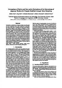
3. Results The baculovirus expression system previously described was further refined by the use of HighFive insect cells which resulted in a 2-fold higher level of expression of COX-2 then previously reported [24]. Purification of the enzyme (Table I) was achieved folloy. ing detergent extraction of the microsomes. Inspection of SDS-PAGE gels of the purification steps (Fig. I) revealed that the enzyme was visible in the crude microsomal fraction as a broad band of 73 kDa which represented -4% of the total protein. BOG detergent was effective in extracting the enzyme from microsomes with good recovery of activity and with a noticeable enrichment of the 73-kDa band as Judged by SDS-PAGE. A 68-kDa band was apparent in the non-extracted microsomal fraction (lane 2, Fig. la) which was recognized by anti-COX-2 antibodies using Western analysis (Fig. Ib). Since
Table I PurificatIOn of recombinant COX-2 FractIOn
Total actlVlty (,umollmin)
Total protein (mg)
SpeCific activity
Recovery (%)
Punfication enrichment (fold)
Mlcrosomes Detergent extract Insoluble mlcrosomes DEAE pool Purified Lentil-Lectin unbound
494 444 6 300 188 71
511 III 275 52 8 28
I 4 0 6 25 2
100 90 I 61 38 14
I 4 0 6 26 3
PunficatlOn COX-2 from insect cells IS described m Section 2. Samples from each step were collected and analysed for protein by Lowry assay and for COX-2 activity by oxygen uptake (Section 2). SpeCific activity is from the oxygen consumplion assay. The results are an average of four preparations with generally good reproducibility between runs. PeroXidase assay (not shown) gave results Similar to oxygen consumptIOn results.
L.P. Wellllogle et al.lFEBS Letters 371 (1995) 315-320
A.
3
2
4
317
5
94-
+-COX2
6743-
3121 -
B. 2
~~~II c.
3
4
I II
M~. 2
5
6
I II
3
Fig 1 SDS-PAGE and Western analysis of COX-2 purified from baculovlrus-infected HighFive cells. Recombinant COX-2 was purified from Insect cells (Section 2) as outlined In Table I. (A) Purification fractIOns were run on a 12% SDS-PAGE gel then stained by Coomassie brilliant blue. Lane L crude mlcrosomes; lane 2. pellet after detergent extraction; lane 3. soluble phase of detergent extracted micro somes; lane 4. p001 from DEAE-Sepharose column; lane 5. Lentil-LectinAgarose-punfied enzyme. The punfied enzyme in this preparation appears as a major band With a molecular weight of 73 kDa and a minor COX-2 band at 65 kDa (see text). (B) Identification of two COX-2 species In nllcrosomes: three samples were run on 12% SDS-PAGE gels and tested: left. crude micro somes; center. detergent insoluble microsome pellets; and nght detergent extracts. The analysis was done either by staining gels With Coomassie brilliant blue (left) or by Western analysis with a COX-specific antibody (right). The two major bands of COX-2 are indicated as a 73-kDa band extracted by detergent and a 68-kDa Insoluble band. (Cl Endoglycosidase treatment of COX-2: purified enzyme was treated with endoglycosidase F and run on a SDSPAGE gel and stained with Coomassie bnlliant blue. Lane I was punfied COX-2 control. lane 2 was COX-2 treated with endoglycosidase F In non-denaturing buffer and lane 3 was COX-2 treated with endoglycosldase F In a denaturing buffer containing SDS (SectIOn 2). In thiS preparation. both bands of COX-2 shift to lower molecular weight; the 73 kDa shifting to 68 kDa and the minor 65 kDa shifting to 63 kDa. See the test for details The relative positIOn for molecular weight standards IS Indicated In the figure as molecular weight x 10- 3 .
maintain the detergent concentration at 0.9% in order to prevent contaminating proteins from adhering to COX-2 and to prevent the enzyme from aggregating. The enzyme was stored at -80°C and little loss in activity was noticed over 6 months. COX-2 activity migrated on a S300 Sephacryl size-exclusion column (Fig. 2, tubes 27-32) in a manner indistinguishable from native COX-I (not shown) and similar to an aldolase standard (molecular weight 154 kDa). As discussed earlier [19,28], this migration corresponds to an enzyme dimer. Some higher molecular weight material eluted at the void volume (Tube 20-22). Rechromatography of the dimer pool (not shown) gave a profile similar to the original column run, containing both dimer and higher aggregate. The result underscores the tendency of COX-2 to aggregate even in detergent. Upon storage of enzyme samples at 4°C for up to I week, higher molecular weight bands became evident by SDS-PAGE. The slight tailing shoulder of the dimer peak in Fig. 2 may represent a small fraction of monomeric COX-2 and this tail was similarly evident upon rechromatography. indicating it was created from the dimer dissociating into monomers during or before the column run. Enzyme activity co-migrated principally with the dimer peak and a slight activity was seen in the higher molecular weight peak. Sequence analysis of the purified COX-2 gave the predicted N-terminal of Ala-Asn-Pro-Cys-, indicating that the 17 aminoacid signal sequence had been efficiently removed. From the SDS-PAGE analysis we estimate the enzyme to be -98% pure. The chief visible contaminant (typically < 5% of the major band) was a band which ran just below the main COX-2 band. has an approximate molecular weight 65 kDa and is likely to be a proteolytic degradation product produced during purification. Endoglycosidase treatment of purified COX-2 with either endoglycosidase F or H resulted in a protein which migrated considerably faster as judged by mobility on SDS-PAGE (Fig. Ic) and having an approximate molecular weight of68 kDa. (It is worth noting that the resulting band co-migrated with the COX-2 material not extracted by detergent from micro somes as seen in Fig. la, lane 3.) When COX-2 was treated with endoglycosidase F either in buffer or buffer supplemented with Fig 2
Size ExclUSion Chromatography
00280 Vo
...
0006
67
... ...
V,
Vop!
...
0005
80
0004
60
0003
40
0002 20 0001 10
the band is not extracted from the microsomes by BOG and since no activity remains in the extracted membranes, the protein presumably represented an improperly folded or denatured COX-2 fraction. The non-extracted band has the same migration as COX-2 which had been treated with endoglycosidase to remove N-linked carbohydrate (see below). The overall yield from microsomes to pure enzyme was -40%. It was essential to
154
60
Fig. 2. S300 Sephacryl SIZe-exclusion chromatography ofCOX-2. After punficatlOn of COX-2 by Lentil-Lectin-Agarose chromatography. the sample was concentrated with an Amicon filtration deVice then the enzyme was passed through a S300 Sephacryl gel filtratIOn column. Fractions were analysed for activity as described in section 2. ActivIty IS expressed In the bargraph as Fop,/IO,u1 of sample (nmol/min) ActiVity corresponded to the main peak with slight actlVlty aSSOCiated With the high molecular weight peak. The late-running peak IS probably non-protein as no activity migrated With thiS peak.
L P W 400 to I. The molecular difference between the various Ser-516-containing peptides in Table 3 (peptides M, o and R) is not clear but may be due to variable posttranslational modification, such as phosphorylation or myristoylation. Kinetic analysis of recombinant COX-2 was performed using both oxygen consumption and peroxidase assays with generally good agreement. As indicated in Table 4, recombinant COX-2 has kinetic properties similar to native COX-I and COX-2. Extent assays using limiting substrate demonstrated the lllcorporation of two molecules of oxygen/molecule of arachidonic acid substrate (not shown). Various fatty acid substrates were utilized by COX-2 with different efficiencies and the order of substrate preference was close to that expected for COX-I (manuscript, in prep.). The addition of heme IS essential to
The development of isoform-specific inhibitors is theoretically possible and yet very few isoenzyme-specific inhibitors have been described. In contrast, numerous examples of specific inhibitors of receptor isotypes exist, such as agents which selectively inhibit the various adrenergic receptor subtypes. One reason for this contrast is that tools have not previously been available to pursue this goal. As one of the first cases where isoform-specific inhibitors are claimed, several recent reports [12,13,29-33] have documented selective COX-2 inhibitors. Presumably, other examples will follow as various targets are defined and evaluated at the molecular level. The expression and purification of human COX-2 has allowed us to attempt scale-up purification and analysis of the recombinant COX-2 enzyme (see also [22,28,34]). COX-2 tends to aggregate; size-exclusion chromatography indicated the majority of the enzyme migrated as a protein dimer, however, even with BOG detergent in excess of its critical micellular concentration, some tendency for the enzyme to form larger aggregates was seen. When dealing with novel expression systems, the potential for heterogeneity of expressed protein is enormous [35]. Pro site analysis of the primary sequence of human COX-2 predicts 4 potential N-Iinked glycosylation sites, 13 potential phosphorylation and 9 potential myristoylation sites. COX-I has three N-linked glycosylation sites all of which are glycosylated [14,19,36] and each of these sites are conserved in COX-2. The fourth site ofCOX-2 Asn-580 was shown by mutation analysis [36] to be incompletely glycosylated in a murine recombinant COX-2 expressed in COS-I cells. In the present study, Asn-130 was shown by HPLC analysis and peptide mapping to contain N-Iinked carbohydrate. Although it is uncertain from the present work whether Asn-53, Asn-396 and Asn-580 were glycosylated, from the changes in molecular weight after endoglycosidase treatment, we speculate that at least two sites are glycosylTable 3 Selective labeling of Ser-516 by aspmn Peak number
cpmJ,Ug
A B C D E F
P
546 1088 63 2700 0 21 0 0 299 366 1260 1831 30440 6979 103860 7113
Q R
35750
G H I J K L M
N
o
Sequence
Assignment F32
(FI8) F6- GP2 F22 FI2 F5 or F(l8119) PIDP + EMSAE GLMGN EMSAE
EMSAE
FlO, F28/29
F30/F(?) F28/29 F28/29
The peaks shown in Fig. 3 were counted for radlOactlVlty. Essentially, all the radioactivity was found III peaks which included the F29 peptide and, therefore, exclUSively located in Ser-516 (see text).
L.P. Wennogle et al. I FEBS Letters 371 (1995) 315-320
320 Table 4 Comparison of kinetic data for COX isoforms Isoform COX-I COX-2 COX-2
Species ovine ovme human
Km (uM)
Vmdl;
Spec. activity
(nmol/min)
(nmol/min/mg)
6 5 3
100 90 80
30 6 25
The expenments were conducted at 37°C in 0.1 M Tns-HCL 2 mM phenol, pH 7.7. The enzyme was added to start the reaction. The optimal velocity of oxygen was measured and used to determme the kinetic constants by Hanes's analysIs.
ated in the major band (> 90% of detergent extracted material). The inactive COX-2 68K band which is not extracted by BOG detergent may represent enzyme which was not properly folded because it did not enter the posttranslational modification pathway upon protein synthesis and, therefore, was not glycosylated and further modified. Aspirin modified Ser-S16 of COX-2, a residue which corresponds to Ser-S30 of COX-l [14,19]. The results support the concept that the two isoforms are structurally similar. A similar result was found when heme binding was investigated. Careful titration of the enzyme resulted in a one-to-one titration of enzyme monomers with added heme (ses also [26,37]). Cyclooxygenase and peroxidase activities of the recombinant enzyme were carefully evaluated after heme reconstitution. The enzyme showed similar kinetic properties to ovine COX-l and the same substrate preference as COX-I. COX-2 oxygen consumption activity is inhibited by aspirin with a change in formation of IS-HETE as recently demonstrated [20,22]. With this body of evidence for similarity of structural and kinetic data between COX-l and COX-2, coupled with the near identity of amino acids lining the substrate binding and catalytic site, it would appear to be a difficult task to exploit molecular differences between the two enzymes. Interes!ingly, proposed isoform-specific agents have revealed different kinetics of inhibition. This information may indicate that subtle amino acid differences of a more peripheral nature along the enzyme binding/reaction pathway may be exploited for specific inhibitor development. It is hoped that by developing a detailed understanding of this enzyme isoform pair, an era of isoenzyme-specific inhibitors may be forthcoming.
References [I] Hla, T. and Neilson. K. (1992) Proc. Natl. Acad. Sci USA 89, 7384-7388.
[2] Jones, DA et al. (1993) J. BIOI. Chern. 268, 9049-9054~ [3] O'Banion, M.K., Winn, YD. and Young, D.A (1992) Proc. Natl. Acad. SCI. USA 89, 4888-4892. [4] PhilliPS, TA. et al. (1993) J. Leukoc. BioI. 53. 411-419. [5] Takahashi. Y. et al. (1992) J. Nutr. Sci. Vltaminol. 134-137. [6] Fletcher, B.S. et al. (1992) J. BIOI. Chern. 267,4338-4344. [7] Sirois, J. and Richards, J.S. (1991) J. BioI. Chern. 267, 63826388. [8] Xle, WL. et al. (1991) Proc. Natl. Acad. SCI. USA 88, 26922696. [9] Kujubu, D.A. et al. (1991) J. BioI. Chern. 266. 12866--12872. [10] Meade. EA, Smith, WL. and DeWitt, D.L (1993) J. Lipid Mediat. 6, 119-129. [II] Vane, J.R. et al. (1994) Proc. Natl. Acad. Sci. USA 91, 2046--2050. [12] Isakson. P (1994) Am. Chern. Soc. Natl. Meet., Washington, DC. Derwent Conf. Fast-Track 2, 161. [13] Reitz. D.B. et al. (1994) Am. Chern. Soc. Natl. Meet., Washington, DC. Derwent Conf. Fast-Track 2, 162. [14] DeWitt, D.L. et al. (1990) 1. BioI. Chern. 265, 5192-5198. [15] Merlie, J.P. et al. (1988) J. BIOI. Chern. 263, 3550-3553. [16] Yokoyama, c.. Takai. T and Tanabe, T (1988) FEBS Lett. 231 347-351. [17] Roth, G.J., Machuga, E.T. and Ozols, J. (1983) Biochemistry, 22, 4672-4675. [18] Lecomte, M. et al. (1994) J. BioI. Chern. 269. 13207-13215. [19] Chen. Y.P, Bienkowski, M.J. and Marnett. L.J. (1987) J. BioI. Chern. 262. 16892-16899. [20] Holtzman, M.J., Turk. J. and Shornick. L.P. (1992) J BioI. Chern. 267, 21438-21445. [21] Meade. EA, Smith, WL. and DeWitt, D.L. (1993) J. BioI. Chern. 268. 6610-6614. [22] O'Neill, G.P. et al. (1994) Mol. Pharmacol. 45, 245-254. [23] Cromlish, WA. et al. (1994) Arch. Biochem. BlOphys. 314, 193199. [24] Miller, D.B. et al. (1994) Biochem. Biophys. Res. Commun. 201, 356--362. [25] Rome, L.H. and Lands, WE. (1975) Proc. Natl. Acad. SCI. USA 72, 4863-4865. [26] Kulmacz, R.J. and Lands. WE. (1984) J. BioI. Chern. 259. 63586363. [27] Ali, S.L. (1976) J. Chromatogr. 126,651-663. [28] Percival, M.D. et al. (1994) Arch. BlOchem. BlOphys. 315,111-118. [29] Chan, C -c. et al. (1994) XII Intr. Congr. Pharmacol., Montreal, July 1994, PIO.U8. Derwent Conf. Fast-Track 2, 266. [30] Copeland, R A et al. (1994) Proc. Natl. Acad. Sci. USA 91,1120211206. [31] Mitchell, J.A. et al. (1993) Proc. Natl. Acad. SCI. USA 90. 1169311697 [32] Barnett, J. et al. (1994) BlOchim. Biophys. Acta 1209. 130-139. [33] Futaki, N. et al. (1993) Gen. Pharmacol. 24, 105-110. [34] Glerse. J.K. et al (1994) Am. Soc. Biochem Mol. BIOI. Annu. Meet. Abstr. 1994. 1203. [35] O'Reilly, D.R., Miller, L.K. and Vuckow, VA. (1992) Baculovlrus ExpreSSIOn Vectors: A Laboratory Manual. pp. 216--236. [36] Otto, J.c., DeWitt, D.L. and Smith, WL. (1993) J. BioI. Chern. 268. 18234-18242. [37] Roth, G.J .. Machuga, E.T. and Stnttmatter, P. (1981) J BioI Chern. 256, 10018-10022.