culate that -7-10 mg of HCA VI is daily swallowed to the gastrointestinal canal. Together with the mucosally located HCA H, this isoenzyme is perhaps also involved in the regulation of pH and buffer capacity in the esophageal and gastrointestinal tract. This hypothesis is supported by our finding that HCA VI retains its enzymatic activity after exposure to pH 2.2 (S. Parkkila et al., unpublished observations). We expect that the role of HCA VI in the esophagus and gastrointestinal canal will be effectively studied in the future by using the sensitive and robust method we developed for measuring salivary HCA VI concentrations in patients with various gastrointestinal disorders. We thank Aino Kuha for skillful technical
assistance.
References 1. Maren TH. Carbonic anhydrase: chemistry, physiolor, and inhibition [Review]. Physiol Rev 1967;47:595-781. 2. Carter MJ. Carbonic anhydrase: isoenzymes, properties, distribution,and functional significance [Review]. Biol Rev 1972;47: 465-513. 3. Tashian RE. The carbonic anhydrases: widening perspectives on their evolution, expression and function [Review]. BioEssays
1989;1O:186-92. 4. Fernley RT, Wright RI), Coghian JP. A novel carbonic anhydrase from the ovine parotid gland. FEBS Lett 1979; 105:299-302. 5. Fernley RT, Darling PE, Aldred P, Wright RI), Coghlan JP. Tissue and species distribution of the secreted carbonic anhydrase isoenzyme. Biochem J 1989;259:91-6. 6. Psrkkila 5, Kaunisto K, Rajaniemi L, Kumpulainen T, Jokinen K, Rajaniemi H. Immunohistochemical localization of carbonic anhydrase isoenzymes VI, II, and i in human parotid and submandibular glands. J Histochem Cytochem 1990;38:941-7. 7 Parkkila S, Kaunisto K, Rajaniemi H. Location of the carbonic anhydrase isoenzymesVI and II in human salivary glands by imxnunohistochemistiy. In: Botr#{233} F, Gros G, Storey BT, eds. Carbonic anhydrase: from biochemistry and genetics to physiolor and clinical medicine. Weinheim: VCH Verlags, 199 1;254-7.
8. Ogawa Y, Chang C-K, Kuwahai-a H, Hong S-S. Toyosawa S, Yagi T. Immunoelectron microscopy of carbonic anhydrase isozyme VI in rat submandibular gland: comparison with isozymes I and II. J Hiatochem Cytochem 1992;40:807-17. 9. Feldstein JB, Silverman DN. Purification and characterization of carbonic anhydrase from the saliva of the rat. J Biol Chem 1984;259:5447-53. 10. Murakami H, Sly WS. Purification and characterization of human salivary carbonic anhydrase. J Biol Chem 1987;262:1382-8. 11. Fernley RT, Wright RI), Coghian JP. Complete amino acid sequence of ovine salivary carbonic anhydrase. Biochemistry 1988; 27:2815-20. 12 Fernley RT, Wright ED, Coghlan JP. Radioimmunoassay of carbonic anhydrase VI in saliva and sheep tissues. Biochem J 1991;274:313-6. 13. Yoshimura H, Iwasaki H, Nishioka T, Matsunioto S. Role of carbonic anhydrase in the bicarbonate excretion from salivary glands and mechanism of ionic excretion. Jpn J Physiol 1959;9:106-23. 14. Fernley RT. Non-cytoplasmic carbonic anhydrases [Review]. TIBS 1988;13:356-9. 15. Smith I, Light.stone PJ, Perry JD. Separation of human silksiline phosphatases by electrophoresis on acrylamide disc gels. Clin Chiin Acta 1968;19:499-505. 16. Parkkila A-K, Parkkila 5, Juvonen T, Rajarnemi H. Carbonic anhydrase isoenzymes II and I are present in the zona glomerulosa cells of the human adrenal gland. Histochemistry 1993;99:37-41. 17. Psrkkila S, Parkkila A-K, Kaunisto K, Waheed A, Sly WS, Rajaniemi H. Location of a membrane-bound carbonic anhydrase isoenzyme (CA W) in the human male reproductive tract. J Histochem Cytochem 1993;41:751-7. 18. Ericsson Y. Clinical investigations of the salivary buffering action. Acta Odontol Scand 1959;17:131-65. 19. Altman Dli Practical statistics for medical research, 1st ed. London:Chapman and Hall, 1991:528-9. 20. Diamandis EP, Christopoulos TK Europium chelate labels in time-resolved fluorescence immunoassays and DNA hybridization
assays.Anal Chem 1990;62:1149A-57A. 21. Hemmilfl IA. Applications [Review]. In: Winefordner JD,
of fluorescence in immunoassays
KolthofflM,eds. Chemical analysis.
New York: Wiley-Interscience, 1991:205-22. 22. Szab#{243} I. Carbonic anhydrase activity in the saliva of children and its relation to caries activity. Caries Res 1974;8:187-91.
CUN. CHEM. 39/10, 2157-21 62 (1993)
Competitive Assay to Improve the Specificity of Detection of Single-Point Mutations in a1-Antitrypsin Deficiency Andar
Gunneberg,’
Graeme
Scobie,2
Karen
Hayes,2
and
Noor Kalsheker2’3
Allele-specific oligonucleotides are used widely for the detection of single point mutations in genes. A modification of this assay based on competition has been devel-
polymerase
oped for detection of the Z mutation of a1-antitrypsin (a1-AT). The normal a1-AT allele is referred to as M, and the Z mutation arises from a single base substitution. Amplified DNA products corresponding to homozygous
incubation with unlabeled M-speciflc oligonucleotide was followed by hybridization with radiolabeled Z-speciflc oh-
‘Department of Clinical Chemistry, Southmead Hospital, Westbuzy-on-Trym, Bristol BS1O 5NB, UK 2Department of Clinical Chemistry, University Hospital, Queen’s Medical Centre, Nottingham NG7 2UH, UK. 8Address correspondence to this author. Fax 44-602-709167; E-mail
MPZ
[email protected]. 16, 1993; accepted May 5, 1993.
Received February
M, heterozygous MZ, and homozygous
Z obtained by the chain reaction were incubated with a twofold
molar excess of unlabeled oligonucleotide prior to hybridization with a radiolabeled oligonucleotide. Thus, initial
and vice versa. This assay increased the specificityof single-point mutation detection three- to four-
gonucleotide,
fold. Furthermore, specific hybridization was obtained at a lower temperature as a consequence signal-to-noise ratio.
of improving
the
Indexing Terms: genotyping ohgonucleotides DNA hybridization polyinerase chain reaction autoradiography .
.
-
CUNICAL CHEMISTRY, Vol. 39, No. 10, 1993
2157
a1-Antitrypsin (a1-AT) is a serum protease inhibitor produced predominantly by hepatocytes.4 In a1-AT deficiency the plasma concentration of the inhibitor falls below critical amounts, leading to unimpeded tissue destruction by enzymes such as neutrophil elastase. This enzyme is released during inflammation, and in the lung can destroy the lung architecture and cause pulmonary emphysema (1). a1-AT deficiency occurs in about 1 in 3000 individuals of northern European origin (2). A major cause of this deficiencyis a single-point mutation (Glu-342 GAG -* Lys AAG) referred to as the Z mutation (2). In heterozygotes (MZ), the plasma concentrations of a1-AT are -60% and in homozygotes 1520% of the mean concentration found in the normal M genotype. The Z mutation is also associated with accumulation of a1-AT in hepatocytes, which results in liver disease in 10-15% of affected individuals (2, 3). Although a1-AT protein types can be identified by isoelectric focusing of serum, the banding pattern is sometimes difficult to interpret, especially in heterozygotes, and storage artifacts may appear (4). Protein typing by isoelectric focusing may thus require confirmation by direct identification of the genotypes, which can be done with DNA from blood or other biological samples, e.g., tissue biopsies. Genotyping by the use of allele-specific oligonucleotides for the Z mutation has been reported (5), as has the direct sequencing of amplified DNA fragments obtained by the polymerase chain reaction (PCR) (6), but the latter is time consuming and labor intensive and requires expertise. Synthetic oligonucleotides have been used widely for the detection of single-point mutations in DNA associated with disease, because they are convenient and make it possible to analyze a large number of samples. The use of PCR has facilitated the use of oligonucleotides in single-point mutation detection. The specificity of this approach is determined by the degree of complementarity, such that even a single base-pair mismatch is less stable than one that shows a perfect match, and it is possible to distinguish between a normal and mutant sequence by using empirically determined hybridization conditions. A method of genotyping 4Nonstandard abbrevzat, . a1-AT, a,-antitrypsin; PCR, polymerase chain reaction SSPE, saline-sodium phoephate-EIYFA buffer; PNK, polynucleotide kinase; and SDS, sodium dodecyl sulfate.
5’
________
Exonsl-IV
patients with a1-AT deficiency with radiolabeled allelespecific oligonucleotides has been described (6). The intensity of signals obtained from labeled bound allelespecific oligonucleotideprobes depends on the specific activity of the label,the severity of the washing conditions, the length of exposure of the film to the probe, and the amount of target sequence. Thus, it is desirable to find ways of improving the signal-to-noise ratio.
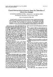
MaterIals and Methods Procedures Amplification of DNA fragments. The location of the fragments amplified and the position of the mutation are highlighted in the schematic of the AAT gene (Figure 1). DNA was prepared from peripheral blood by previously described methods (7).Exon V, containing the normal M sequence and Z mutation, was amplified by using -1 g of genomic DNA and the following oligonucleotides: 5’ end of exon V. 5’ TGT CCA CGT GAG CCT TGC TCG AGG CCT GGG 3’; 3’ end of exon V. 5’ GGA CCA GCT CAA CCCTTC TTT AAT GTC ATC 3’. We added 5 L of DNA to 3 /LL (100 pmol) of each oligonucleotide, 5 L of dNTPs (2mmol/L), 5 L of lOx Taq polymerase buffer (lx is 10 mmol/L Tris-HC1, pH 8.8, containing 50 mmol/L KC1, 1.5 mmol/L MgC12, and 10 g/L nonionic detergent; Northumbrian Biologicals, UK), 1 jL (2500 kU/L) of Taq polymerase, and 28 L of Analar-grade H20 to make up to 50 L. The followingcyclewas repeated 35 times on a Techne heating block denaturation at 91 #{176}C for 1.5 mm, annealing at 60#{176}C for 1.5 mm, and extension at 73#{176}C for 2 mm, with a final extension step at 73#{176}C for 10 min. Dot-blotting the DNA fragments. We vortex-mixed 10 .tL of PCR product (about 100 mg(L) with 200 L of 0.4 mol/L NaOH at room temperature for 15 a and spotted 50 p1 (about 250 ng) onto a nylon membrane (Zetaprobe; Bio-Rad Instruments, Hemel Hempstead, UK), using a Perspex manifold designed for this purpose. A piece of filter paper was moistened with 2 x salinesodium phosphate-EDTA (SSPE) buffer (lx is 150 mmol/L NaC1, 10 mmol/L sodium phosphate, and 1 mmol/L EDTA, pH 7.4) and placed over the lower grid of the manifold. Appropriate sizes of nylon membrane were cut, moistened with 2 x SSPE, and placed on top of the filter paper. The upper grid was then placed on top of
:::
I[onvJi
Glu 342 (N)
*
5’ 3’
amplification
end of #{149}XOn V: 5 md of ezon V: 5
3’
Lys 342 (Z)
priamrs:
TOT CCA COT GAG CCT TOC TCG AGO CCT 000 3 OGA CCA OCT CAl CCC rrc TTT AlT OTC ATC 3
Ag. 1.SchematIcofpaitofthea1-antltrypsln gene,showing relative positions ofamplification primersand position oftheZ mutation In exon V The geneconsists of five axons;the Initiation codon ATG is in exon II 2158
CUNICAL CHEMISTRY, Vol. 39, No. 10, 1993
the nylon membrane, and the excess fluid removed by suction. To demonstrate the effect of loading different amounts of DNA target sequence, we applied 10-fold (25 ng), 100-fold (2.5 ng), and 500-fold (0.5 ng) dilutions of the PCR products to the membrane. Labeling the probes. The following allele-specific oligonucleotides were used as probes: M probe, 5’ ACC ATC GAC GAG AAA GGG ACT 3’; Z probe, 5’ ACC ATC GAC AAG AAA GGG ACT 3’. We incubated 5 pL of [y-32P]ATP (50 DCi), specific activity >5000 kCilmol, at 37#{176}C for 30 mm with 1.25 pL (6.8 ng or 10 pmol) of M or Z probes, 1.75 p1 of H20, 1 p1 of lOx polynucleotide kinase (PNK) buffer (0.5 mol/L Tris-HC1, pH 7.6, containing 0.1 mol/L MgC12, 50 mmol/L dithiothreitol, 1 mmol/L spermidine, and 1 mmol/L EDTA), and 1 pL (1000 kU/L) of PNK After incubation, the PNK was inactivated by heating at 85#{176}C for 10 mm. Competition assay. The principle of the method is illustrated in Figure 2. Prehybridization was carried out with all the membranes at 52#{176}C, each in 10 mL of 5x SSPE containing 10 gIL sodium dodecyl sulfate (SDS), and 500 mgIL herring sperm DNA. After 1 h, 2.5 p1 (13.6 ng or 20 pmol) of unlabeled oligonucleotide corresponding to Z or M probe was added to the 10 mL of preincubation fluid such that the membrane to be probed with radiolabeled Z probe was preincubated with unlabeled M probe and vice versa. All the membranes were then incubated for another hour at 52#{176}C. Hybridization was then conducted only in the presence of radiolabeled probe at 52#{176}C for 1 h. A twofold molar excess of unlabeled probe was chosen, having been found to work optimally.
STEP I
CCLI
Washing off nonspecifically bound labeled probe. The initial low-stringency wash consisted of two 30-min washes at 23#{176}C with 100 mL of 2x SSPE-1 gIL SDS, followed by a 15-mm wash at 52#{176}C with 100 mL of 5x SSPE-SDS. The high-stringency wash at 62#{176}C, with 100 mL of 5x SSPE-SDS, was continued for 20 mm to discriminate between M and Z target sequences without competition hybridization. The stringency of washes was reduced by lowering the temperature for the final wash and maintaining the salt concentration. A number of different temperatures was tested before selecting the final conditions for the high-stringency washes. Autoradiography. The filters were exposed to preflashed Fuji Ri blue-sensitive film for 4-16 h at room temperature with CaWO4 intensifying screens. The exposure time was adjusted according to the specific activity of the radiolabeled probe. The intensity of the signals was scanned by densitometry (Quick Scan densitometer; Helena Laboratories, Beaumont, TX), at a alit width of 4 mm,to determine the linearity of the signal strength with respect to exposure time. For the data reported in this paper the signal strength was well within the linear range of the x-ray film. A linear increase in signal strength was obtained up to about 24 h. Subjects
Samples from six patients with known genotypes and three patients with unknown genotypes were analyzed. In some of the patients the genotypes were confirmed by direct sequencing of the amplified products. Results Single-point mutations can be detected with allelespecific oligonucleotides by adjusting the temperature
N PROSE STEP 1 COLI
#{149}1
‘rTnT 2
N PROSE
-
k’
CTC
ALLELE
‘g 5’
N ALLELE
iTEP- 2
NOTZ
PROSE STEP 2
*A5.
NOT 2 PROOf
I
Thfl 2
5’
ALLELE N ALLELE
STEP 3 STEP
3
a
‘-AAS-
2
Tfl1E
ALLELE
5’
N ALLELE
A
B
FIg. 2. PrInciple of the competitive assay (A) Unlabeled N probe bInds lees stron9ly to Z target sequence because of the single base-pair mismatch and is displaced by labeled Z probe; (B) unlabeled M probe binds more strongly to N target sequence and thusreducesnonspecific binding of the labeled Z probe
CUNICAL CHEMISTRY, Vol. 39, No. 10, 1993
2159
LOW STRINGENCY WASH
M N PROBE
Z PROBE
3.
HIGH STRINGENCY WASH 20 MIN
M
M
MZ
Z
N
MZ
Z
Z
a.. #{149} #{234}.
M PROBE
N
Ag.
MZ
HIGH STRINGENCY WASH 5 MIN
MZ
MZ
#{149} #{149} #{149}
Z
N
MZ
Z PROBE
0#{149}
Z N PROBE
Z
0
Z PROBE
Effects of stringency and duration of wash on signal strength of the assay the nonspecific binding of M probe to Z target sequence (and of Z probe to M target sequence) was substantially reduced. By densitometric scanning of the signals and comparing the M Z ratios with and without competition, a three- to fourfold increase in specificity was obtained with competition over that of the assay without competition (Figure 4). Moreover, there was an apparent lack of discrimination at low stringency without competition. Figure 5 shows the effect of loading different amounts (250, 25, 2.5, and 0.5 ng) of target sequence onto the
and duration of the washing conditions without competition (Figure 3). These conditions were determined empirically. After 5 mm of the high-stringency wash, the binding of M probe to Z homozygote target sequence and of Z probe to M target sequence was reduced (Figure 3). After 20 mm of the high-stringency wash, the M probe bound to M and MZ target sequence only, whereas the Z probe bound only to Z and MZ target sequence. These results were obtained for equivalent amounts of loaded target sequence. With competition, even after a low-stringency wash, N
M PROBE
MZ
Z
M M PROBE
-
+
MZ
Z
#{149}. #{149}1
M
Z PROBE
-
MZ
Z
#{149} . ..
M
Z PROBE
MZ
Z
+
0 H probe without
competition
N probe with competition
A N target
Z target
sequence
sequence
2160
N target sequence
CUNICALCHEMISTRY,Vol. 39, No. 10, 1993
Z
target
sequence
Fig. 4. Effect of competition on signal strength at low stringency (A) Membrane hybridized without (-) and with (+) competition
by N-and Z-speclflc probes; (B) scan of the signals to determine specificity
N
11 Too oo 1 To
N
N PROBE
N’’
1 To Too oo
N’’
To Too oo
N
MZ
N PROBE
MZ
+
z
z
N
11 Too oo 1 To
M
Z PROBE
MZ
N
Z PROBE
MZ
+
z
z
FIg. 5. Effect of loading different amounts of target sequence with and without competition at low stringency was loaded Induplicate; membranes were hybridized without (-) and with (+) competition byM and Z specific probe. N = neat; 1/10,
Target sequence 1/600
=
10-, 100-, and 500-fold dilutions, respectively
membranes.
Without competition, the M probe bound equally to M, Z, and MZ target sequences at low stringency, although there was less nonspecific binding by the Z probe to M target sequence at 100-fold (2.5 ng) and 500-fold (0.5 ng) dilutions (Figure 5). The use of competition resulted in much less nonspecific binding even at low stringency; i.e., M probe bound much less strongly to Z target sequence and Z probe less strongly to M target sequence. A reduction in exposure time should make it possible to use even less stringent conditions to obtain obvious differences in signal intensity between perfectly matched and single-base mismatched oligonucleotide probes to target sequence. Discussion oligonucleotides is a simple single-point mutations. However, the method requires careful control of the reaction conditions. The introduction of a competition step significantly improves the specificity of the reaction and makes it possible to use less stringent conditions to detect single-point mutations. Nonetheless, false-positive signals may be obtained because of loading of different amounts of DNA. Given identical conditions with regard to the specific activity of the labeled probe, the length of exposure of the x-ray film, and the stringency of the wash, differences in the amount of PCR product can significantly affect the signal strength. Differences in the amount of target sequence loaded can therefore lead to difficulties in interThe use of allele-specific
and effective method for screening
pretation. A competitive
assay
has been tried with labeled and
‘/100,
and
oligonucleotides used simultaneously in the hybridization reaction, although a 20-fold molar excess of the unlabeled allele-specific oligonucleotide was re-
unlabeled
quired (R. B. Wallace, personal communication). In our method, sequential hybridization with unlabeled and labeled allele-specific oligonucleotides allowed a twofold molar excess of the unlabeled allele-specific oligonucleotide to be used for absolute discrimination. A further possibility is to extend the competition assay to both the prehybridization and hybridization steps. Preliminary data suggest that the results obtained with the preincubation step followed by simultaneous incubation with labeled and unlabeled oligonucleotides in the hybridization reaction does not improve the specificity significantly when compared to a single preincubation step, and there is a small reduction in sensitivity. A general requirement for the use of oligonucleotide probes is to define precise conditions for the posthybridization washes to achieve discrimination between normal and mutant sequences. The abifity to relax the conditions slightly by using a competition step and yet improve the specificity should make it easier to determine these conditions. This approach could be extended to a number of techniques that involve the use of synthetic oligonudeotides.For example, in situhybridizationto detect specific genes in tissues and library screening could be improved by reducing the nonspecific binding that occurs with such procedures. This could be achieved by deliberately synthesizing mismatched oligonucleotides. Another alternative is to use tetramethyl ammonium chloride, which reduces the disparity between GC and CUNICAL CHEMISTRY, Vol. 39, No. 10, 1993
2161
AT base pairs, so that hybridization becomes purely a function of the length of the oligunucleotide (8). The combined use of a competition assay with tetramethyl ammomum chloride may make it possible to obtain near-universal
conditions
for specific single-point
muta-
References 1. Laurel CB, Eriksson S. The electrophoretic alpha-i-globulin pattern of serum in alpha-l-antitrypsin deficiency. Scand J Clin Lab Invest 1963;15:132-40. 2. Cox DW. Alpha-i-antitrypain deficiency. In: Scriver CR, Beaudet AL, Sly WS, et al., ode. The metabolic basis of inherited disease, 6th ed. New York McGraw-Hill, 1989:2409-37. 3. Anonymous. Alpha-i-antitrypsin, Z, and the liver [editorial]. Lancet
1992;340:402-3. N. Techniques
Association
of Clinical Pathologists
Broadsheet
113,
London: British Medical Association, 1985:1-il. 5. Newton CR, Kalsheker N, Graham A, Powell S, Gammack A, Riley J, et at Diagnosis of alpha-l-antitrypsin deficiency by enzymatic amplification of human genomic DNA and direct sequencing of polymerase chain reaction products. Nucleic Acids Res 1988;17:8233-43.
tion detection.
4. Kalsheker
variants.
for the study of alpha-1-antitrypsin
6 Bruun Petersen K, Kolvraa S, Bolund L, Bruun Petersen G, Koch J, Gregersen N. Detection of alpha-1-antitrypsin genotypes by analysis of amplified DNA sequences. Nucleic Acids Res 1988; 16:352. 7. Kunkel LM, Smith DK, Boyer SH, Borgoanker DS, Wachtel 58, Miller OJ, et al. Analysis of Y chromosome specific reiterated DNA in chromosome variants. Proc Natl Acad Sd USA 1977;74:1245-9. 8. Wood WI, Gitschier J, Lasky LA, Lawn EM. Base compositionindependent hybridization in tetramethyl ainmonium chloride: a method for oligonucleotide screening of highly complex gene libraries. Proc Nati Aced Sci USA 1985;82:1585-8.
CUN. CHEM. 39/1 0, 2162-2165 (1993)
Quantification of the Cross-Link Pentosidine in Serum from Normal and Uremic Subjects Masaakl Takahashl,”3 Kazuhiro Kushida,’ Kouichi Kawana,’ Chiaki Ishihara,’ Masashi Denda,’ Tetsuo Inoue,’ and Kentaro Iloriuchi2 Pentosidine is a fluorescent cross-link compound that accumulates in human tissuesfrom uremicand diabetic patients. Using SP-Sephadex C-25 pretreatment before reversedphase HPLC, we developeda method for quantifying pentosidine in the acid hrolysate of serum. We examinedconcentrationsof pentosidinein serum from 98 patients with end-stagerenal disease requinng hemodialysisand from 33 normalcontrol subjects. The mean (± SD) concentration of pentosidine was significantly greater in serum from uremic patientsthan that from control subjects (1267 ± 695 nmol/L vs 77 ± 40 nmol/L, P = 0.0001). There was a significant correlation betweenserum pentosidine concentrations and age in control subjects (r = 0.453, P