We have recently shown that antibody- induced blockade of C5a, C5a receptors, or IL-17A greatly reduced the harmful outcomes of sepsis. In the current study ...
The FASEB Journal article fj.11-183236. Published online April 8, 2011.
The FASEB Journal • Research Communication
Complement dependency of cardiomyocyte release of mediators during sepsis Gelareh Atefi,*,1 Firas S. Zetoune,*,1 Todd J. Herron,†,‡ Jose´ Jalife,†,‡ Markus Bosmann,* Rami Al-Aref,* J. Vidya Sarma,* and Peter A. Ward*,2 *Department of Pathology, †Cardiovascular Research, Department of Internal Medicine, and ‡ Department of Molecular and Integrative Physiology, University of Michigan Medical School, Ann Arbor, Michigan, USA ABSTRACT We have recently shown that antibodyinduced blockade of C5a, C5a receptors, or IL-17A greatly reduced the harmful outcomes of sepsis. In the current study, normal cardiomyocytes from young (300 g) male Sprague-Dawley rats responded in vitro to C5a (ED50ⴝ55 nM) with release of IL-6 and TNF␣, peaking between 2 to 8 h. Neutralizing antibodies to mouse C5a or IL-17A (ED50ⴝ40 g for each, based on improved survival) reduced spontaneous in vitro release of cardiosuppressive cytokines and chemokines in cardiomyocytes obtained from mice with polymicrobial sepsis. A non-neutralizing C5a antibody had no such effects. Cardiomyocytes from septic mice (C57Bl/6) showed increased mRNA for TNFR1, IL-6 (gp80), and C5aR at 6 h after sepsis. Cardiomyocytes from septic C5aRⴚ/ⴚ or C5L2ⴚ/ⴚ mice did not show spontaneous in vitro release of cytokines and chemokines. These data suggest that cardiomyocytes from septic mice release suppressive cytokines in a C5a-, C5aR-, and IL-17A-dependent manner, followed by mediator reactivity with receptors on cardiomyocytes, resulting in defective contractility and relaxation. These data may be relevant to a strategy for the treatment of heart dysfunction developing during sepsis.—Atefi, G., Zetoune, F. S., Herron, T. J., Jalife, J., Bosmann, M., Chen, A., Al-Aref, R., Sarma, J. V., Ward, P. A. Complement dependency of cardiomyocyte release of mediators during sepsis. FASEB J. 25, 000 – 000 (2011). www.fasebj.org
Key Words: cytokines 䡠 cytokine receptors 䡠 C5a 䡠 IL-17A 䡠 cardiomyopathy
There is generally accepted evidence that humans in sepsis often exhibit a hyperdynamic cardiovascular state that is associated with biventricular dilatation, high cardiac output, and reduced systemic vascular resistance (1, 2). Such responses appear to be reversible if survival occurs, with little or no evidence of residual structural damage to the heart (3, 4). There are suggestions that sepsis in humans and in animals is linked to the presence of circulating cytokines (TNF␣, IL-1, and IL-6) that have cardiosuppressive effects, defined by their in vitro ability to cause defective contractility and relaxation of cardiomyocytes (CMs) (5). There are 0892-6638/11/0025-0001 © FASEB
at least 3 hypotheses to explain heart dysfunction in sepsis: loss of energetic function in CMs due to defective electron transport in mitochondria, resulting in inadequate levels of ATP (reviewed in refs. 6 –9); circulating and, perhaps, intracardiac cytokines causing impairment of CM contractility and relaxation (10 –16); and complement activation during sepsis, resulting in generation of the powerful anaphylatoxin C5a, which can react with its receptor, C5aR, on CMs to induce defects in CM contractility and relaxation (4, 17–19). Following sepsis in rodents and humans or acute thermal trauma in rodents, there is accumulating evidence for the presence of cardiosuppressive cytokines in serum, which, when added in vitro to CMs, results in defects in contractility and relaxation. In the case of septic human serum, such effects have been indirectly linked to the presence of TNF␣, IL-1, IL-6, and IL-10 (11–13). TNF␣ and IL-1 have been shown to impair contractility of electrically stimulated human atrial heart strips (12), while repetitive subcutaneous injection of IL-6 into rats causes heart dilatation and reduced cardiac output (13). Suppression of rat CM contractility can be induced by TNF␣, IL-1, or human septic shock serum, with reversal of CM dysfunction in the copresence of TGF (14). In rodents exposed to acute thermal injury of skin or after infusion of endotoxin (LPS), CMs released TNF␣ that was associated with NFB activation (16). There have been suggestions that there may be synergy in the copresence of different cardiosuppressive cytokines (12). Such data suggest that sepsis, burn injury, and endotoxemia cause the appearance of cardiosuppressive cytokines in serum and that under certain conditions, CMs may produce and release similar cytokines that could interact with receptors on CMs, causing defects in contractility and relaxation. We have recently reported that in cecal ligation and puncture (CLP)-induced sepsis in rats, there are pro1
These authors contributed equally to this work. Correspondence: The University of Michigan Medical School, Department of Pathology, 1301 Catherine Rd. Box 5602, Ann Arbor, MI 48109-5602, USA. E-mail: pward@ umich.edu doi: 10.1096/fj.11-183236 2
1
found contractility defects in CMs that seem to be linked to the presence of C5a interacting with its receptor, C5aR. C5aR is up-regulated on CMs following CLP (17). C5a interaction with C5aR on normal CMs in vitro resulted in substantial defects in CM contractility and relaxation. Such responses were exaggerated when CLP CMs were used. Furthermore, when C5a was blocked in vivo with neutralizing antibody in CLP rats, CMs from these animals demonstrated no defects in vitro in spontaneous contractility or relaxation (17). In the current study, we show that CLP CMs contain and release cardiosuppressive cytokines in a C5a-, IL-17A-, and C5aR-dependent manner, suggesting a possible linkage between these molecules and the ensuing cardiac dysfunction of sepsis.
region was neutralizing for C5a, but this was not the case for the IgG antibody to the N-terminal region of C5a. Details of these antibodies are provided elsewhere (19, 20). In CLP mice, the ED50 (providing 50% survival) for the antibody to the C-terminal peptide of C5a was 40 g i.v. (administered at the time of CLP; ref. 20), while all CLP mice given preimmune rabbit IgG were dead before d 3 (20). The rat neutralizing mAb to mouse IL-17A was purchased from eBioscience (San Diego, CA, USA). The ED50 of this antibody in CLP mice was 50 g i.v., producing 50% survival 5 d after CLP, whereas only 10% of mice survived when given a control mouse IgG (21). Finally, 80 –90% CLP mice that received 40 g i.v. of affinity-purified IgG to the N-terminal region of C5a, which is a non-neutralizing antibody to C5a, were dead by 72 h (19). Mice were sacrificed by abdominal aortic exsanguination at 6, 12, 24, and 48 h after CLP. CM isolation and responses to C5a
MATERIALS AND METHODS Experimental animals Specific pathogen-free, 8- to 9-wk-old male wild-type (WT) mice (Jackson Laboratories, Bar Harbor, ME, USA), C5⫺/⫺ and C5⫹/⫹ mice (congenic strains B10.D2/oSn and B10.D2/ nSn; Jackson Laboratories), C5aR⫺/⫺, and C5L2⫺/⫺ mice (breeding stock from Dr. C. Gerard, Boston Children’s Hospital, Boston, MA, USA) were used. All mice were on a C57BL/6 background. Young adult male Sprague-Dawley rats (Harlan, Indianapolis, IN, USA) weighing ⬃300 g were used where indicated. Animals were housed in a specific pathogenfree environment and allowed to acclimate to their surroundings for 1 wk prior to use. Standard mouse and rat chow and water were available to animals during the course of the experiment. All experiments were performed in accordance with the guidelines set forth by the U.S. National Institutes of Health for care and use of animals. Approval for experimental protocol was obtained from the University Committee on Use and Care of Animals at the University of Michigan Medical School. Sepsis model and antibodies employed Intraperitoneal injections of ketamine and xylazine were used for anesthesia and analgesia. Experimental sepsis was induced using the moderately severe form of CLP, as described elsewhere (18). Briefly, after an abdominal midline incision, the cecum was exposed, ligated (involving 75% total cecum), and punctured through and through with a 21-gauge needle. A small portion of feces was expressed through the puncture sites. After repositioning of the bowel, the abdomen was closed in layers using surgical sutures (Ethicon Inc., Somerville, NJ, USA) and metallic clips. Before and after the surgery, animals had unrestricted access to food and water. To induce the intermediate-grade CLP, 40% of the cecum was ligated. All mice received fluid resuscitation (1.0 ml lactated Ringer’s solution), which was given subcutaneously every 12 h starting at the time of sham surgery or CLP, for a total of 48 h. Some mice received an intravenous infusion at the time of CLP of 40 –50 g preimmune rabbit IgG, anti-C5a affinity-purified IgG, or rat mAb to mouse IL-17A. As indicated, the rabbit affinity-purified IgG to C5a was directed toward the C-terminal peptide region of C5a (residues 58 – 77) or toward the N-terminal region (residues 1–16). These antibodies were reactive with both rat and mouse C5a (19, 20). The affinity-purified IgG antibody against the C-terminal 2
Vol. 25
July 2011
CMs were isolated from left ventricular walls of rats (Sprague Dawley, young adult males, 300 g) using a commercial kit (Adumyts Cardiomyocyte Isolation Kit, Cellutron Life Technology, Highland Park, NJ, USA) according to the manufacturer’s instructions. Rats were treated with anticoagulant (500 U heparin i.p.) 30 min before heart isolation. Exsanguination was induced by transection of the distal abdominal aorta. Thereafter, 5.0 ml sterile HBSS was injected into the left ventricle in order to clear blood from the aorta and its tributaries. The heart was removed and placed in ice-cold HBSS. The heart was then placed in a 5.0 ml A1 solution, and the ventricles were finely minced with surgical scissors. The minced tissue in A1 solution was then incubated at 37°C for 12 min on a gentle shaker. Then, the minced tissue was allowed to settle, and the supernatant fluid was gently aspirated. Thereafter, 5.0 ml A2 solution (containing hyaluronidase and collagenase II) was added to the minces and the tubes placed on a shaker for 10 min at 37°C. Thereafter, the minced tissue was subjected to centrifugation at 800 g. The supernatant fluids were gently aspirated, and 1.0 ml solution A3 was added to the pellets. Following this, 5.0 ml of the A3 solution was admixed, and the tissue was incubated on a shaker for 10 min at room temperature, followed by centrifugation. These steps were repeated 5 times. The minced tissue was finally suspended in 0.5 ml (mouse) or 5.0 ml (rat) HBSS. CMs were counted in a hemocytometer. The number of CMs used for a given experiment is described in Results. In another set of experiments, mouse CMs from 25-g C57Bl/6/J mice were isolated as indicated above. In such cases there were 10-fold reductions in units of heparin employed (30 U) and 5-fold reductions in volume (0.5 ml) of solutions (A1, A2, and A3) employed. Preparation of mouse CMs CLP or sham-operated mice were treated with the anticoagulant heparin sodium (250 U i.p.). After 15 min, mice were deeply anesthetized using ketamine and xylazine, and hearts were immediately exposed via median sternotomy. Heart perfusion was initiated via the right ventricle by injecting 10 ml Kerbs-Henseleit buffer (containing, in mM: 118 NaCl, 4.7 KCL, 21 NaHCO3, 1.8 CaCl2, 1.2 MgSO4, 1.2 KH2PO4, and 11 glucose, pH 7.40), supplemented with 5 mM 2-aminoethanesulfonic acid (taurine) and calcium (1.2 mM). The solution was injected into the right ventricle, and thence into the pulmonary arteries and finally into the systemic circulation. Blood and perfusion fluid were drained from the inferior vena cava. Hearts were then rapidly removed and placed in 4°C Kerbs-Henseleit buffer without calcium, rinsed, and
The FASEB Journal 䡠 www.fasebj.org
ATEFI ET AL.
dissected into small pieces. CM isolation was performed using the Cellutron kit. Then CMs were filtered through a 100-m cell strainer (Fisher Scientific, Pittsburgh, PA, USA) and incubated with 5 ml of 6% BSA-KHB containing 300 M, 600 M, and 1.2 mM calcium for 45 min (15 min for each calcium concentration). CMs were counted using a hemocytometer. CMs with rod-like shapes with clearly defined and sharp striations were counted, whereas cells with membrane blebbing, loss of striations, or rounded cells were not counted. Cell suspensions with viability of ⬎75% were used for all subsequent experiments. The purity of CM suspensions and possible contamination with leukocytes was assessed with Giemsa staining, using cytospin preparations (data not shown). Subsequently, cells were cultured in 6-well (3.5-cm diameter) tissue culture plates (at 37°C, 5% CO2) in medium (2 ml of medium containing 5⫻104 CMs/well) supplied by Cellutron containing antibiotic-antimycotic (Invitrogen, Carlsbad, CA, USA) overnight. The supernatant fluids were analyzed by ELISA for mediators. Plasma collection Whole blood was collected by abdominal aortic puncture using 1.0 ml syringes containing anticoagulant citrate dextrose (ACD; Baxter, Deerfield, IL, USA) in a 9:1 (blood/ ACD) ratio at different time points after CLP. Samples were centrifuged (2000 rpm, 10 min, 4°C); plasma was obtained and immediately stored at ⫺80°C until further analysis. RNA isolation and detection of C5aR, C5L2, IL-6R and TNF␣R mRNA in CMs by real-time quantitative PCR analysis Mouse hearts were obtained 0, 6, 12, and 24 h after CLP or sham procedure (laparotomy only), and CMs were isolated as described earlier. Total RNA was isolated using the TRIzol method (Life Technologies, Carlsbad, CA, USA) according to the manufacturer’s instructions. Digestion of any contaminating DNA was achieved by treatment of samples with RNasefree DNase (Promega, Madison, WI, USA). Reverse transcription was performed with 1 g RNA using TaqMan Reverse Transcription Reagents (Applied Biosystems, Carlsbad, CA, USA) according to the manufacturer’s protocol. Real-time PCR was performed with primers as indicated below and in Niederbichler et al. (17). Primers for C5aR: forward, 5⬘-GAAGCGGCAACCTGGGGATGT-3⬘; reverse, 5⬘-CGTCTGGCTCGAAGGCTGTCAC-3⬘. Primers for C5L2: forward, 5⬘-CTGGGCCTCTTGCTGACTGTGC-3⬘; reverse, 5⬘-GCCCCAGGAAGCCAAAGAGGA-3⬘. Primers for TNFRI: forward, 5⬘-GCTGACCCTCTGCTCTACGAA3⬘; reverse, 5⬘-GCCATCCACCACAGCATACA-3⬘. Primers for IL-6 receptor (gp80): forward, 5⬘-CCAACCACGAAGGCTGTGCT-3⬘; reverse, 5⬘-GCTCCACTGGCCAAGGTCAA-3⬘. Primers for housekeeping gene GAPDH: forward, 5⬘-TACCCCCAATGTGTCCGTCGTG-3⬘; reverse, 5⬘-CCTTCAGTGGGCCCTCAGATGC-3⬘. Real-time PCR reactions were prepared in duplicate with a 50 l reaction using PCR Master Mix SYBR Green (Applied Biosystems). A 30-min incubation at 48°C was followed by a “hot start” for 10 min (at 95°C), 40 cycles being used for amplification with a melting temperature of 95°C (15 s) and an annealing/extending temperature of 60°C (1 min), follow by melting curve generation starting at 60°C with a gradual increase in the temperature to 95°C in 0.2°C steps. An amplification plot was generated using 2-fold dilution of the cDNA generated from a known (1 g) of mRNA. The cycle threshold (CT) was set above the baseline fluorescence. Plotting the log dilution vs. CT value generated a standard curve. Quantification of C5aR, C5L2, IL-6R (gp80), TNFR1, MEDIATOR RELEASE FROM SEPTIC CARDIOMYOCYTES
and GAPDH in the sample was determined using standard curves. Purity of the products was assessed by generating melting curves. Furthermore, the PCR products were run out on the 1% gel to confirm that the amplicons generated were single bands at the expected size (unpublished data). The C5aR, C5L2, IL-6R (gp80), TNFR1 ratio to GAPDH was then plotted for the various time points in CLP and sham-treated mice. Real-time PCR was performed using the 7500 real-time PCR system (Applied Biosystems). CT values, standard curves, and melting curves were generated using the software provided by the manufacturer (Applied Biosystems). Heart homogenate protocol Hearts from mice were harvested, rinsed, and trimmed of fat and atria in Krebs-Henseleit buffer without calcium and kept at 4°C. Each heart was then homogenized in 1.2 ml of RIPA lysis buffer (Upstate, Temecula, CA, USA), which contained a protease inhibitor cocktail (Roche, Indianapolis, IN, USA), until no large tissue pieces were present (⬃1 min). The homogenates were kept on ice for 15 min, and then sonicated for 10 s (using a Branson sonicator, power setting 4, 40% duty level; Branson Ultrasonics, Danbury, CT, USA). Samples were centrifuged at 12,000 g for 10 min, and the supernatants obtained were stored immediately at ⫺80°C until further analysis. Cytokine/chemokine measurements CMs and plasma samples were collected as described above. Mouse ELISA kits (R&D Systems, Minneapolis, MN, USA) were used to measure IL-1, TNF␣, IL-6, MIP-1␣, and MIP-2. The mediators MCP-1, KC, IL-10, and IL-1 were measured by ELISA using the Bio-Plex 200 System (Bio-Rad, Hercules, CA, USA). Measurements were performed according to the manufacturer’s protocols. Statistical analysis All values are expressed as means ⫾ se. Data were analyzed with a 1-way ANOVA, and individual group means were then compared with a Student-Newman-Keuls test. Differences were considered significant at values of P ⬍ 0.05.
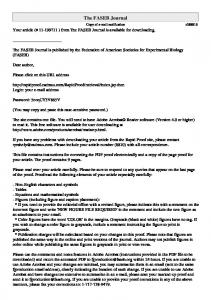
RESULTS Features of isolated CMs and CLP hearts Figure 1A shows the typical morphology of isolated CMs, demonstrating a CM from a sham-treated rat heart, the features of which have been described elsewhere (22). Immunostaining was performed with primary antibody against ␣-actin and Texas Red-conjugated secondary antibody, revealing the repetitive pattern of actin protein. Such CMs demonstrate spontaneous contractions (not shown). Figure 1B shows the time course for release of IL-6 in sham-treated rat CMs (2⫻106) exposed to 100 nM recombinant rat (rr) C5a for various periods of time. Release of IL-6 peaked at 2 h following addition of 100 nM C5a and persisted through the 8-h interval. At 24 h of incubation with C5a, IL-6 levels had fallen back to baseline levels. Figure 1C, D features CMs (2⫻106) from sham-treated rat hearts responding to rr C5a (8 h incubation at 37°C) in 3
Figure 1. A) Normal rat CM immunostained with primary antibody against ␣-actin and Texas-Red conjugated secondary antibody. B) Time course for release of IL-6 from rat CMs (2⫻106) exposed to 100 nM recombinant rat C5a at 37°C. C, D) Dose responses for sham rat CMs (2⫻106) incubated with HBSS (none) or recombinant rat C5a (40 –100 nM) for 12 h at 37°C, following which supernatant fluids were evaluated by ELISA for TNF␣ (C) and IL-6 (D). For each bar, n ⫽ 5. E) Frozen sections of rat heart obtained before CLP (panels 1–3) or 24 h after CLP and stained for TNF␣ (panel 4) or IL-1 (panel 5). In panel 1, the primary antibody (to TNF␣) was omitted.
a dose-dependent manner (ED50⫽55 nM), with progressive release of TNF␣ and IL-6. Release of TNF␣ appeared to peak between 80 –100 nM C5a; in the case of IL-6, peak release followed a similar pattern. Figure 1E represents immunostaining of frozen sections of rat hearts, featuring sham-treated CMs at ⫻25 (Fig. 1E1–3) and CLP hearts 24 h after CLP at ⫻100 (Fig. 1E4, 5). At 0 h (Fig. 1E1), no staining for TNF␣ was found when the secondary antibody was omitted. Frozen sections of sham-treated CLP hearts revealed constitutive staining for TNF␣ (Fig. 1E2) and, at higher resolution (⫻100), intense cytoplasmic staining for TNF␣ in hearts 24 h after CLP (Fig. 1E4). The striations indicate that TNF␣ was present within CMs. Constitutive staining for IL-1 revealed cytoplasmic granular and cytoplasmic staining in sham-treated hearts (Fig. 1E3) and more intense granular and cytoplasmic staining in CMs of CLP hearts (at 24 h; Fig. 1E5). Similar results were obtained for IL-6 (data not shown). Accordingly, CLP was associated with accentuated presence of cytokines in CMs.
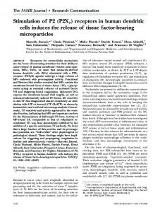
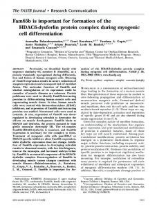
enates (Fig. 3) from CLP mice, a consistent pattern emerged in which cytokines and chemokines peaked at 24 h after CLP, followed by a decline at 48 and 72 h. In heart homogenates, TNF␣ was present in very low amounts compared to other mediators (Fig. 3). Mediator levels were higher in plasma than in heart homogenates, but this may be an artifact, since mediator levels in heart homogenates were expressed as picograms per gram total protein. It is possible that some contributions from contaminating inflammatory cells could have occurred in the homogenates. Figure 4 shows that spontaneous mediator release from cultured CMs obtained from CLP WT mice occurred as early as 8 –12 h after CLP, with peak release at 24 h, followed by a drop to baseline levels 48 h after CLP. Once again, TNF␣ levels in supernatant fluids were very low but showed a similar time course in which the peak was at 24 h after CLP.
Time courses for mediator presence in mouse plasma, heart homogenates and CM culture fluids after CLP
Since IL-6, TNF␣, and IL-1 have been designated as cardiosuppressive cytokines (12, 14 –16), it is possible that their release from CLP CMs results in a positive feedback, with released cytokines reacting with upregulated receptors on surfaces of CMs. As shown in Fig. 5, mRNA expression was measured in control WT mouse CMs obtained before CLP and 6, 12, and 24 h after CLP. Real-time PCR was used. As shown in Fig. 5A, mRNA for mouse C5aR rose almost 3-fold by 6 h after CLP and then fell back to baseline by 12 h, while mRNA for C5L2 showed a very small increase (nonsignificant)
As indicated in the introduction, the literature frequently emphasizes IL-1, TNF␣, and IL-6 as cardiosuppressive cytokines. However, a series of other cytokines and chemokines were also studied in the context of CLP, since many are proinflammatory mediators thought to have adverse effects in the setting of the cytokine storm. In plasma (Fig. 2) and in heart homog4
Vol. 25
July 2011
Increased mRNAs for cytokine and C5a receptors in mouse CMs after CLP
The FASEB Journal 䡠 www.fasebj.org
ATEFI ET AL.
Figure 2. Presence of cytokines and chemokines in plasma from sham-treated and WT mice at 12, 24, 48, and 72 h after CLP. A) IL-6. B) TNF␣. C) IL-1. D) MIP-1␣. E) MIP-2. F) MCP-1. G) KC. H) IL-10. For each bar, n ⱖ 5.
at 6 h, falling back to basal levels (Fig. 5B). In the case of IL-6R (gp80), like the data for C5aR (Fig. 5A), mRNA levels peaked at 6 h after CLP and fell back to basal levels 12 and 24 h after CLP (Fig. 5C), while mRNA for TNFR1 was elevated 6 h after CLP and persisted 12 and 24 h after CLP (Fig. 5D). The upregulation of C5aR, IL-6R, and TNF␣R1 following CLP could set the stage for cytokine release from CMs, as a result of autocrine/paracrine interaction of mediators with upregulated CM receptors, in a manner that could further accentuate the release of cardiosuppressive cytokines from CMs. Alternatively, interaction of cytokines with upregulated receptors on CLP CMs might activate signaling pathways that cause contractility and relaxation of CMs.
Requirements for C5a receptors, C5a, and IL-17A for cytokine/chemokine release from CLP CMs These studies were performed using CMs from WT and various knockout (KO) mice (all on a C57Bl/6 background) 24 h after CLP, at which time there was a peak in spontaneous in vitro release of cytokines and chemokines from CMs (Fig. 4). The data in Fig. 6 show the effects of the presence or absence on/in CMs of C5aR or C5L2 on spontaneous mediator release from CMs (24 h after CLP) as compared to release from WT CMs under similar conditions. For each of the 8 mediators evaluated, there were global reductions in KO (C5aR⫺/⫺, C5L2⫺/⫺) CMs, with mediator levels falling to levels found in sham-treated CMs (Fig. 6), suggesting that mediator release is dependent on the combined
Figure 3. Presence of cytokines and chemokines in heart homogenates from sham-treated and WT mice at 12, 24, 48, and 72 h after CLP. A) IL-6. B) TNF␣. C) IL-1. D) MIP-1. E) MIP-2. F) MCP-1. G) KC. H) IL-10. Data are expressed as amount of mediator (pg/ml) per gram total protein. For each bar, n ⱖ 5. MEDIATOR RELEASE FROM SEPTIC CARDIOMYOCYTES
5
Figure 4. Release of mediators from sham-treated or CLP WT CMs obtained at 8, 12, 24, and 48 h after CLP. A) IL-6. B) TNF␣. C) MIP-1␣. D) MIP-2. CMs were cultured for 18 h at 37°C. For each bar, n ⱖ 5.
engagement of both C5aR and C5L2. Even the antiinflammatory mediator, IL-10, fell to baseline levels in C5a receptor KO CMs (Fig. 6H). These data are reminiscent of effects of dual blockade or absence in vivo of C5L2 and C5aR in CLP mice, in which plasma levels of cytokines and chemokines after CLP were almost abolished compared to effects of single-receptor blockade or absence of C5a receptors (4, 10, 18, 20, 21). Such data are consistent with the concept that mediator release from CMs after CLP is dependent on combined and perhaps sequential engagement of C5aR and C5L2. Other patterns of note in Fig. 6 suggest that the least abundant cytokines and chemokines released from WT CLP CMs are TNF␣, IL-1, and IL-10, each being produced in amounts ⬍ 70 pg/ml, while other products (IL-6, MIP-1␣, MIP-2, MCP-1, and KC) were produced in amounts ⬎250 pg/ml. In Fig. 7, CMs were obtained from WT mice that had undergone sham surgery or CLP 24 h earlier. As indicated, mice were treated at the time of CLP with 50 g i.v. control IgG vs. the same amount of rat neutralizing mAb to mouse IL-17A (23) or 40 g affinitypurified rabbit anti-mouse C5a directed against the C-terminal region (as described above and in ref. 21). The IC50 values for anti-C5a and anti-IL-17A IgG for enhanced survival after CLP were 40 g for each antibody. Neutralization of IL-17A greatly improved survival in CLP WT mice and profoundly depressed the cytokine storm in plasma. WT CLP CMs (2⫻106 at 24 h) were incubated in vitro at 37°C for 18 h to assess spontaneous release of mediators. The supernatant fluids were subsequently analyzed for cytokines (TNF␣ and IL-6) and chemokines (MIP-1␣ and MIP-2). As shown in Fig. 7A, TNF␣ levels were low in supernatant fluids from sham-treated CMs (13 pg/ml) and only doubled in CMs from CLP (24 h) mice treated with normal IgG. In vivo treatment of WT CLP mice with anti-C5a (neutralizing IgG reactive with the C-terminal region) modestly reduced TNF␣ release (nonsignifi6
Vol. 25
July 2011
cant); anti-IL-17A had no effect on TNF␣ release from CLP CMs, but the amount of TNF␣ released was very low. However, treatment of mice with either neutralizing antibody to C5a or IL-17A caused IL-6 levels to drop to baseline levels (Fig. 7B). Anti-C5a reduced MIP-1␣ release from CLP CMs, while anti-17A also caused reductions to baseline levels (Fig. 7C). The pattern for MIP-2 release (Fig. 7D) was very similar to that for MIP-1␣ (Fig. 7C). Collectively, the data suggest that in vivo neutralization of either C5a or IL-17A reduces in vitro release of cytokines and chemokines from CLP CMs, with the exception of limited effects on TNF␣ release, similar to what we have noted before when plasma levels of TNF␣ were evaluated after CLP in WT mice were treated with either anti-C5a or antibodies to C5aR or C5L2 (20). Precisely how C5a and IL-17A enhance cytokine and chemokine release from CLP CMs remains to be determined, and whether they act in sequence has not been evaluated. In data not shown, 50 g affinity-purified non-neutralizing rabbit IgG (reactive with the N-terminal region of C5a) and a neutralizing IgG (reactive with the C-terminal region of C5a) were employed immediately after CLP induction in WT C57Bl/6 mice. The non-neutralizing antibody had no inhibitory effects on mediator release, while the neutralizing antibody caused 73, 56, and 95% reductions in release of IL-6, MIP-1␣, and MIP-2, respectively.
DISCUSSION As indicated above, there is evidence in both human sepsis and experimental sepsis (such as endotoxemia, infusion of live Escherichia coli, or CLP) that complement activation is occurring, followed by the cytokine storm, which is defined by the presence of numerous proinflammatory cytokines and chemokines in plasma. This event often evolves into multiorgan failure, although the extent
Figure 5. mRNA expression based on real-time PCR for C5a receptors (C5aR, C5L2), IL-6R (gp80) and TNFR1, in shamtreated (control) WT mouse hearts and in hearts at 6, 12, and 24 h following CLP. A) C5aR. B) C5L2. C) IL-6R. D) TNFR1. For each bar, n ⫽ 3 separate samples.
The FASEB Journal 䡠 www.fasebj.org
ATEFI ET AL.
Figure 6. Release of mediators from CMs obtained from WT mice before (sham) and 24 h after CLP, as well as from CLP mice that were C5aR⫺/⫺ or C5L2⫺/⫺. A) IL-6. B) TNF␣. C) IL-1. D) MIP-1␣. E) MIP-2. F) MCP-1. G) KC. H) IL-10. For each bar, n ⫽ 5.
to which the presence of numerous proinflammatory mediators in plasma can be directly linked to organ injury and failure is not clear. In the case of IL-1, TNF␣, and IL-6 (as well as IL-8 in humans), these cytokines have been described as having cardiosuppressive effects. The term cardiosuppressive refers to the fact that these cytokines, when added in vitro to CMs, cause reversible contractile and relaxation defects. Such evidence has been obtained by the use of recombinant cytokines, as well as by neutralization or removal of cytokines from septic human sera (5, 11–15). In the current report, sham-treated rat CMs responded to C5a in vitro in a dose-dependent manner (Fig. 1), resulting in release of proinflammatory cytokines (TNF␣ and IL-6). CMs obtained from CLP-treated rodents spontaneously released a variety of proinflammatory cytokines and chemokines in vitro (Fig. 4). There appears to be a linkage between C5a, C5a receptors (C5aR and C5L2), and IL-17A and spontaneous release in vitro of cytokines and chemokines from CLP CMs based
Figure 7. Cytokines (TNF␣, IL-6) and chemokines (MIP-1␣, MIP-2) released from sham-treated mouse CMs and from 24 h CLP CMs obtained from WT mice administered control IgG, anti-C5a (40 g), or anti-IL-17A IgG (50 g), all administered i.v. at time 0. A) TNF␣. B) IL-6. C) MIP-1␣. D) MIP-2.For each bar, n ⱖ 5. MEDIATOR RELEASE FROM SEPTIC CARDIOMYOCYTES
on in vivo blockade of C5a or IL-17 and the use of C5a receptor-KO mice (Figs. 6, 7). Up-regulation in CLP CMs of receptors (for C5a, TNF␣, and IL-6; Fig. 5) might potentiate the autocrine/paracrine effects of cytokines released from CLP CMs, based on mediator release described in Figs. 4 and 6 – 8. After in vivo blockade (neutralization) of C5a or IL-17A, in vitro release of cytokines and chemokines from CLP mouse CMs was dramatically reduced except for TNF␣, which was present at very low levels (Fig. 7). Release of TNF␣ and IL-1 was low (20 and 9 pg/ml, respectively) as compared to IL-6 and other proinflammatory cytokines and chemokines, which were released in much higher quantities (100 –1000 pg/ml; Fig. 6). These data suggest that the presence of C5a and IL-17A is necessary for full release of mediators from CLP CMs (Fig. 7). The role of IL-17A in the cytokine storm after CLP has been documented recently, based on in vivo neutralization of IL-17A in CLP WT mice (21). This resulted in profound reductions in plasma levels of proinflammatory cytokines and chemokines and increased levels of the anti-inflammatory cytokine IL-10. Such data suggest, in the setting of CLP, that C5a and IL-17A are upstream “master switches” that govern the cytokine storm. Our current data demonstrate release of a variety of proinflammatory cytokines and chemokines from CMs obtained from CLP mice. Such release occurred in a manner that also required availability of both C5aR and C5L2 (Fig. 6). Collectively, these findings suggest that maximal mediator release from CMs after CLP requires IL-17A as well as C5a and both C5a receptors. These findings may have a parallel with our recent study in which optimal survival in CLP mice required absence or blockade of both C5aR and C5L2, which resulted in ablation of the cytokine storm (20). Such data indicate that development of the cytokine storm requires engagement of both C5aR and C5L2, perhaps in a sequential manner. Figure 8 shows an overview of the findings presented in the current report. It appears that CLP activates 7
Figure 8. Proposed mechanisms of septic cardiomyopathy based on the data presented herein.
complement systemically, resulting in generation of C5a, which interacts with C5aR (and perhaps C5L2) on CMs to bring about contractility defects and development of heart failure (17). In parallel, C5a interactions with C5aR and C5L2 on CMs cause release from CMs of TNF␣ and IL-6. Simultaneously, up-regulation of receptors on CLP CMs occurs, including C5aR, TNFR1, and IL-6R (Fig. 5). This would allow positive feedback interaction of released cytokines with their receptors on CMs, resulting in contractility and relaxation defects. Whether C5a independently reacts directly with C5aR (and perhaps C5L2) to cause electrophysiological defects or has effects on CMs that are linked to release of cardiosuppressive cytokines from CMs remains to be determined. It might be noted that ischemic injury to the heart activates gene machinery that results in production of a host of complement proteins that may set the stage for a complement storm in the myocardium (23). Such data suggest that, under certain conditions, CMs can express complement proteins that can ultimately produce complement activation products, C5a and C5b-9, both of which have been linked to cardiac dysfunction after acute ischemic injury (4, 17, 23–25). We have recently suggested that once a pathological process is triggered in the heart, its outcomes may be greatly amplified by C5-derived complement activation products locally generated (24). As indicated above, reversible cardiac dysfunction clearly occurs after sepsis in both humans and animals. The basis for this complication is debatable. Some possibilities are described above. Loss of CM ATP levels has been associated with activation of the DNA repair enzyme poly(adenosine diphosphate-ribose) polymerase (PARP), which results in consumptive depletion of ATP (26). Loss of mitochondrial ATP may be linked to broad cellular dysfunction that is commonly seen in the shock syndrome. Since there is evidence that heart tissue obtained from autopsy specimens from humans with sepsis shows the presence of activated PARP (26), this could cause ATP depletion, leading to the onset of 8
Vol. 25
July 2011
mitochondrial energetic dysfunction in the heart in sepsis. Inhibition of PARP has been described to be cardioprotective after acute myocardial ischemia (27). Our own group has demonstrated that CLP in mice and rats leads to C5a generation and up-regulation of C5aR on CMs (Fig. 5 and refs. 17, 20). Blockade of C5a with neutralizing antibody was highly protective in the setting of CLP (28) and prevented the development of defects in both contractility and relaxation of CMs (17). Furthermore, since sham-treated CMs constitutively express C5aR, in vitro addition of C5a to CMs induced defects in contractility and relaxation (17). Finally, cardiosuppressive cytokines have been described by several groups in the context of sepsis or endotoxemia. These cytokines cause development of contractile defects in CMs. Such cytokines may be serum derived or directly produced in the heart and include TNF␣, IL-6, and IL-1 (Fig. 1 and as described above). Others have suggested that, at least in human septic serum, IL-6, IL-8, and IL-10 may be important cardiosuppressive factors (11). On the basis of such reports, addition of any of these recombinant cytokines to sham-treated CMs in vitro will induce contractile defects. It seems highly likely that under such conditions cardiosuppressive cytokines may affect CM function via interaction with cytokine receptors on CMs. Furthermore, some reports suggest that neutralization or removal of these cytokines reduces the adverse effects of septic serum on CMs (see above). The accumulating evidence that CLP CMs release a variety of cytokines and chemokines, together with evidence that cardiosuppressive cytokines can be released from CMs after endotoxemia or acute burn injury of skin (29, 30), suggests that intramyocardial production of such cytokines may result in selfinflicted harm to the heart during sepsis. This could be referred to as a myocardial cytokine storm. Obviously, the plethora of cytokines with cardiosuppressive effects suggests that targeting individual cytokines for blockade will not likely be an effective strategy for preventing or treating cardiac dysfunction in sepsis as related to cytokines. Rather, targeting one of the master switches of cytokine expression in sepsis (C5a or IL-17A) or relevant receptors may be a more efficacious intervention. This work was sponsored by U.S. National Institutes of Health grants GM-61656, GM-29507, and HL-31963 to P.A.W. and NHLBI-T32-HL007517-28 to G.A.
REFERENCES 1.
Rabuel, C., and Mebazaa, A. (2006) Septic shock: a heart story since the 1960s. Intensive Care Med. 32, 799 – 807 2. Clowes, G. H., Jr., Vucinic, M., and Weidner, M. G. (1966) Circulatory and metabolic alterations associated with survival or death in peritonitis: clinical analysis of 25 cases. Ann. Surg. 163, 866 – 885 3. Babuin, L., and Jaffe, A. S. (2005) Troponin: the biomarker of choice for the detection of cardiac injury. CMAJ 173, 1191–1202 4. Rittirsch, D., Flierl, M. A., and Ward, P. A. (2008) Harmful molecular mechanisms in sepsis. Nat. Rev. Immunol. 8, 776 –787
The FASEB Journal 䡠 www.fasebj.org
ATEFI ET AL.
5.
6. 7.
8.
9. 10. 11. 12.
13.
14.
15. 16.
17.
18. 19.
Parrillo, J. E., Burch, C., Shelhamer, J. H., Parker, M. M., Natanson, C., and Schuette, W. (1985) A circulating myocardial depressant substance in humans with septic shock. Septic shock patients with a reduced ejection fraction have a circulating factor that depresses in vitro myocardial cell performance. J. Clin. Invest. 76, 1539 –1553 Crouser, E. D. (2004) Mitochondrial dysfunction in septic shock and multiple organ dysfunction syndrome. Mitochondrion 4, 729–741 Gellerich, F. N., Trumbeckaite, S., Opalka, J. R., Gellerich, J. F., Chen, Y., Neuhof, C., Redl, H., Werdan, K., and Zierz, S. (2002) Mitochondrial dysfunction in sepsis: evidence from bacteraemic baboons and endotoxaemic rabbits. Biosci. Rep. 22, 99 –113 Brealey, D., Brand, M., Hargreaves, I., Heales, S., Land, J., Smolenski, R., Davies, N. A., Cooper, C. E., and Singer, M. (2002) Association between mitochondrial dysfunction and severity and outcome of septic shock. Lancet 360, 219 –223 Harrois, A., Huet, O., and Duranteau, J. (2009) Alterations of mitochondrial function in sepsis and critical illness. Curr. Opin. Anaesthesiol. 22, 143–149 Flierl, M. A., Rittirsch, D., Huber-Lang, M. S., Sarma, J. V., and Ward, P. A. (2008) Molecular events in the cardiomyopathy of sepsis. Mol. Med. 14, 327–336 Joulin, O., Petillot, P., Labalette, M., Lancel, S., and Neviere, R. (2007) Cytokine profile of human septic shock serum inducing cardiomyocyte contractile dysfunction. Physiol. Res. 56, 291–297 Cain, B. S., Meldrum, D. R., Dinarello, C. A., Meng, X., Joo, K. S., Banerjee, A., and Harken, A. H. (1999) Tumor necrosis factor-alpha and interleukin-1beta synergistically depress human myocardial function. Crit. Care Med. 27, 1309 –1318 Janssen, S. P., Gayan-Ramirez, G., Van den Bergh, A., Herijgers, P., Maes, K., Verbeken, E., and Decramer, M. (2005) Interleukin-6 causes myocardial failure and skeletal muscle atrophy in rats. Circulation 111, 996 –1005 Kumar, A., Paladugu, B., Mensing, J., and Parrillo, J. E. (2007) Transforming growth factor-beta1 blocks in vitro cardiac myocyte depression induced by tumor necrosis factor-alpha, interleukin-1beta, and human septic shock serum. Crit. Care Med. 35, 358–364 Maass, D. L., White, J., and Horton, J. W. (2002) IL-1beta and IL-6 act synergistically with TNF-alpha to alter cardiac contractile function after burn trauma. Shock 18, 360 –366 Maass, D. L., Hybki, D. P., White, J., and Horton, J. W. (2002) The time course of cardiac NF-kappaB activation and TNF-alpha secretion by cardiac myocytes after burn injury: contribution to burn-related cardiac contractile dysfunction. Shock 17, 293–299 Niederbichler, A. D., Hoesel, L. M., Westfall, M. V., Gao, H., Ipaktchi, K. R., Sun, L., Zetoune, F. S., Su, G. L., Arbabi, S., Sarma, J. V., Wang, S. C., Hemmila, M. R., and Ward, P. A. (2006) An essential role for complement C5a in the pathogenesis of septic cardiac dysfunction. J. Exp. Med. 203, 53– 61 Rittirsch, D., Huber-Lang, M. S., Flierl, M. A., and Ward, P. A. (2009) Immunodesign of experimental sepsis by cecal ligation and puncture. Nat. Protoc. 4, 31–36 Huber-Lang, M. S., Sarma, J. V., McGuire, S. R., Lu, K. T., Guo, R. F., Padgaonkar, V. A., Younkin, E. M., Laudes, I. J., Riede-
MEDIATOR RELEASE FROM SEPTIC CARDIOMYOCYTES
20.
21.
22.
23.
24. 25.
26.
27.
28.
29. 30.
mann, N. C., Younger, J. G., and Ward, P. A. (2001) Protective effects of anti-C5a peptide antibodies in experimental sepsis. FASEB J. 15, 568 –570 Rittirsch, D., Flierl, M. A., Nadeau, B. A., Day, D. E., HuberLang, M., Mackay, C. R., Zetoune, F. S., Gerard, N. P., Cianflone, K., Kohl, J., Gerard, C., Sarma, J. V., and Ward, P. A. (2008) Functional roles for C5a receptors in sepsis. Nat. Med. 14, 551–557 Flierl, M. A., Rittirsch, D., Gao, H., Hoesel, L. M., Nadeau, B. A., Day, D. E., Zetoune, F. S., Sarma, J. V., Huber-Lang, M. S., Ferrara, J. L., and Ward, P. A. (2008) Adverse functions of IL-17A in experimental sepsis. FASEB J. 22, 2198 –2205 Herron, T. J., Vandenboom, R., Fomicheva, E., Mundada, L., Edwards, T., and Metzger, J. M. (2007) Calcium-independent negative inotropy by beta-myosin heavy chain gene transfer in cardiac myocytes. Circ. Res. 100, 1182–1190 Singh, M. V., Kapoun, A., Higgins, L., Kutschke, W., Thurman, J. M., Zhang, R., Singh, M., Yang, J., Guan, X., Lowe, J. S., Weiss, R. M., Zimmermann, K., Yull, F. E., Blackwell, T. S., Mohler, P. J., and Anderson, M. E. (2009) Ca2⫹/calmodulin-dependent kinase II triggers cell membrane injury by inducing complement factor B gene expression in the mouse heart. J. Clin. Invest. 119, 986 –996 Ward, P. A. (2009) The sepsis seesaw: seeking a heart salve. Nat. Med. 15, 497– 498 Buras, J. A., Rice, L., Orlow, D., Pavlides, S., Reenstra, W. R., Ceonzo, K., and Stahl, G. L. (2004) Inhibition of C5 or absence of C6 protects from sepsis mortality. Immunobiology 209, 629 – 635 Soriano, F. G., Nogueira, A. C., Caldini, E. G., Lins, M. H., Teixeira, A. C., Cappi, S. B., Lotufo, P. A., Bernik, M. M., Zsengeller, Z., Chen, M., and Szabo, C. (2006) Potential role of poly(adenosine 5⬘-diphosphate-ribose) polymerase activation in the pathogenesis of myocardial contractile dysfunction associated with human septic shock. Crit. Care Med. 34, 1073–1079 Pacher, P., and Szabo, C. (2007) Role of poly(ADP-ribose) polymerase 1 (PARP-1) in cardiovascular diseases: the therapeutic potential of PARP inhibitors. Cardiovasc. Drug Rev. 25, 235–260 Czermak, B. J., Sarma, V., Pierson, C. L., Warner, R. L., Huber-Lang, M., Bless, N. M., Schmal, H., Friedl, H. P., and Ward, P. A. (1999) Protective effects of C5a blockade in sepsis. Nat. Med. 5, 788 –792 Ballard-Croft, C., Carlson, D., Maass, D. L., and Horton, J. W. (2004) Burn trauma alters calcium transporter protein expression in the heart. J. Appl. Physiol. 97, 1470 –1476 Thompson, M., Kliewer, A., Maass, D., Becker, L., White, D. J., Bryant, D., Arteaga, G., Horton, J., and Giroir, B. P. (2000) Increased cardiomyocyte intracellular calcium during endotoxin-induced cardiac dysfunction in guinea pigs. Pediatr. Res. 47, 669 – 676 Received for publication February 18, 2011. Accepted for publication March 31, 2011.
9