Complement-Dependent Acute-Phase Expression of C-Reactive Protein and Serum Amyloid P-Component1 Alexander J. Szalai,2* Frederik W. van Ginkel,† Yue Wang,* Jerry R. McGhee,† and John E. Volanakis*‡ The acute-phase response (APR) is regulated by TNF-␣, IL-1, and IL-6 acting alone, in combination, or in concert with hormones. The anaphylotoxin C5a, generated during complement activation, induces in vitro the synthesis of these cytokines by leukocytes and of acute-phase proteins by HepG2 cells. However, there is no clear evidence for a role of C5a or any other complement activation product in regulation of the APR in vivo. In this study, using human C-reactive protein (CRP) transgenic mice deficient in C3 or C5, we investigated whether complement activation contributes to induction of the acute-phase proteins CRP and serum amyloid P-component (SAP). Absence of C3 or C5 resulted in decreased LPS-induced up-regulation of the CRP transgene and the mouse SAP gene. Also, LPS induced both the IL-1 and IL-6 genes in normocomplementemic mice, but in complement-deficient mice it significantly induced only IL-6. Like LPS injection, activation of complement by cobra venom factor led to significant elevation of serum CRP and SAP in normocomplementemic mice but not in complement-deficient mice. Injection of recombinant human C5a into human CRP transgenic mice induced the IL-1 gene and caused significant elevation of both serum CRP and SAP. However, in human CRP transgenic IL-6-deficient mice, recombinant human C5a did not induce the CRP nor the SAP gene. Based on these data, we conclude that during the APR, C5a generated as a consequence of complement activation acts in concert with IL-6 and/or IL-1 to promote up-regulation of the CRP and SAP genes. The Journal of Immunology, 2000, 165: 1030 –1035.
T
issue injury results in a systemic reaction termed the acute-phase response (APR)3 (1), that includes changes in the serum levels of several plasma proteins produced primarily by hepatocytes (2). Regulation of the APR is largely mediated by the proinflammatory cytokines TNF-␣, IL-1, and IL-6 acting alone, in combination, or in concert with various steroid hormones (3–5). A convenient murine model for studying induction of acute-phase protein genes is i.p. injection of LPS (6), which reproducibly elicits increased expression of proinflammatory cytokines (6, 7). LPS also activates the classical and alternative pathways of complement (8, 9), leading to generation of the bioactive peptides C3a and C5a (10). The anaphylotoxin C5a elicits from target cells a wide range of activities (10), including the synthesis of TNF-␣, IL-1-, and IL-6 by leukocytes (11–13), which amplify the inflammatory process. The biological activity of C5a is mediated through the C5a receptor (C5aR, CD88), a G-protein-coupled seven-transmembrane-domain protein expressed on cells of myeloid origin (10, 14), and also on nonmyeloid cells in the liver and lung (15–17). In mice, hepatic expression of C5aR is increased by *Division of Clinical Immunology and Rheumatology and †Department of Microbiology, University of Alabama, Birmingham, AL 35294; and ‡Biomedical Sciences Research Center “A. Fleming,” Vari, Greece Received for publication January 4, 2000. Accepted for publication April 25, 2000. The costs of publication of this article were defrayed in part by the payment of page charges. This article must therefore be hereby marked advertisement in accordance with 18 U.S.C. Section 1734 solely to indicate this fact. 1 This research was supported in part by National Institutes of Health Grant AI 42183 (to A.J.S.). 2 Address correspondence and reprint requests to Dr. Alexander J. Szalai, Division of Clinical Immunology and Rheumatology, Department of Medicine, University of Alabama, Birmingham, AL 35294-0006. E-mail address:
[email protected] 3 Abbreviations used in this paper: APR, acute-phase response; C3D, C3 deficient; C5aR, C5a receptor (CD88); C5D, C5 deficient (DBA/2J mice); CRP, C-reactive protein; CRPtg, human CRP transgenic C57BL/6J mice; IL-6D, IL-6 deficient; rC5a, recombinant human C5a; SAP, mouse serum amyloid P-component; CoVF, cobra venom factor.
Copyright © 2000 by The American Association of Immunologists
administration of LPS (17). Furthermore, C5a has been shown to induce the synthesis and secretion of acute-phase proteins by HepG2 cells (16, 17). Thus, it seems likely that by binding to C5aR on liver cells and/or by stimulating secretion of proinflammatory cytokines by myeloid cells, C5a generated as a consequence of complement activation contributes to regulation of the APR. The pentraxins C-reactive protein (CRP) and serum amyloid P-component (SAP) (18, 19) are evolutionarily conserved, Ca2⫹binding proteins that participate variably in the APR in different species (3). For example, CRP is a major acute-phase protein in humans (20), while in the mouse it is a trace plasma component and only a minor acute-phase protein (21–23). In contrast, SAP is highly inducible during the APR in mice but not in humans (24). Numerous studies using freshly isolated human hepatocytes and hepatoma cell lines have established that IL-6 is the major inducer of the CRP gene and that IL-1 and glucocorticoids act in synergy with IL-6 to enhance CRP gene induction (25–29). Similar studies using primary mouse hepatocytes (30) have shown that the mouse SAP gene can be induced directly by either IL-1 or IL-6. We have shown (31) that in mice constitutive and IL-6-induced acute-phase expression of a human CRP transgene requires testosterone, whereas testosterone does not influence expression of the mouse SAP gene, which requires IL-1 plus IL-6 for acute-phase expression. Horowitz et al. (32) reported an association between in vivo activation of the alternative complement pathway by inulin and increased serum CRP levels in rabbits, but the observation was not pursued further. Shortly thereafter, Pepys and Rogers reported (33) that in mice depleted of complement by i.p. injection of cobra venom factor (CoVF), induction of SAP by LPS was unaffected. However, the impact of complement activation by CoVF per se on SAP expression was not addressed. Recently, we reported (34) that 24 h after injection of CoVF into human CRP transgenic (CRPtg) mice, serum levels of human CRP and mouse SAP are elevated. 0022-1767/00/$02.00
The Journal of Immunology In the present study, we generated by selective breeding CRPtg mice deficient in C3 (C3D) or C5 (C5D) to investigate directly the contribution of these complement proteins to the induction of the CRP and SAP genes during the APR. We show that absence of either C3 or C5 attenuates the response of the human CRP transgene and mouse SAP gene to LPS. Injection of CoVF alone led to significant elevation of serum CRP and SAP in normocomplementemic mice, but not in either complement deficient strain. Importantly, injection of recombinant human C5a (rC5a) mimicked the effect of complement activation by CoVF, i.e., it caused elevation of serum CRP and SAP. Injection of rC5a into C3D or C5D mice also elicited elevation of CRP and SAP. In complementdeficient mice, the reduced acute-phase expression of the CRP and SAP genes after LPS administration was paralleled by the absence of significant elevation of serum IL-1. Conversely, induction of the CRP and SAP genes in these mice by injection of rC5a was associated with elevation of serum IL-1, but not of IL-6. Nevertheless, an absolute requirement for IL-6 in the induction of CRP and SAP by rC5a was indicated by the failure of rC5a to induce either gene in IL-6-deficient (IL-6D) mice. The combined data support the notion that C5a generated during complement activation contributes to up-regulation of the CRP and SAP genes during the APR. C5a does not act directly to mediate expression of these acute-phase genes, rather its effect is mediated in concert with IL-6 and/or of IL-1.
Materials and Methods Animals All mice used in this study were fed and watered ad libitum and barrier maintained under a 12-h light-dark cycle according to protocols established by the Animal Resources Program at the University of Alabama at Birmingham. We have previously described the establishment of a breeding colony of CRPtg C57BL/6J congenic mice (35). CRPtg mice carry a 31-kb ClaI fragment of human genomic DNA comprised of the CRP gene, 17 kb of 5⬘-flanking sequence, and 11.3 kb of 3⬘ flanking sequence (36). After injection of LPS into CRPtg mice, peak levels of serum IL-1 and IL-6 are attained by 2 h, followed by a human CRP response with peak serum levels reached by 18 h (31). The generation and genetic background of IL-6D and complement C3D mice has also been described (37, 38). IL-6D mice are homozygous for a disruption of the fourth exon of the murine IL-6 gene and produce no serum IL-6 after LPS injection (37). C3D mice produce no serum complement C3 due to targeted disruption of the murine C3 gene promoter (38) and lack the ability to generate C3-convertases through the alternative pathway and C5-convertases through either the classical or alternative pathway. DBA/2J mice are complement C5D due to a spontaneous mutation in exon 7 of the murine C5 gene (39). Female CRPtg mice were crossed with IL-6D males to produce hybrids, and CRPtg hybrids were backcrossed to the IL-6D parent to generate CRPtg/IL-6D mice. The same breeding system was used to generate CRPtg/C3D and CRPtg/C5D mice. C3D and IL-6D mice were backcrossed to C57BL/6J for at least five generations before hybridization with CRPtg. Mice were screened for presence of the CRP transgene and for inheritance of the C3 or C5 mutant alleles using PCRs specific for human CRP (40), mouse C3 (38), or mouse C5 (39). IL-6D, C3D, and C5D vs sufficient progeny were obtained in the expected Mendellian ratios. The CRP transgene was responsive to induction by LPS in all three types of CRPtg F1 hybrids. Male F2 mice (8 –12 wk old) were used in experiments, and all appeared normal and healthy.
Administration of LPS, CoVF, and rC5a LPS from Eschericia coli (serotype 026:B6) was purchased from SigmaAldrich (St. Louis, MO), resuspended in sterile 0.9% NaCl, and injected i.p. at a dose of 25 g per mouse. CoVF from Naja naja (Quidel Corporation, San Diego, CA) was dissolved in sterile pyrogen-free 0.9% NaCl (Abbott Laboratories, North Chicago, IL) and also injected i.p. at 25 g per mouse. N. naja CoVF is known to support the formation of potent C3- and C5-convertases (41, 42). We have shown (34) that single i.p. injection of 25 g CoVF reduces mouse serum C3 to ⬍3% of initial values within 4 h and that the hypocomplementemia lasts several days. rC5a (Sigma-Aldrich) was reconstituted in 0.25% (w/v) BSA in sterile pyrogen-free 0.9% NaCl and injected i.p. at 2.5–20 g/mouse. rC5a has been shown to acti-
1031 vate various mouse cells including mast cells (43), astrocytes (44), and monocytes and macrophages (10, 45). Limulus amebocyte assays confirmed that CoVF and rC5a was not contaminated with endotoxin.
Measurement of serum IL-1, IL-6, CRP, and SAP Sera from blood samples (50 l) collected before and 2 and 18 h after injection of stimulants were used to measure the concentration of IL-1 and IL-6 (2 h sera) and CRP and SAP (18 h sera). Mouse IL-1 and IL-6 ELISAs were performed exactly as described (31, 37) using rat mAb MP520F3 and biotinylated mAb MP5-32C11 (PharMingen, San Diego, CA) for IL-6 detection and rat mAb 13A10 and biotinylated mAb 13D11 (Biosource International, Camarillo, CA) for IL-1 detection. Peroxidase-labeled goat anti-biotin (Vector Laboratories, Burlingame, CA) was used as the reporter, and recombinant mouse IL-1 and recombinant mouse IL-6 (Genzyme, Cambridge, MA) were used to generate standard curves. The lower limit of detection in each case was 10 pg of cytokine/ml. CRPtg/ IL-6D mice were identified by absence of detectable IL-6 in sera collected 2 h after LPS injection (31). ELISA for CRP used sheep anti-human CRP serum (Cappel, Durham, NC) and anti-CRP mAb HD2-4 (46) as the capture and detection Ab, respectively, and affinity-purified human CRP (47) as the standard. The assay does not detect mouse CRP and has a lower limit of detection of 20 ng of human CRP per ml of mouse serum. ELISA for mouse SAP was performed as described (48) using sheep and rabbit antiSAP serum as the capture and detection Ab, respectively, and mouse SAP reference standards all from Calbiochem-Novabiochem (San Diego, CA). The lower limit of detection is 25 g of SAP per ml serum.
Statistical analysis All values are reported as the mean ⫾ SEM of at least three experiments. Comparisons of means were performed using Student’s t tests with p ⬍ 0.05 considered significant.
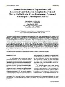
Results To investigate the possible role of complement in up-regulation of the CRP transgene and SAP gene during the APR, CRP and SAP serum levels were measured in age-matched male CRPtg/C3D and CRPtg/C5D mice before and after LPS injection. Normocomplementemic CRPtg and CRPtg/IL-6D mice were used as controls. Injection of LPS caused an ⬃12-fold increase of serum human CRP in normocomplementemic CRPtg mice but only an ⬃4-fold increase in CRPtg/C3D and CRPtg/C5D mice (Fig. 1A). The difference between normocomplementemic and complement-deficient mice was significant. Concomitant induction of the mouse SAP gene followed the same pattern, i.e., serum SAP was increased ⬃20-fold in normocomplementemic controls as compared with ⬃10-fold in complement-deficient mice. However, the difference in SAP increase between the two groups did not achieve statistical significance (Fig. 1B). In accordance with our earlier report (31), the CRP transgene was unresponsive and the SAP gene was induced only ⬃2.5-fold by LPS in CRPtg/IL-6D mice (Fig. 1, A and B). These data suggest that an intact complement system is required for full LPS-induced acute-phase expression of the CRP transgene and probably also of the SAP gene. Because IL-6 is absolutely required for LPS induction of the CRP transgene and IL-1 together with IL-6 is required for full LPS induction of the SAP gene (31), we investigated whether induction of these cytokines by LPS is altered in complement-deficient mice. As shown in Fig. 1C, LPS administration evoked significant elevation of serum IL-6 in all mice carrying a normal IL-6 gene, although levels were significantly lower in C5D compared with normocomplementemic ( p ⫽ 0.007) or C3D ( p ⫽ 0.014) mice (Student’s t tests). In contrast, LPS caused a significant increase of serum IL-1 only in normocomplementemic animals (Fig. 1D). Thus, a normal complement system is necessary for LPS induction of the IL-1 but not of the IL-6 gene. The combined data suggested that complement activation products act together with IL-6 and probably also IL-1 to enhance induction of the CRP transgene by LPS. In the case of SAP, the
1032
COMPLEMENT-DEPENDENT REGULATION OF CRP AND SAP
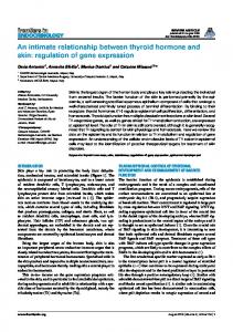
FIGURE 2. Effect of CoVF injection on serum levels of the acute-phase proteins CRP (A) and SAP (B). CRPtg mice and CRPtg mice deficient in C3, C5, or IL-6 were injected i.p. with 25 g of CoVF. Human CRP and mouse SAP were quantitated in sera before and 18 h after CoVF injection, and the results (mean ⫹ SEM for at least three experiments) are expressed on the vertical axis as fold increases over baseline values. The total number of mice analyzed in each group is indicated above the columns in A. The asterisks indicate fold increases significantly greater than unity (p ⬍ 0.05; Student’s t tests).
FIGURE 1. Effect of LPS injection on serum levels of CRP (A), SAP (B), IL-6 (C), and IL-1 (D). CRPtg mice and CRPtg mice deficient in C3, C5, or IL-6 were injected i.p. with 25 g of LPS. Human CRP and mouse SAP were quantitated in sera collected before and 18h after LPS injection, and IL-1 and IL-6 were quantitated before and 2 h after injection. The total number of mice analyzed in each group is indicated above the columns in A. The results (mean ⫹ SEM for at least three experiments) are expressed on the vertical axis as fold increases over baseline values of CRP and SAP and as 2-h serum concentrations of IL-6 and IL-1. Single asterisks indicate a statistically significant difference in fold increases of CRP or SAP compared with CRPtg mice, and double asterisks indicate a significantly greater than constitutive level of IL-6 or IL-1 (ⴱ, p ⬍ 0.05; ⴱⴱ, p ⬍ 0.005; Student’s t tests). The average constitutive level of each cytokine was 186 ⫾ 41 pg IL-1/ml of serum (all mice combined) and 9.6 ⫾ 1.4 ng IL-6/ml (excluding IL-6D mice).
enhancing effect of complement activation was less pronounced and probably could also be attributed to interaction with IL-6 and IL-1. To further investigate the effects of complement activation on the APR, we measured the cytokine and acute-phase protein responses of mice injected with the potent complement activator CoVF. As shown in Fig. 2, injection of CoVF in normocomplementemic mice reproducibly induced significant elevation of both CRP and SAP. Neither acute-phase protein was elevated substan-
tially following CoVF injection in C3D or C5D mice, indicating that CoVF-induced up-regulation was mediated by complement activation products and not directly by CoVF or by a possible contaminant in the CoVF preparation. Injection of CoVF did not induce the IL-1 or the IL-6 gene (data not shown) and failed to up-regulate CRP and SAP in IL-6D mice. The results are consistent with the view that complement activation products act in concert with IL-6 to enhance the APR. Because C5a has been shown to induce expression of some acute-phase proteins in vitro (17) and it cannot be produced by C5D mice and likely is not generated in CoVF-injected C3D mice, we tested directly if C5a can induce CRP and SAP acute-phase responses. Injection of rC5a into CRPtg mice elicited significant elevation of both CRP and SAP in a dose-dependent manner (Fig. 3). In fact, at the highest dose used (20 g of rC5a) the magnitude of the CRP and SAP responses (about 2- and 12-fold, respectively) were comparable to those elicited by CoVF (Fig. 2, A and B). Furthermore, injection of rC5a elicited significant elevation of serum IL-1 but not IL-6 (Fig. 4). Conclusive evidence that C5a participates in acute-phase regulation of the CRP transgene and SAP gene was obtained by experiments using C3D and C5D mice. Injection of rC5a in either strain evoked a small but significant elevation of serum CRP (Fig. 5A). A parallel elevation of SAP did not achieve statistical significance (Fig. 5B). Although serum IL-6 was not increased after injection of rC5a, a requirement for the cytokine was indicated by the failure of rC5a to cause an increase in serum CRP in IL-6D mice (Fig. 5A).
The Journal of Immunology
1033
FIGURE 5. Serum concentration of human CRP (A) and mouse SAP (B) in C3D (䡺), C5D (f), and IL-6D (E) CRPtg mice measured before and 18 h after injection i.p. of 20 g of rC5a. The asterisks indicate a significant elevation of serum CRP above preinjection levels (p ⬍ 0.05; Student’s t tests). FIGURE 3. Dose-response of CRP (A) and SAP (B) induction by rC5a. CRPtg mice were injected with the indicated amounts of rC5a suspended in 0.25% BSA, 0.9% NaCl. Human CRP and mouse SAP were quantitated in sera collected before and 18 h after injecting rC5a. Results (mean ⫹ SEM for three experiments) are expressed on the vertical axis as fold increases over baseline values. The total number of mice analyzed in each group is indicated above the columns in A. The asterisks indicate a fold increase significantly greater than unity (p ⬍ 0.05; Student’s t tests).
Discussion The APR is an essential manifestation of innate host defense against pathogens, its protective effect being dependent upon recognition of conserved repeating microbial structural elements (e.g., endotoxin, teichoic acids, or surface polysaccharides) by acute-
FIGURE 4. Changes in concentration of serum cytokines induced by rC5a. CRPtg mice were injected with 20 g of rC5a. IL-6 (A) and IL-1 (B) were quantitated in sera collected before (basal) and 2 h after rC5a injection (rC5a-induced). Representative results (mean ⫹ SEM) from one of three experiments using five mice are shown. The asterisk indicates a significantly greater than basal concentration of IL-1 (p ⬍ 0.05; Student’s t tests).
phase proteins (e.g., CRP, mannose-binding lectin, LPS-binding protein) (1–3, 49). Also, certain acute-phase proteins are known to influence the initiation and type of adaptive immune responses (50, 51). Thus, identifying the factors that regulate synthesis of acutephase proteins is an important step toward understanding host defense mechanisms as well as the pathophysiology of infectious and certain autoimmune diseases. A widely used in vivo model to study the APR is injection of LPS into mice (6), a treatment that elicits a constellation of inflammatory responses that mimics the endotoxin-induced APR in humans (52). Extensive studies in both species reveal that LPS-induced expression of acute-phase protein genes is largely controlled by IL-6, IL-1, and hormones (2, 49). Combinations of these mediators have additive, inhibitory, or synergistic effects on the APR (2), and studies using cytokine-deficient mice (37, 53–55) show that the APR is not dependent on a single cytokine or hormone but uses multiple overlapping and redundant pathways. The data we have generated using LPS-injected CRPtg mice and their IL-6D counterparts (Fig. 1) reinforce these concepts and are in agreement with our previous finding that in male CRPtg mice IL-6 is essential for acute-phase induction of CRP while maximal induction of SAP requires both IL-6 and IL-1 (31, 35). A novel finding of the present study is that in complement-deficient mice receiving LPS, up-regulation of CRP and SAP is blunted (Fig. 1, A and B) despite significant up-regulation of serum IL-6 (Fig. 1C). Furthermore, following LPS injection there is no significant elevation of IL-1 in the sera of complement-deficient mice (Fig. 1D). As it is known that in addition to being a strong inducer of cytokine synthesis (7) LPS is an efficient activator of the complement alternative and classical pathways (8, 9), we speculated that LPS-mediated complement activation contributed to the observed up-regulation of the IL-1, CRP, and SAP genes. To further test this concept, we measured the APR of mice injected with CoVF, a protein that activates the alternative complement pathway (41, 42). We found that CoVF injection caused significant elevation of both CRP and SAP serum levels in CRPtg mice, but not in their C3D, C5D, or IL-6D counterparts (Fig. 2). Furthermore, CoVF induced CRP and SAP without eliciting significant increase of serum IL-1 or IL-6. The combined results strongly support the concept that complement activation and up-regulation of serum CRP and SAP are causally related, as originally proposed by Horowitz et al. (32).
1034 Because there was no substantial difference between C3D and C5D mice in terms of their attenuated acute-phase responses to either LPS or CoVF (Figs. 1 and 2), we reasoned that probably the crucial defect was their common inability to generate C5a during complement activation. Direct proof for this hypothesis was provided by experiments using rC5a. Injection of rC5a led to significant elevation of CRP and SAP serum levels in CRPtg mice in a dose-dependent manner (Fig. 3). Also, injected rC5a elicited significant elevation of serum CRP in both C3D and C5D mice (Fig. 5). Injection of rC5a also induced significant elevation of IL-1 but not of IL-6 (Fig. 4). However, the CRP transgene was not responsive to rC5a in IL-6D mice (Fig. 5). Thus, the C5a effect on CRP expression apparently requires the presence of IL-6, albeit in low concentrations such as those expected to be expressed constitutively in CRPtg mice. It has been shown that in vitro C5a induces the synthesis and release of leukocyte-derived cytokines (11–13) known to participate in the regulation of the APR (3–5) and the expression of the acute-phase proteins ␣1-antitrypsin, ␣1-antichymotrypsin, C3, and complement factor B by HepG2 cells (16, 17). The current investigation using CRPtg mice has provided the first direct evidence that C5a contributes to the regulation of the APR in vivo. The significance of these findings is underlined by the fact that complement activation is an early consequence of most if not all forms of tissue injury that cause an APR. Included are bacterial infections, burns, ischemic necrosis, and immune-complex-mediated injury. Therefore, we propose that C5a generated as a consequence of complement activation following tissue damage or necrosis cooperates with proinflammatory cytokines and stress hormones to ensure maximal acute-phase expression of CRP, SAP, and perhaps also additional acute-phase proteins. Thus, complement activation products are an integral component of the highly complex network of mediators that interact to ensure appropriate expression of genes during the APR.
Acknowledgments We thank Mark A. McCrory for his expert technical assistance.
References 1. Kushner, I. 1982. The phenomenon of the acute phase response. Ann. NY Acad. Sci. 389:39. 2. Gabay, S., and I. Kushner. 1999. Acute-phase proteins and other systemic responses to inflammation. N. Engl. J. Med. 340:448. 3. Kushner, I., and A. Mackiewicz. 1993. The acute phase response: an overview. In Acute Phase Proteins: Molecular Biology, Biochemistry, and Clinical Applications. A. Mackiewicz, I. Kushner, and H. Baumann, eds. CRC Press, Boca Raton, FL, p. 3. 4. Baumann, H., G. P. Jahreis, and K. K. Morella. 1990. Interaction of cytokine- and glucocorticoid-response elements of acute phase plasma protein genes. J. Biol. Chem. 265:22275. 5. Baumann, H., and J. Gauldie. 1990. Regulation of the hepatic acute phase plasma protein genes by hepatocyte stimulating factors and other mediators of inflammation. Mol. Biol. Med. 7:147. 6. Won, K., S. P. Campos, and H. Baumann. 1993. Experimental systems for studying hepatic acute phase response. In Acute Phase Proteins: Molecular Biology, Biochemistry, and Clinical Applications. A. Mackiewicz, I. Kushner, and H. Baumann, eds. CRC Press, Boca Raton, FL, p. 255. 7. Luster, M. I., D. R. Germolec, T. Yoshida, F. Kayama, and M. Thompson. 1994. Endotoxin-induced cytokine gene expression and excretion in the liver. Hepatology 19:480. 8. Morrison, D. C., and L. F. Kline. 1977. Activation of the classical and properdin pathways of complement by bacterial lipopolysaccharides (LPS). J. Immunol. 118:362. 9. Reid, R. R., A. P. Prodeus, W. Khan, T. Hsu, F. S. Rosen, and M. C. Carroll. 1997. Endotoxin shock in antibody-deficient mice: unraveling the role of natural antibody and complement in the clearance of lipopolysaccharide. J. Immunol. 159:970.
COMPLEMENT-DEPENDENT REGULATION OF CRP AND SAP 10. Ember, J. A., M. A. Jagels, and T. E. Hugli. 1998. Characterisation of complement anaphylotoxins and their biological responses. In The Human Complement System in Health and Disease. J. E. Volanakis and M. M. Frank, eds. Marcel Dekker, Inc., New York, p. 241. 11. Okusawa, S., K. B. Yancey, J. W. M. van der Meer, S. Endres, G. Lonnemann, K. Hefter, M. M. Frank, J. F. Burke, C. A. Dinarello, and J. A. Gelfand. 1988. C5a stimulates secretion of tumor necrosis factor from human mononuclear cells in vitro. J. Exp. Med. 168:443. 12. Okusawa, S., C. A. Dinarello, K. B. Yancey, S. Endres, T. J. Lawley, M. M. Frank, J. F. Burke, and J. A. Gelfand. 1987. C5a induction of human interleukin 1: synergistic effect with endotoxin or interferon-␥ . J. Immunol. 139: 2635. 13. Scholz, W., M. R. McClurg, G. J. Cardenas, M. Smith, D. J. Noonan, T. E. Hugli, and E. L. Morgan. 1990. C5a-mediated release of interleukin 6 by human monocytes. Clin. Immunol. Immunopath. 57:297. 14. Nataf, S., N. Davoust, R. S. Ames, and S. R. Barnum. 1999. Human T-cells express the C5a receptor and are chemoattracted to C5a. J. Immunol. 162:4018. 15. McCoy, R. L., D. L. Haviland, E. P. Molmenti, T. Ziambaras, R. A. Wetsel, and D. H. Perlmutter. 1995. N-formylpeptide and complement C5a receptors are expressed in liver cells and mediate hepatic acute phase gene regulation. J. Exp. Med. 182:207. 16. Buchner, R. R., T. E. Hugli, J. A. Ember, and E. L. Morgan. 1995. Expression of functional receptors for human C5a anaphylotoxin (CD88) on the human hepatocellular carcinoma cell line HepG2: stimulation of acute-phase protein-specific mRNA and protein synthesis by human C5a anaphylotoxin. J. Immunol. 155:308. 17. Haviland, D. L., R. L. McCoy, W. T. Whitehead, H. Akama, E. P. Molmenti, A. Brown, J. C. Haviland, W. C. Parks, D. H. Perlmutter, and R. A. Wetsel. 1995. Cellular expression of the C5a anaphylotoxin receptor (C5aR): demonstration of C5aR on nonmyeloid cells of the liver and lung. J. Immunol. 154:1861. 18. Osmand, A. P., B. Friedenson, H. Gewurz, R. H. Painter, T. Hofmann, and E. Helton. 1977. Characterisation of C-reactive protein and the complement subcomponent C1t as homologous proteins displaying cyclic pentameric symmetry (pentraxins). Proc. Natl. Acad. Sci. USA 74:739. 19. Pepys, M. B., M. L. Baltz, F. C. de Beer, R. F. Dyck, S. Holford, S. M. Breathnach, M. M. Black, C. R. Tribe, D. J. Evans, and A. Feinstein. 1982. Biology of serum amyloid P component. Ann. NY Acad. Sci. 389:286. 20. Agrawal, A., J. M. Kilpatrick, and J. E. Volanakis. 1993. Structure and function of human C-reactive protein. In Acute Phase Proteins: Molecular Biology, Biochemistry, and Clinical Applications. A. Mackiewicz, I. Kushner, and H. Baumann, eds. CRC Press, Boca Raton, FL, p. 79. 21. Bodmer, B., and R. Siboo. 1977. Isolation of mouse C-reactive protein from liver and serum. J. Immunol. 118:1086. 22. Whitehead, A. S., K. Zahedi, M. Rits, R. F. Mortensen, and J. M. Lelias. 1990. Mouse C-reactive protein: generation of complementary DNA clones, structural analysis, and induction of messenger RNA during inflammation. Biochem. J. 266:59. 23. Siboo, R., and E. Kulisek. 1978. A fluorescent immunoassay for the quantification of C-reactive protein. J. Immunol. Methods 23:59. 24. Le, P. T., M. T. Muller, and R. F. Mortensen. 1982. Acute phase reactants of mice. I. Isolation of serum amyloid P-component (SAP) and its induction by a monokine. J. Immunol. 129:665. 25. Zhang, D., S.-L. Jiang, D. Rzewnicki, D. Samols, and I. Kushner. 1995. The effects of interleukin-1 on C-reactive protein expression in Hep3B cells is exerted at the transcriptional level. Biochem. J. 310:143. 26. Depraetere, S., J. Willems, and M. Joniau. 1991. Stimulation of CRP secretion in HepG2 cells: cooperative effect of dexamethasone and interleukin 6. Agents Actions 34:369. 27. Toniatti, C., R. Arcone, B. Majello, M. Ganter, G. Arpaia, and G. Ciliberto. 1990. Regulation of the human C-reactive protein gene: a major marker of inflammation and cancer. Mol. Biol. Med. 7:199. 28. Ganter, U., R. Arcone, C. Toniatti, G. Morrone, and G. Ciliberto. 1989. Dual control of C-reactive protein gene expression by interleukin-1 and interleukin-6. EMBO J. 8:3773. 29. Ganapathi, M. K., D. Rzewnicki, D. Samols, S.-L. Jiang, and I. Kushner. 1991. Effect of combinations of cytokines and hormones on synthesis of serum A amyloid and C-reactive protein in Hep 3B cells. J. Immunol. 147:1261. 30. Lin, B.-F., N.-O. Ku, K. Zahedi, A. S. Whitehead, and R. F. Mortensen. 1990. IL-1 and IL-6 mediate increased production and synthesis by hepatocytes of acute-phase reactant mouse serum amyloid P-component (SAP). Inflammation 14:297. 31. Szalai, A. J., F. W. van Ginkel, S. A. Dalrymple, R. Murray, J. R. McGhee, and J. E. Volanakis. 1998. Testosterone and IL-6 requirements for human C-reactive protein gene expression in transgenic mice. J. Immunol. 160:5294. 32. Horowitz, J., J. E. Volanakis, and R. E. Stroud. 1979. Rabbit CRP production after inulin injection. Proc. Soc. Exp. Biol. Med. 160:222. 33. Pepys, M. B., and S. L. Rogers. 1980. Complement-independence of the acutephase production of serum amyloid P-component (SAP) in mice. Br. J. Exp. Path. 61:156. 34. Szalai, A. J., D. E. Briles, and J. E. Volanakis. 1996. Role of complement in C-reactive-protein-mediated protection of mice from Streptococcus pneumoniae. Infect. Immun. 64:4850. 35. Szalai, A. J., D. E. Briles, and J. E. Volanakis. 1995. Human C-reactive protein is protective against fatal Streptococcus pneumoniae infection in transgenic mice. J. Immunol. 155:2557. 36. Ciliberto, G., R. Arcone, E. F. Wagner, and U. Ru¨ther. 1987. Inducible and tissue-specific expression of human C-reactive protein in transgenic mice. EMBO J. 6:4017.
The Journal of Immunology 37. Dalrymple, S. A., L. A. Lucian, R. Slattery, T. McNeil, D. M. Aud, S. Fuchino, F. Lee, and R. Murray. 1995. Interleukin-6-deficient mice are highly susceptible to Listeria monocytogenes infection: correlation with inefficient neutrophilia. Infect. Immun. 63:2262. 38. Circolo, A., G. Garnier, W. Fukuda, X. Wang, T. Hidvegi, A. J. Szalai, D. E. Briles, J. E. Volanakis, R. A. Wetsel, and H. R. Colten. 1999. Genetic disruption of the murine complement C3 promoter region generates deficient mice with extra-hepatic expression of C3 mRNA. Immunopharmacol. 42:135. 39. Wetsel, R. A., D. T. Fleischer, and D. L. Haviland. 1990. Deficiency of the murine fifth complement component (C5): a 2-base pair gene deletion in a 5⬘exon. J. Biol. Chem. 265:2435. 40. Nunomura, W., Y. Takakuwa, and T. Higashi. 1994. Changes in serum concentration and mRNA level of rat C-reactive protein. Biochim. Biophys. Acta 1227:74. 41. Zabern, I. V., B. Hinsch, H. Przyklenk, G. Schmidt, and W. Vogt. 1980. Comparison of Naja n. naja and Naja h. haje cobra-venom factors: correlation between binding affinity for the fifth component of complement and mediation of its cleavage. Immunobiology 157:499. 42. Van den Berg, C., P. C. Aerts, and H. Van Dijk. 1991. In vivo anticomplementary activities of the cobra venom factors from Naja naja and Naja haje. J. Immunol. Methods 136:287. 43. Lim, H. W., D. He, S. Esquenazi-Behar, K. B. Yancey, and N. A. Soter. 1991. C5a, cutaneous mast cells, and inflammation: in vitro and in vivo studies in a murine model. J. Invest. Dermatol. 97:305. 44. Osaka, H., A. McGinty, U. E. Ho¨epken, B. Lu, C. Gerard, and G. M. Pasinetti. 1999. Expression of C5a receptor in mouse brain: role in signal transduction and neurodegeneration. Neuroscience 88:1073. 45. Zwirner, J., T. Werfel, H.-C. Wilken, E. Thiele, and O. Go¨tze. 1998. Anaphylotoxin C3a but not C3a (desArg) is a chemotaxin for the mouse macrophage cell line J774. Eur. J. Immunol. 28:1570.
1035 46. Kilpatrick, J. M., J. F. Kearney, and J. E. Volanakis. 1982. Demonstration of calcium-induced conformational change(s) in C-reactive protein by using monoclonal antibodies. Mol. Immunol. 19:1159. 47. Volanakis, J. E., W. L. Clements, and R. E. Schrohenloher. 1978. C-reactive protein: purification by affinity chromatography and physicochemical characterization. J. Immunol. Methods 23:285. 48. Taktak, Y. S., and B. Stenning. 1992. Solid phase enzyme immunoassays for the quantification of serum amyloid P (SAP) and complement component 3 (C3) proteins in acute-phase mouse sera. Horm. Metab. Res. 24:371. 49. Suffredini, A. F., G. Fantuzzi, R. Badolato, J. J. Oppenheim, and N. P. O’Grady. 1999. New insights into the biology of the acute phase response. J. Clin. Immunol. 19:203. 50. Fearon, D. T., and R. M. Locksley. 1996. The instructive role of innate immunity in the acquired immune response. Science 272:50. 51. Medzhitov, R., and C. A. Janeway, Jr. 1997. Innate immunity: impact on the adaptive immune response. Curr. Opin. Immunol. 9:4. 52. Suffredini, A. F., and N. P. O’Grady. 1999. Pathophysiologic responses to endotoxin in humans. In Endotoxin in Health and Disease. H. Braude, D. C. Morrison, S. M. Opal, and S. N. Vogel, eds. Marcel Dekker, New York p. 830. 53. Kopf, M., H. Baumann, G. Freer, M. Freundenberg, M. Lamers, T. Kishimoto, R. Zinkernagel, H. Bluethmann, and G. Ko¨hler. 1994. Impaired immune and acute-phase responses in interleukin-6-deficient mice. Nature 368:339. 54. Zheng, H., D. Fletcher, W. Kozak, M. Jiang, K. H. Hofmann, C. A. Conn, D. Soszynski, C. Grabiec, M. E. Trumbauer, A. Shaw, et al. 1995. Resistance to fever induction and impaired acute-phase response in interleukin-1 -deficient mice. Immunity 3:9. 55. Leon, L. R., W. Kozak, J. Peschon, and M. J. Kluger. 1997. Exacerbated febrile response to LPS, but not turpentine, in TNF double receptor-knockout mice. Am. J. Physiol. 272:R563.