Computational experiments with a threedimensional model of the Cochlea Edward Givelberg Division of Computer Science, University of California, Berkeley
[email protected]
Julian Bunn Center for Advanced Computing Research, California Institute of Technology, Pasadena
[email protected]
Abstract: We present results from a series of compute-intensive simulation experiments employing a realistic and detailed three-dimensional model of the human cochlear macro-mechanics. Our model1 uses the immersed boundary method2,3 to compute the fluid-structure interactions within the cochlea. It is a three-dimensional model based on an accurate cochlear geometry obtained from physical measurements. It includes detailed descriptions of the elastic material components immersed in the fluid, and is based on the previously developed immersed boundary method for elastic shells 4. The basilar membrane is modeled by a fourth-order partial differential equation of shell theory. The results reproduce the basic well known characteristics of cochlear mechanics and constitute a successful initial step in model validation. 1. Introduction The development of powerful computers increases the significance of computational modeling as a tool of research in cochlear mechanics. The construction of cochlear models using a combination of asymptotic analysis and computing methods has been extensively explored over five decades. Three-dimensional models were considered by Steele and Taber5, de Boer6, Kolston7,8 and Parthasarati, Grosh and Nuttal9. We present results of a series of computer simulation experiments with a realistic and detailed three-dimensional model of the human cochlear macro-mechanics. By cochlear macromechanics we mean the mechanics of a passive physical system consisting of elastic material components interacting with a viscous incompressible fluid; the system does not include details of the organ of Corti nor a model of the active mechanism of the cochlea. More precisely, this system is a model consisting of the bony walls of the scala tympani and the scala vestibuli, the cochlear partition (comprising the basilar membrane and the bony shelf), and the oval and the round windows, each covered with elastic membranes. The study of the mechanics of this system amounts to the study of the motion of the basilar membrane in response to excitation of the oval window. This system can be completely described by a set of partial differential equations defined on a domain of three-dimensional space. Such a description is not unique. While the viscous, incompressible fluid is canonically described by the Navier-Stokes equations, there is no unique and obvious choice for the description of the elastic components of the cochlea. We use the immersed boundary method2,3 to compute the fluid-structure interactions. The immersed boundary method is a general method for modeling systems with elastic tissue immersed in a viscous, incompressible fluid. It was originally developed by Peskin and McQueen to study blood flow in the heart. The method has been applied to a variety of bio-
1
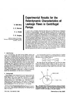
fluid and engineering problems. Our implementation of the immersed boundary method allows great flexibility in the choice of the models for the elastic components. The present model1 is based on the immersed boundary method for elastic shells 4 . In particular, the basilar membrane is modeled as an isotropic elastic shell by a fourth-order partial differential equationi. 2. The Model While the immersed boundary method enables construction of realistic models, it often leads to very intensive computing requirements, which can only be satisfied by the development of specialized algorithms for execution on systems with multiple CPUs. This is certainly true in the case of the cochlea. We have used a 32 processor shared-memory HP “Superdome” computer to construct and execute a cochlea model code that is based on a 2563 -point computational grid for the fluid, and several grids that model the elastic materials within the cochlea, a total of approximately 750,000 points. The geometric model of the cochlear anatomy is based on measurements that include the position of the center line of the basilar membrane, its width and the cross-sectional area of the scalae. There are six surfaces in this model: the basilar membrane, the spiral bony shelf, the tubular walls of the scala vestibuli and the scala tympani and the semi-elliptical walls sealing the cochlear canal and containing the oval and the round window membranes (see Fig1).
Fig 1 A rendering of the geometric model of the cochlea with several parts of the outer shell removed in order to expose the cochlear partition, consisting of the narrow basilar membrane and the bony shelf. The round window is located directly below the oval window and in this picture it is partially obscured by the cochlear partition. The basilar membrane is modeled as an elastic shell following the prototype tested earlier10. It is 3.5 cm long and we assume its width grows linearly from 0.0015 cm at the stapes to 0.0052 cm at the helicotrema. The elastic force that the membrane exerts on the surrounding fluid is given by a fourth order partial differential operator acting on the vector field that measures the displacement of the membrane from its elastic equilibrium. We have assumed that the Lamé coefficients of the shell are constant, and the variable compliance of the basilar membrane is i
In the future this model will be replaced by a more accurate orthotropic shell model.
2
due to its changing width and thickness. We have used this shell equation to estimate the thickness necessary to achieve the increasing compliance measured by von Bekesy. Indeed, with only a slight variation in the thickness the estimated compliance per unit length increases as
e ? x , where ? ? 1 ? 0. 7 cm.
The oval window and the round window membranes are also modeled as elastic shells, but unlike in the case of the basilar membrane, the compliance of each of these shells is constant throughout. The windows are geometrically identical: each is modeled by a rectangular grid, all of whose points outside a given circle are fixed. Hence each such grid represents a circular plate within a rectangular piece of bone. 3. Numerical Experiments Our experiments typically consisted of applying a pure tone input of given frequency at the stapes and a study of the subsequent motion of the basilar membrane. The input at the stapes was simulated by specifying an external periodic force vector field on the oval window grid. A very small time step of 30 nano-seconds was chosen to guarantee numerical stability and good detail. The choice of the time step was made as a result of the convergence study of the system4 . Our initial aim in the numerical experiments was to reproduce the qualitative characteristics of cochlear mechanics predicted by asymptotic analysis and previously reported computational models. Consequently, we have attempted to capture the steady state response of the basilar membrane to pure tones and in each of our experiments we have run the system for the duration equivalent to several input cycles. For example, for a 10 kHz input tone we have run the system for more than 30,000 time steps, equivalent to 0.9 milli-seconds of total simulated time. On 16 processors of the HP SuperDome this computation required more than 20 hours to complete. Every 10th time step of the simulation the program generated an output file containing the instantaneous position of the computational grids. The total amount of storage required for a single experiment is measured in tens of Gigabytes of disk space. Correspondingly, the 2 kHz experiment required five times as much computational time and storage. 4. Results
Our experiments reproduced the basic characteristic features of cochlear mechanics. Initially, we verified that all of our experiments remained in the system's linear regime, i.e. the input force at the stapes was kept very small, resulting in basilar membrane displacements on the order of nano-meters. Indeed, increasing the force by a factor of 10 resulted in basilar membrane displacements almost exactly ten times bigger. Since in each experiment the system was started from rest, we have observed initial oscillatory transient response, which was followed by the smoothing of the traveling wave. In each instance, in response to a pure tone input frequency, we have observed a traveling wave propagating from the stapes in the direction of the helicotrema. The amplitude of the wave gradually increases until it reaches a peak at a characteristic location along the basilar membrane, and this location depends on the input frequency. The speed of the wave is sharply reduced as it propagates further along the basilar membrane. These features can be observed in the following animation (Fig 2) of the results of a simulation experiment with an 8 kHz driving force.
3
Fig 2 Animation of the 8 kHz pure tone experimental results Mm. 1. Fig 2 (41MBytes) [http://pcbunn.cacr.caltech.edu/cochlea/tall.mpeg]
The wave propagation and its abrupt shutdown are even more clearly illustrated by the motion of the points of the middle line of the basilar membrane in the animation shown in Fig 3. The animation also demonstrates the transient response of the model. The calculation of the wave envelope begins after the transient response has subsided.
Fig 3 Animation of the motion of the centre line of the basilar membrane, in the 8kHz tone experiment Mm.2.(7MBytes) [http://pcbunn.cacr.caltech.edu/cochlea/Animated_8kHz_LineX10.avi]
4
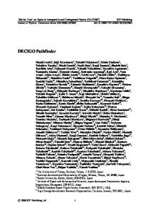
As expected, we observe that the higher the input frequency, the closer the peak of the wave is to the stapes: the wave envelopes of the steady state solutions for experiments with four different frequencies are shown in Fig 4.
Fig 4 Steady state envelopes of the basilar membrane displacement, for experiments with 16 kHz, 8 kHz, 5 kHz and 4 kHz tones. Since experiments at low frequencies require much longer compute times, our results are currently limited to frequencies = 2kHz. At the other extreme, model simulations of the response of the high frequency tuned region of the basilar membrane appear to suffer from insufficient resolution. By frequency-selecting the middle region of the basilar membrane we are able to generate the cochlear tuning curves shown in Fig 5.
Fig 5 Tuning curves for the human cochlea, derived from our experiments
5
5. Summary and Conclusions We have presented results of several large-scale computer simulation experiments using a detailed three-dimensional model of cochlear macro-mechanics. Our results demonstrate the basic characteristic features of cochlear mechanics. Extensive testing of the model reveals some limitations of the method, which requires significant computing resources. We are currently developing a distributed algorithm to efficiently deploy a large numb er of processors concurrently to tackle this simulation. We believe that the results presented here justify this approach, and we expect the algorithms together with a numerical method of higher order accuracy will remove the current limitations, and will make it possible to enrich the model with more detail, for instance by allowing the incorporation of a model of the organ of Corti. Acknowledgements We thank Santiago Lombeyda for creating the three-dimensional animation presented in this paper, and Sarah Emery Bunn for assistance in preparation of the manuscript. 6 6 References 1
E. Givelberg and J.Bunn, “A comprehensive three-dimensional model of the cochlea”, To be published in J.Comp.Phys. 2 S. Peskin and D.M. McQueen, “A general method for the computer simulation of biological systems interacting with fluids,” in Proc. Of SEB Symposium on Biological Fluid Dynamics, Leeds, England(1994). 3 C.S. Peskin, “The immersed boundary method,” Acta Numerica, 11, 479--517 (2002). 4 E. Givelberg, “Modeling elastic shells immersed in fluid,” Submitted for publication 5 C.R. Steele and L.A. Taber, “Comparison of wkb calculations and experimental results for threedimensional cochlear models.” J. Acoust. Soc. Am., 65, 1007--1018 (1979). 6 B.P. de Boer, “Solving cochlear mechanics problems with higher order differential equations.” J. Acoust.Soc.Am., 72, 1427--1434 (1982). 7 P.J. Kolston, “Finite element micromechanical modeling of the cochlea in three dimensions,” J.Acoust.Soc.Am., 99, 455--467 (1996). 8 P.J. Kolston, “Comparing in vitro, in situ and in vivo experimental data in a three dimensional model of mammalian cochlear mechanics,” Proc. Natl..Acad.Sci. USA, 96, 3676--3681 (March 1999). 9 A.A. Parthasarati, K. Grosh, and A.L. Nuttall, “Three-dimensional numerical modeling for global cochlear dynamics,” J. Acoust. Soc. Am., 107, 474--485 (2000). 10 E. Givelberg, Modeling Elastic Shells Immersed in Fluid , Ph.D. thesis, New York University (1997).
6