tDivision of Biology, California Institute of Technology, Pasadena, California 91125. Submitted January 22, 1993; Accepted March 5, 1993. We have developed ...
Molecular Biology of the Cell Vol. 4, 469-482, May 1993
Computer Simulation of the Phosphorylation Cascade Controlling Bacterial Chemotaxis Dennis Bray,* Robert B. Bourret,t# and Melvin I. Simont *Department of Zoology, University of Cambridge, Cambridge CB2 3EJ, United Kingdom; and tDivision of Biology, California Institute of Technology, Pasadena, California 91125 Submitted January 22, 1993; Accepted March 5, 1993
We have developed a computer program that simulates the intracellular reactions mediating the rapid (nonadaptive) chemotactic response of Escherichia coli bacteria to the attractant aspartate and the repellent Ni2+ ions. The model is built from modular units representing the molecular components involved, which are each assigned a known value of intracellular concentration and enzymatic rate constant wherever possible. The components are linked into a network of coupled biochemical reactions based on a compilation of widely accepted mechanisms but incorporating several novel features. The computer motor shows the same pattern of runs, tumbles and pauses seen in actual bacteria and responds in the same way as living bacteria to sudden changes in concentration of aspartate or Ni2+. The simulated network accurately reproduces the phenotype of more than 30 mutants in which components of the chemotactic pathway are deleted and/or expressed in excess amounts and shows a rapidity of response to a step change in aspartate concentration similar to living bacteria. Discrepancies between the simulation and real bacteria in the phenotype of certain mutants and in the gain of the chemotactic response to aspartate suggest the existence of additional as yet unidentified interactions in the in vivo signal processing pathway. INTRODUCTION
Coupled protein phosphorylations are a universal mechanism of intracellular signaling and the basis of many complex nets of reactions in eucaryotic cells. Such networks are poorly understood, despite their importance, because of the large number of components involved, and the multiple links that exist between them. Not only are quantitative data on the individual reactions lacking, but we also lack a vehicle by which we can comprehend the implications of such data and verify the overall performance of the entire network. Without making many detailed calculations, it will be difficult to predict how the network performs under a particular set of starting conditions, whether it will ever show oscillatory or chaotic behavior, and how the performance of the network will be affected by genetic modifications. The rational solution to this problem is to employ a computer simulation that can be used to catalogue biochemical data, test current hypotheses of mechanism, and make predictions of performance under a virtually f Present address: Department of Microbiology and Immunology, University of North Carolina, Chapel Hill, NC 27599.
©) 1993 by The American Society for Cell Biology
limitless variety of conditions. However, there have been very few attempts to develop quantitative models of this sort for cell signaling pathways involving coupled phosphorylation reactions. Early steps in this direction include models of the spatial propagation of oscillations in Ca2+ concentration in a variety of cells (reviewed by Dupont and Goldbeter, 1992) and the initial response to light of vertebrate photoreceptors (Lamb and Pugh, 1992). For the vast majority of cell signaling pathways, there is insufficient information to construct a meaningful simulation. Without knowing most of the relevant intracellular concentrations and enzymatic rate constants in a pathway, computer simulations can only be phenomenological representations having little analytical power or predictive force at the molecular level. One of the few signaling pathways for which there is presently sufficient information to attempt a detailed computer simulation is that underlying the chemotactic response in coliform bacteria. This entails a small and defined set of intracellular proteins which, through coupled phosphorylation and methylation reactions, enable the bacterium to respond in an informed, if not intelligent, way to its chemical environment. All of the intracellular proteins involved in this response have 469
D. Bray et al.
been sequenced and purified. Many of the enzymatic reactions they catalyze have been analyzed kinetically. Mutants lacking identified proteins either singly or in combination have been isolated in large numbers and their chemotactic responses documented. There is, in short, a uniquely large and complete set of data, unmatched in any other cell signaling system, against which a model can be tested. We have therefore undertaken to simulate the biochemical reactions of bacterial chemotaxis in a computer program, which we hope will eventually reproduce all the salient biochemical, genetic and behavioral observations made on living bacteria. As the first step toward this aim we describe in this paper the simulation of the chain of signals from the cell surface receptor to the flagellar motor that mediate the rapid, excitatory response to specific attractants and repellents. The slower adaptation of the system due to receptor methylation, the kinetics of which have been modeled previously (Goldbeter and Koshland, 1982; Asakura and Honda, 1984), is not part of the present model.
MATERIALS AND METHODS Signal Pathway To provide a background to the model described in this paper, we now review briefly the protein reactions that carry the chemotactic signal from the membrane receptor to the flagellar motor. The chemotactic response of coliform bacteria such as Escherichia coli and Salmonella typhimurium depends on the ability to modulate swimming behavior in response to external stimuli. This is achieved with linked chains of protein phosphorylation, which carry signals from the membrane receptors to the flagellar motor (Figure 1) (Bourret et al., 1991; Stock et al., 1991, 1992). Although many details are still debated, it is widely accepted that binding of an attractant or repellent molecule to a receptor changes the rate of autophosphorylation of an intracellular protein kinase, denoted CheA' (Borkovich et al., 1989; Borkovich and Simon, 1990; Ninfa et al., 1991). The phosphorylated form of CheA (CheAp) transfers its phosphate group to a second protein CheY (Hess et al., 1988c; Wylie et al., 1988). The product of this phosphotransferase reaction, CheYp, interacts with the flagellar motor so as to increase clockwise rotation, which produces "tumble" swimming behavior. The default state of the motor in the absence of CheYp is counterclockwise rotation, which results in "run" swimming behavior (Larsen et al., 1974; Parkinson, 1978). Two other components involved in this direct or "excitation" response are CheW, which appears to mediate the effect of receptor on CheA phosphorylation (Borkovich et al., 1989; Borkovich and Simon, 1990; Ninfa et al., 1991) and CheZ, which counteracts the effect of CheYp by dephosphorylation (Hess et al., 1988a,c). Flagellar rotation is also modified on a slower time scale by the methylation of receptor proteins by a methyltransferase CheR (Springer and Koshland, 1977), and by the removal of these methyl groups by the methylesterase CheB (Stock and Koshland, 1978). Like CheY, the methylesterase CheB is regulated by phosphotransfer from CheAp (Hess et al., 1988c; Lupas and Stock, 1989; Stewart et al., 1990). Our model concerns the chain of coupled protein phosphorylations that carry signals from the Tar receptor to the flagella motor, not the modification of the Tar receptor by methylation, which forms the
lAbbreviations used: A, CheA; asp, aspartate; B, CheB; M, flagellar motor; ni, Ni2" ion; p, phosphorylated form of indicated protein; R, CheR; T, Tar; W, CheW; Y, CheY; Z, CheZ.
470
(sp)
Figure 1. Signal transduction pathway in bacterial chemotaxis. Symbols within circles denote the Che proteins, in some cases phosphorylated, which carry the chemotactic signals from the receptor protein Tar to the flagellar motor. Solid arrows represent phosphorylation reactions. Dashed arrows indicate regulatory interactions. The methylation reactions catalyzed by CheR and CheB (open arrows) are not included in the computer simulation. basis of the adaptation of this response. In consequence, we do not expect the model to show the slow recovery of swimming behavior characteristic of wild-type bacteria, but it should be able to reproduce the short-term, impulsive responses of wild-type and mutant bacteria. It should also reflect the short-term behavior of mutants in which the demethylating enzyme CheB has been removed or overexpressed, because the latter protein forms part of the flux of phosphate groups in the excitatory pathway.
Theory In this section we present the algorithms used to represent the signaling reactions and the assumptions inherent in their use. Information on the binding of aspartate or Ni2" to the Tar receptor is relayed through the cytoplasm by changes in the concentrations of various phosphorylated protein species. It is convenient to divide the individual steps in this cascade into binding steps, in which two molecular components associate or disassociate, and reaction steps in which a phosphate group is added or removed from a protein. Each step is represented by an equation which is evaluated repeatedly at intervals of time At, which is typically 5 ms. Experimentally, the response of bacteria to a step change in attractant or repellant concentration occurs in -0.2 s (Segall et al., 1982) and evidence of a delay due to diffusion has been obtained only in abnormally long filamentous mutants (Segall et al., 1985). No data is available regarding on and off rates for individual binding reactions. Given the observed rapidity of signal propagation through the entire pathway, and for the sake of computational stability, we therefore make the simplest assumption, namely that individual binding steps reach equilibrium within each iteration cycle. Note that the concentrations of binding species will nevertheless change with time due to the phosphorylation and dephosphorylation reactions described below. Binding of a small molecular weight ligand L that is in large excess to a protein receptor R, specifically the binding of aspartate or Ni2+ to Tar in one of its complexed forms, is described by the simple binding equation
[RL] = [Ro]( Molecular Biology of the Cell
Bacterial Chemotaxis Simulation where [Ro] and [Lo] are the total concentrations of receptor and ligand, respectively, [RL] is the concentration of the protein-ligand complex, and Kd is the dissociation constant of the complex. All other binding steps in the simulation involve the association of cytoplasmic proteins at comparable, and usually low, concentrations and for this we use the relationship: [RL] = 0.5{[Ro] + [Lo] + Kd - V([Ro] + [Lo] + Kd)2 - 4[Ro] X [Lo]} where [Ro] and [Lo] are arbitrarily assigned to the two proteins (see Segel, 1975). Reaction steps are not assumed to run to completion within At and are integrated by successive steps of the simulation. For protein autophosphorylations we use the simple Michaelis-Menten relationship kcat X [ATP]l V
J=[l ]
[ATP] + Km
J
where v is the rate of formation of the phosphorylated species in moles per second, [E] is the free enzyme concentration, [ATP] is the concentration of ATP, k_cat is the catalytic rate constant and Km is the Michaelis constant (Michaelis and Menten, 1913; Fersht, 1984). For all other reactions we assume that the concentrations of reacting species are small, so that the reactions are simply described by v=
(k.cat/Km) [E] X [S]
1.0
coL 0.6
-
0 0
*~0.4-
o
10
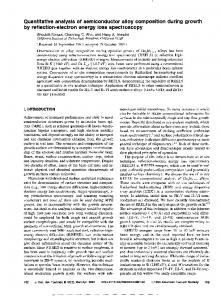
20
30
CheYp (nM) Figure 3. Proportion of time spent in run (l), tumble (0), and pause (A) modes predicted for the flagellar motor model, with the resting concentration of CheYp in a wild-type bacterium set to 9 nM.
or, for autodephosphorylations v=
klcat [E]
Flagellar Motor Unlike all other components of the simulation, the flagellar motor is not a single molecular species. It is a complex structure, built from approximately 40 distinct proteins that rotates either clockwise or counterclockwise at speeds of .300 cycles/s (significantly less when the bacterium is tethered to a surface) (Jones and Aizawa, 1991). The direction of rotation is controlled by the phosphorylated form of CheY through an interaction with the switch components (FliG, FliM, and FliN proteins) of the motor Uones and Aizawa, 1991). The interaction appears to be a simple binding of CheYp rather than a phosphotransfer reaction (Bourret et al., 1990). It was shown previously by Block et al. (1982, 1983) that a simple two-state model in which run and tumble states differ in the binding of a single ligand has similar stochastic properties to the runs and
Different states of the flagellar motor (M) are determined by the binding of CheYp (Yp): ccw rotation (run) State 'T M ccw rotation (run) State '3' MYp zero rotation (pause) State '5' MYpYp (tumble) cw rotation State '7' MYpYpYp (tumble) State '9' MYpYpYpYp cw rotation Occupation of the different states is governed by reversible equilibria: Ki = kil/klr M + Yp q-b MYp K2 = k2l/k2r MYp + Yp W MYpYp K3 = k3d/k3r MYpYp + Yp W MYpYpYp MYpYpYp + Yp W.& MYpYpYpYp K4 = k4l/k4r
Apgarent dissociation constants for the different states are determined by two constants. a and K:
Ki = ic/4 K2 = 2alc/3 K3 = 3a21c/2 K4 = 4a3K
Figure 2. Multiple equilibria between the flagellar motor complex (M) and the phosphorylated form of CheY (Yp) used to calculate motor behavior. Vol. 4, May 1993
tumbles of the flagellar motor. In the present simulation, this model has been extended to incorporate two additional features. One is the cooperativity between CheY levels and flagellar motor response seen in mutant strains in which CheY is over-expressed to different levels (Kuo and Koshland, 1989). The effective species is now thought to be CheYp. The other feature added to the motor model is the capacity for pausing, which has been reported by Eisenbach and colleagues to be an inherent part of the behavioral repertoire of coliform bacteria (Lapidus et al., 1988; Eisenbach et al., 1990). These observations are incorporated into a model in which the motor complex, denoted M, binds sequentially up to four CheYp molecules thereby producing five distinct molecular species. Two of these (M and MYp) rotate counterclockwise, another two (MYpYpYp and MYpYpYpYp) rotate clockwise, and one species (MYpYp) is stationary, corresponding to the pausing of the motor (Figure 2). In a population of bacteria, the rotational bias (defined as the fraction of time spent in the counterclockwise mode) is therefore given by the following:
bias
=
M + MYp M + MYp + MYpYp + MYpYpYp + MYpYpYpYp
As in the earlier model of Block et al. (1982, 1983), transitions between each pair of states are governed by paired first-order rate constants, such as kl, and k2rYp (which is pseudo first-order at a given concentration of Yp). For an individual motor, these rate constants represent the probabilities per unit time of terminating the current state. The Adair-Pauling model of a multisite allosteric enzyme allows dissociation constants for each binding step to be calculated from two parameters-the dissociation constant of binding of the first binding site (K) and the factor (a) by which the affinity of successive binding sites change (Segel, 1975). We have assumed that the flagellar motor is similarly well-behaved and calculated K and a from an arbitrarily assigned value for the resting (unstimulated) concentration of CheYp and from the observed proportion of time spent in runs, pauses, and tumbles of an unstimulated, wild-type bacterium (Lapidus et al., 1988; Eisenbach et al., 1990). In trial simulations we found that the resting concentration of CheYp could be varied over a wide range without affecting overall performance. In the absence of experimental data bearing on this value, we selected 9 nM, which in combination with the best available values of reaction rates, produces an approximately wild-type rotational bias for mutant strains lacking either Tar, CheW, and CheZ; Tar and CheZ; or CheW and CheZ (Liu and Parkinson,
471
D. Bray et al.
the next time interval of dt s, is k2rYpdt. The probability that a motor that is currently turning counterclockwise (that is, in either state "1" or state "3") will stop (move to state "5") in the next time interval is given by rA
600-
(2[pd
k2[Ypdt [M][MYp] + [MYp]J
0 O
4000
z
200-
ihUihimmimimmm.m...... 0
2
4
6
8
10
Run Length (sec)
Similar expressions exist for the termination of tumbles and pauses. Values of the rate constants k2,, k3l, k3r, and so on were estimated from the dissociation constants of individual reactions (K1 = kl,/kl etc.) in conjunction with the experimentally observed proportions of runs, tumbles, and pauses seen in unstimulated wild-type bacteria. Probabilities of transition between the different states of the motor model were derived from these rate constants. Then the history of an individual flagellar motor was simulated by starting it in state 1 (see Figure 2) and allowing a random number generated within the program to determine, at each cycle, whether the motor changed in state 3. If and when the motor arrived at state 3, then a similar procedure determined whether, in successive cycles, it changed to state 5 or back to state 1. In this way the stochastic behavior of a single flagellar motor was stimulated. The performance of wild-type, unstimulated motors modeled according to this prescription, is closely similar to that described experimentally, as illustrated in Figure 4 (see also Figure 6).
Numerical Integration
z 0
,0
z 0
2
4
6
8
10
Tumble Length (sec) Figure 4. Durations of runs and tumbles predicted for a wild-type unstimulated bacterium using a random number generator. The last category in each histogram represents all episodes > 10 s in length. Results from 5 X 10⁶ simulation cycles are displayed, totaling 2595 runs and 2093 tumbles. There were also 4683 pauses during the same period distributed as follows: 0-0.4 s, 4250; 0.4-0.8 s, 403; 0.8-1.2 s, 29; 1.2-1.6 s, 1; >1.6 s, 0. The run and tumble distributions conform closely to experimentally determined values (see Block et al., 1983). 1989). This choice results in values of a = 0.14, and K = 2.25 X 10⁻⁷ M. Figure 3 shows the changes in proportion of time spent in runs, tumbles, and pauses as CheYp changes in concentration. The distribution corresponds to positive cooperativity between CheYp and the rotational bias with a Hill coefficient of about 3.0. The multiple equilibria shown in Figure 2, which represent the steady state population levels of the different rotational forms, can also be used to reproduce the stochastic behavior of a single motor, as might be observed in a bacterium tethered to a coverslip with a single flagellar hook (Silverman and Simon, 1974). A similar calculation, although simpler, was made by Block et al. (1982) in respect to their 2-state model. Thus with reference to Figure 2, the probability that a given motor currently in state "3" will change to state "5" in 472
The simulation uses a procedure in which each binding or reaction step is taken in turn and the concentrations of the reactants and products are updated according to the appropriate algorithm. Changes within a reaction step are predicted by the Euler method. We selected this rudimentary integration procedure rather than one of the many more sophisticated routines available because it makes it easy to treat individual reactions separately for display purposes and because of the computational stability it provides. Because each reaction or binding step is taken in turn there is no possibility of extrapolated concentrations exceeding the available material. The program is thus extremely robust, and ligand concentrations from 10-12 to 1M can be applied without causing instability. Similarly, levels of enzymes can be manipulated by "mutation" ad libitum. The primary disadvantage of the method we have selected is its inaccuracy when compared with the more usual methods of numerical integration. As already mentioned, however, the values of binding constants and rate constants available to us are only crude approximations to the actual kinetic parameters within the cell. Our primary purpose here is to summarize the existing body of research in a simple formulation and to test whether it gives approximately the correct response following a variety of mutational modifications, and for this the procedure we have used is adequate. As with any form of numerical integration, there is the opportunity to increase accuracy by reducing the step-size. Tests of the present simulation with wild-type bacteria over a range of repellent and attractant concentrations, showed stable values of rotational bias, with