BIOSIGNAL 2004
Computer Tomography-Based Mathematic Modelling Of The Blood Flow In Human Descendent Aneurysmatic Aorta Tonar Z 1,2, Jeník J 2, T eška V 3, Novák M 4, Ferda J 4 Department of Histology and Embryology, Faculty of Medicine in Pilsen, Charles University in Prague, 2 New Technologies Research Centre, University of West Bohemia in Pilsen, 3 Surgery Clinic, 4 Radiodiagnostic Clinic, University Hospital Pilsen, Czech Republic
[email protected] 1
The aim of the study was to simulate the pulsatile three-dimensional flow through a realistic model of human abdominal aneurysmatic aorta and its branches. We performed the simulations for both Newtonian and non-Newtonian blood analog fluid. The results described flow features with use of velocity magnitudes and wall shear stress distributions. The present numerical code provided a preliminary simulation tool for assessment of risk of growth and rupture of the aneurysm.
1
Introduction
2
Methods
Numerical simulation of the flow in the aortic system [4] proved itself to be a promising method for better understanding of the development of atherosclerotic abdominal aortic aneurysm (AAA) and its dependence on flow structure. Our first task was to create a simplified finite-element grid of the lumen of aneurysmatic aorta and its main visceral and pelvic branches. The second aim was to study the blood flow through this realistic geometry.
The morphology of the computational 3-D tetrahedral grid was obtained from 76 years old patient undergoing computer tomography (CT) angiography of descendent aorta and pelvic arteries because of subrenal AAA (length of 14.5 cm, width 8 cm, parietal thrombus 4 cm thick; inner diameter of aneurysm was 57×41 mm), affecting also both of the common iliac arteries. The data were transferred from a 16-row CT (Somatom Sensation 16, Siemens, Forchheim, Germany) via the DICOM format into software AmiraTM (TGS Europe, Mérignac Cedex, France) and full spatial resolution of the CT data matrix was preserved (512×512 pixels, both slice width and increment of 0.75 mm). The calibrated image data set was segmented semiautonomically with respect to the lumen of aorta, coeliac trunc, superior mesenteric artery, right renal artery, and bifurcation of common iliac arteries. The left renal artery was not included into the model, its filling with radiologic contrast was impaired by atheromatous plaque so the reconstruction of its lumen was not reliable. The model involved 16 vessels with one inlet boundary and 11 outlets. The flow redistribution into the peripheral branches of aorta was guaranteed by boundary condition set on the outlet planes as velocity outlet and the values were adapted according to [6]. The quality of the grid (900 000 tetrahedral elements) was enhanced by adaptive resizing of elements according to vessel diameter and irregular regional shape of the aneurysm (Gambit, Fluent Europe, Sheffield, Great Britain). Progressive improvement and multiple checks of the model were based on advices from vascular surgeon and radiologist. The 3-D mathematic model of flow was based on the numerical finite volume method offered by the package Fluent [7]. First, a laminar viscous model was used together with a steady segregated implicit solver and the blood was modelled as Newtonian incompressible fluid with constant density and constant fluid viscosity [11]. The boundary conditions respected real morphology and the velocity inlet and outflow values were adopted from [8]. Second, there were applied
BIOSIGNAL 2004 unsteady boundary conditions, and the non-Newtonian fluid was simulated as pseudoplastic continuum with apparent viscosity reducing with shear rate (the power-law fluids). The variation of viscosity with shear rate can be expressed by equation (1), where power-law index n = 0.708 (-) expresses pseudoplastic continuum [5]. The consistency index value is k = 0.0862 (kg.sn.2/m). Shear rate γ is formulated by deformation tensor (equation 2). The shear stress tensor represents the non-Newtonian fluid (3). (1) (2) (3)
3
Results
4
Discussion
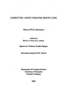
The developed mesh (Fig 1) reflected the most significant features of the vessel wall altered by atherosclerosis and asymmetric aneurysmatic dilatation. The model included huge and medium-sized vessels, with mesh density created adaptively according to the lumen (Fig 2). We found only tiny differences in flow structure between results based on Newtonian and non-Newtonian fluid (model comprised no small vessels). As the blood is a non-Newtonian fluid, we demonstrate the resulting flow characteristics for this option as distribution of the path lines and velocity magnitudes. During the systole, two swirls developed ventrally in the cranial segment of aneurysm and laterally in the right junction of aneurysm neck and sac close to the right renal artery (Fig 3). Both swirls persisted through the whole cardiac cycle (Fig 4) with maximum in end-diastolic phase (Fig 5). The distribution of wall shear stress was computed (Fig 6).
During the computation, we found the spatial resolution of the model to be sufficient even in the very irregular and realistic morphology of aneurysm and branches of aorta. The localization of swirls corresponded to general rules of cardiovascular fluid dynamics [12]. The lateral swirl could be caused by both the effect of right renal artery and local irregular bending of aneurysm neck. In our case of realistic model, the swirls persisted through both systole and diastole. In idealized smooth model of AAA without branching [4], the whirls regressed during the peak systolic flow and (unlike in our model) other swirls originated in the caudal segment of AAA. Our simulation of distribution of wall shear stress in the aneurysmatic sac yielded values comparable to [3]. The 3-D flow field in AAA depends a great deal on the geometry of the vessel, as proved by simulations of flow pattern in hypothetically shaped idealized and asymmetric models under realistic pulsatile flow conditions [3,14]. In realistic models involving abdominal aortic branches, more information on the flow field in bifurcation regions can be acquired. Such information may provide additional insight into hemodynamic factors involved in the predilection of atherosclerotic lesions in AAA development [9]. When modelling vascular networks with multiple branches, outflow boundary conditions play a great role in blood flow distribution. Also the effect of exercise on hemodynamic conditions and flow redistribution in aorta is considerable [10]. Patients with AAA are in danger of aneurysm rupture. Morphological and mechanical risk factors of rupture include e.g. aneurysm diameter and its growth rate, loss of elastin and inflammatory infiltrates leading to mechanical inferiority of the wall, tensile wall stress, shear stress, flow patterns and swirls, blood pressure etc. Our model comprises neither fluid-wall interaction nor mechanics of the vessel wall, both of these factors being of great importance. We are aware of limitations of the current study, because under certain conditions the
BIOSIGNAL 2004 physiologic shear stress on the inner wall can be of minor effect than the tensile stress within the wall due to pressure in an AAA [2]. Simulation of aneurysm fluid dynamics and its effect on aneurysm wall mechanics in realistic 3-D models [1] served already as a guidance to assess the risk of rupture of the aneurysm. Unlike in our study, the authors delt with the aneurysmatic sac only, omitting the haemodynamic influence of arterial branches of precedent and subsequent segment of the aorta. However, the effect of the flow in aorta branches on the flow in the aneurysm might be significant and deserves a further interest. Fillinger et al. [2] analyzed rupture risk over time in patients with AAA under observation, performing nonlinear hyperelastic modelling of aneurysm wall behaviour compared to CT data and blood pressure observation. A noninvasive study of 3-D tensile wall stress was found to be superior to maximum diameter for determining rupture risk. The simulation of wall stress provided significant differences of clinical use for aneurysms that could be safely observed for longer periods or needed surgical repair to prevent rupture within a short time. The effect of 3-D shape appeared to dominate the effect of blood pressure. At present, there is need for predictive models with realistic morphology in order to gather experience with computational simulations correlated to clinical decision making. Our method of finite-element grid construction enables us to consolidate the view of clinical medicine (i.e. vascular surgery), diagnostic imaging methods and computational simulation. It does not describe the very important composition of atherosclerotic vessel wall, which can be extracted from the high-resolution magnetic resonance [13]. Nevertheless, CT angiography remains the most frequent and routine method of aneurysm diagnostics and morphometry in patients undergoing elective surgery for this condition. Young healthy patients with large aneurysms have a risk-benefit ratio that favours vascular or endovascular surgery. The presented method could be useful in patients with boundary value of aneurysm diameter and high surgical risk or in asymptomatic patients with smaller aneurysms (5-6 cm) without rapid expansion. In this patients, the final decision on surgery could be ambiguous because mortality and morbidity of elective surgery are too high when compared to conservative treatment. Also the tensile stress analysis of the aneurysm wall has the potential to aid management in patients who are at high risk for surgery and have aneurysms of a moderate size [2]. Pulsatile flow assessment should help the surgeon to evaluate localization and singificance of swirls in the AAA. Whether simulation of wall shear stress correlates with risk of initial dissection of AAA, remains unsolved. Until now, we are lacking reference papers and case-reports bringing clinical experience with complementary results of simulation of blood flow through the AAA of the same patient.
5
Conclusions
Creating mathematical models based on real morphology provides a tool integrating the view of medical diagnostics, therapy, and modelling of AAA. The present numerical code provides a preliminary simulation tool for assessment of risk of growth and rupture of the aneurysm. The numerical result obtained in this study showed the flow features in a model of AAA with realistic morphology. This information provided an additional insight into hemodynamic factors involved in the predilection of rupture of aneurysm. The approach presented was found to be suitable e.g. for follow-up study of patients observed for the aneurysm growth, where simulations could be correlated with surgeon’s clinical experience.
Acknowledgement
This paper is based upon work sponsored by the Ministry of Education of the Czech Republic under research and development project LN00B084.
BIOSIGNAL 2004
Fig 1. Computational grid of descendent Fig 2. Detail of the grid with adaptive size aorta and its main branches based on CT scan of elements and artificially elongated data, left anterior view of subrenal AAA. endings of the branches (suitable for the simulation), right caudal anterior view.
Fig 4. Distribution of the path lines during Fig 3. Distribution of the path lines during peak systolic flow, velocity magnitude in early diastole, velocity magnitude in color scale (m.s-1), right craniolateral view. color scale (m.s-1), right craniolateral view.
Fig 5. Distribution of the path lines during Fig 6. Wall shear stress at the end of end-diastolic flow, velocity magnitude in systole in color scale (Pa), right craniolateral color scale (m.s-1), right craniolateral view. view, some arterial segments out of scale.
References [1]
[2]
Di Martino ES, Guadagni G, Fumero A, Ballerini G, Spirito R, Biglioli P, Redaelli A. Fluid-structure interaction within realistic three-dimensional models of the aneurysmatic aorta as a guidance to assess the risk of rupture of the aneurysm. Medical Engineering & Physics 2001;23:647-55. Fillinger MF, Marra SP, Raghavan ML, Kennedy FE. Prediction of rupture risk in abdominal aortic aneurysm during observation: Wall stress versus diameter. Journal of
BIOSIGNAL 2004 [3] [4] [5] [6] [7] [8] [9] [10] [11] [12] [13] [14]
Vascular Surgery 2003;37:724-32. Finol EA, Amon CH. Flow-induced wall shear stress in abdominal aortic aneurysms: Part II - pulsatile flow hemodynamics. Computer Methods in Biomechanical and Biomedical Engineering 2002;5:319-28. Finol EA, Keyhani K, Amon CH. The effect of asymmetry in abdominal aortic aneurysms under physiologically realistic pulsatile flow conditions. Journal of Biomechanical Engineering 2003;125:207-17. Hussain MA, Kar S, Puniyani RR. Relationship between power law coefficients and major blod constituents affecting the whole blood viscosity. Journal of Biosciences 1999;24:329-37. Lee D, Chen JY. Numerical simulation of steady flow fields in a model of abdominal aorta with its peripheral branches. Journal of Biomechanics 2002;35:1115-22. Manual Fluent 6.1. Fluent.Inc Europe, Sheffield, Great Britain, 2001. Mills CJ, Gabe IT, Gault JH, Mason DT, Ross J Jr, Braunwald E, Shillingford JP. Blood velocity and pressure wave-forms in the major arteries in man. Clinical Science 1970;38:10-20. Shipkowitz T, Rodbers VGJ, Frazin LJ, Chandran KB. Numerical study on the effect of secondary flow in the human aorta on local shear stresses in abdominal aortic branches. Journal of Biomechanics 2000;33:717-28. Taylor CA, Hughes TJR, Zarins CK. Effect of exercise on hemodynamic conditions in the abdominal aorta. Journal of Vascular Surgery 1999;29:1077-89. Valenta J. Biomechanics. Prague: Academia, 2000. Verdonck P, Perktold K. Intra and Extracorporal Cardiovascular Fluid Dynamics, Vol.2 - Fluid Structure Interaction. Southampton: WIT Press, 2000. Yang F, Holzapfel G, Schulze-Bauer C, Stollberger R, Thedens D, Bolinger L, Stolpen A, Sonka M. Segmentation of wall and plaque in in vitro vascular MR images. International Journal of Cardiovascular Imaging 2003;19:419-428. Yip TH, Yu SCM. Cyclic flow characteristics in an idealized asymmetric abdominal aortic aneurysm model. Journal of Engineering in Medicine 2003;217: 27-39.