On the present work, we propose an algorithm that after unstable segments identification by the first derivative estimates the baseline of antepartum fetal.
Computerized Algorithm for Baseline Estimation of Fetal Heart Rate L Jiménez1, R González1, MJ Gaitán1, S Carrasco1, C Vargas2 1
Universidad Autónoma Metropolitana–Iztapalapa, México D F, México 2 Centro de Investigación Materno Infantil-Gen, México D F, México
Abstract On the present work, we propose an algorithm that after unstable segments identification by the first derivative estimates the baseline of antepartum fetal cardiotachograms. Therefore, 50 records 5 minutes long, derived from abdominal ECG, were processed according to the methods of Dawes, Mantel, and our proposal. Besides visual analysis, statistical differences among methods were established. More appropriate visual fits of the baseline were gotten by our proposal (76%) than Mantel (22%) and Dawes (2%). Since lower mean baseline was computed from Mantel (137.8±8.4 bpm) than from Dawes (140.4r8.4 bpm) and our proposal (140.0r8.4 bpm), different account for accelerations and decelerations were also found. Our proposal showed advantages to estimate the baseline of cardiotachograms as it was properly adjusted at the beginning of the traces, depended of only 15 seconds of valid segments and corresponded more closely to the visual baseline.
1.
Introduction
Precise baseline estimation is the critical step for a proper analysis of the fetal cardiotachogram (CTG); hence, expert definitions of baseline, accelerations and decelerations have been outlined [1,2]. Such definitions, however, have resulted in a circular problem, since baseline is determined previous accelerations and decelerations exclusion, but these are identified previous baseline recognition. Hence, visual interpretation remains as a subjective procedure with significant intra- and interobserver disagreements [3-7], which have not been resolved even when strict definitions are applied [8,9]. Computerized analysis of the CTG has been suggested to eliminate disagreements. However, Bracero et al [9] recently presented results that besides confirmed the intra- and inter-observer disagreements of human experts, showed discrepancies between these and the computer. Moreover, the same authors observed that the computer detected a higher proportion of non-reactive traces which were less associated with proved adverse outcomes.
0276−6547/02 $17.00 © 2002 IEEE
Since the computerized detection of baseline, accelerations and decelerations did not improve the positive prediction, we assume that current approaches for CTG analysis should be reviewed. In fact, although some of them are nowadays used at the bedside, they still showing problems such as a) uncertainty at the beginning of the traces, b) displacement following large swings within the trace, and c) dependency of traces 6 to 10 minutes long at least [10]. Prompted by these observations, the aim of the present work was to propose an algorithm that reduces such problems in antepartum CTGs, in particular when applied to short term records.
2.
Materials
50 antepartum CTGs, derived from abdominal electrocardiograms (ECGa), were recorded from 50 pregnant women attending their pregnancy and labor at the Centro de Investigación Materno Infantil, in Mexico City. After patients gave informed consent according to the Declaration of Helsinki, they were included as participants and their physical and obstetrical characteristics corresponded to women 18 to 34 years old, 57 to 72 Kg of body weight, single pregnancy with low to middle perinatal risk, and 32 to 40 weeks of gestation. As ECGa in this study was not used as clinical test, perinatal outcomes were not considered.
3.
Methods
3.1.
Signal acquisition
ECGa was digitized on a PC with the Acknowledges System MP100 (BIOPAC, CA, USA) at 1.0 kHz, during 5 minutes. Mothers in left lateral decubitus were recorded with bipolar lead by mean of a bioelectric amplifier AB621G (Nikon Kohden, Tokyo, Japan). An AgCl electrode was used as leg reference, while two others were placed at the abdominal maternal surface 18-20 cm distant, previous standard cleansing of the respective skin areas. The positive electrode was fixed on the pubis, while the negative was on and around the umbilical area. These electrode positions guaranteed measurable fetal QRS complexes which were separated from the ECGa by
477
Computers in Cardiology 2002;29:477−480.
resting the maternal QRS complexes [11]. Fetal heart rate (FHR) was computed from the measured beat-to-beat RR intervals, and the derived time series were processed in according to the algorithms of Dawes et al (Dw) [12], Mantel et al (Mn) [10], and our present proposal (Pp).
3.2.
Signal processing and analysis
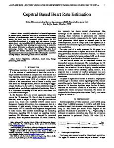
The algorithms of Dw and Mn to determine the FHR baseline were applied as described [10,12], except that the time series of FHR were used as beat-to-beat instead of the average of 3.5 s and 2.5 s usually employed by Dw and Mn, respectively. Our algorithm, Pp, was divided on three steps (Figure 1). The first one made an overall smoothing of the time series based on a moving average of the FHR with a Hanning window of 27 points. On the second step, abrupt changes of the time series were identified by first derivative amplitude (dFHR) and time thresholds. Thus, segments with dFHR surpassing the level of 1.0 beats/minute/second (bpm/s) were eliminated, and the remaining were averaged (P), but only those t15 s long and between P±10 bpm were validated as possible baseline segments. To obtain the final estimation of the baseline, on the third step, the validated segments underwent cubic spline interpolation, linear extrapolation at both ends of the time series when necessary, and a third order zero-phase low-pass filtering with cut off frequency of 0.033 Hz. This filter assumed valid baseline fluctuations as two cycles per minute or lower [1]. Besides a visual analysis, baselines estimated by each method were processed to compute time-domain parameters, in bpm, as Mean (Bmean), Standard Deviation (Bstdev), Minimum Value (Bmin), Maximum Value (Bmax), and Range (Brange). On the frequencydomain, the power spectral density of the baselines was estimated and the bandwidth (Bbw) was computed assuming a stable frequency response when attenuation of –3 decibel was observed. Finally, the effect of the estimated FHR baseline on the number of records withand the total sum of- accelerations and decelerations was evaluated. These events were uniformly defined as differences between the original CTG and the estimated baseline, where the differences indicate a segment of successive values with the same sign that reaches a peak (acceleration) or a nadir (deceleration) of at least 15 bpm in less than 30 s, and has a total duration of at least 15 s.
3.3.
Statistical analysis
For Bmean, Bstdev, Bmin, Bmax, Brange and Bbw, handled as quantitative parameters, the mean and standard deviation were obtained to test statistical differences, whereas occurrence of accelerations and decelerations were described as counting and proportions
Figure 1. Sequence of signal processing to estimate the fetal heart rate (FHR) baseline: a) original signal, b) smoothed signal, c) separation of stable (darker lines) and abrupt changes (lighter lines) segments, d) estimated baseline on the original signal, after cubic spline interpolation, extrapolation and filtering of the stable segments. used to assess inter-observer agreement. Comparisons of quantitative data were done by analysis of residuals and by repeated-measures one-way analysis of variance, followed by post hoc Tukey’s test to pinpoint differences. Inter-observer agreement to detect accelerations and decelerations was assessed by the proportion of agreement (PA) for multiple observers, and the kappa statistic, both statistics with 95% confidence interval (CI) [13]. PA values whose lower limit of CI was above 0.5 indicated significant agreement. For kappa values above 0.75, between 0.4 and 0.75 and below 0.4, agreement
478
beyond chance was considered excellent, fair to good, and poor, respectively. Bias among algorithms were evaluated by the Cochran’s test whose significance was given by F2 statistic. For all hypothesis test, p