HILDE SMITH,'* SIERD BRON,1 JAN VAN EE,2 AND GERARD VENEMA'. Department of Genetics, Center ofBiological Sciences, 9751 NN Haren (Gn),' and ...
Vol. 169, No. 7
JOURNAL OF BACTERIUOLOGY, July 1987, P. 3321-3328
0021-9193/87/073321-08$02.00/0 Copyright © 1987, American Society for Microbiology
Construction and Use of Signal Sequence Selection Vectors in Escherichia coli and Bacillus subtilis HILDE SMITH,'* SIERD BRON,1 JAN VAN EE,2 AND GERARD VENEMA' Department of Genetics, Center of Biological Sciences, 9751 NN Haren (Gn),' and Gist-Brocades N.V., 2600 MA Delft,2 The Netherlands
Received 10 February 1987/Accepted 17 April 1987 To study the diversity and efficiency of signal peptides for secreted proteins in gram-positive bacteria, two plasmid vectors were constructed which were used to probe for export signal-coding regions in Bacilus subtilis. The vectors contained genes coding for extracellular proteins (the aL-amylase gene from Bacilus licheniformis and the j3-lactamase gene from Escherichia colt) which lacked a functional signal sequence. By shotgun cloning of restriction fragments from B. subtilis chromosomal DNA, a great variety of different export-coding regions were selected. These regions were functional both in B. subtlis and in E. coli. In a number of cases where protein export had been restored, intracellular precursor proteins of increased size could be detected, which upon translocation across the cellular membrane were processed to mature products. The high frequency with which export signal-coding regions were obtained suggests that, in addition to natural signal sequences, many randomly cloned sequences can function as export signal.
The ability of bacilli to secrete large amounts of exoenzymes into the culture medium (17) makes these organisms interesting for the synthesis of foreign gene products on an industrial scale. This is one of the reasons why, from an applicational point of view, studies on protein secretion in these organisms are of interest. Research on the mechanism of protein secretion in gram-positive bacteria is also important from a scientific point of view. In both gram-positive and gram-negative bacteria, secreted proteins are initially synthesized with N:terminal signal sequences, which are removed during translocation across the cellular membrane. Signal sequences of gram-positives are usually longer than those of gram-negatives and frequently contain more charged amino acid residues in the N terminus (26). In addition, several gram-positive exoproteins, such as several proteases, are synthesized as even larger precursor forms, called prepropolypeptides (20, 25, 28). To optimize the efficiency of protein secretion by bacilli, a better understanding of the parameters affecting the translocation mechanism(s) is desirable. So far, the approach most frequently used has been to introduce mutations in the various regions of the signal sequence (for review, see reference 19). Particularly in Escherichia coli, this has resulted in valuable information about functional properties of the different regions of the signal peptides. An alternative approach would be to select a considerable number of signal sequences naturally occurring in the chromosome and to compare the properties of the corresponding signal peptides. We have chosen for this approach to study the properties of signal sequences in Bacillus subtilis. The present paper describes an efficient system for the selection of export signal sequences. The approach was to construct two plasmid vectors containing genes coding for extracellular proteins which lacked part of or the entire signal sequence. Since signal sequences are usually functionally interchangeable (3, 15, 23), we expected that cloning in the correct reading frame of DNA fragments coding for a promoter, ribosomal binding site, start codon, and signal sequence would result in the synthesis and export of the protein. Two *
different genes coding for easily assayable extracellular proteins were chosen as signal sequence selection probes: (i) the a-amylase gene from Bacillus licheniformis and (ii) the P-lactamase gene from E. coli. These genes have already been cloned (16; Gist-Brocades N.V., European patent application 8320106.9, 1983) and sequenced (21, 30). The results showed that, by shotgun cloning of restriction fragments from B. subtilis chromosomal DNA in the signal sequence selection vectors, a large variety of different export-coding regions could be selected. These regions appeared to function both in B. subtilis and in E. coli.
MATERIALS AND METHODS Bacteria and plasmids. Table 1 lists the bacterial strains and plasmids used. B. subtilis 8G-5 (amy) was obtained by congression after transformation of competent 8G-5 cells with DNA from B. subtilis 1-85 and selection for his' recombinants. Media and plates. TY medium contained (per liter) 10 g of tryptone, 5 g of yeast extract, and 10 g of NaCl (pH 7.4). Minimal media used in the competence regimen for B. subtilis were as described before (2). Media were supplemented with antibiotics as follows: for B. subtilis, chloramphenicol at 5 ,ug/ml and erythromycin at 1 ,ug/ml; and for E. coli, ampicillin at 25 ,ug/ml and erythromycin at 100 ,ug/ml (unless stated otherwise). (Bio)chemicals. Restriction enzymes, DNA polymerase I, T4 DNA polymerase, and T4 DNA ligase, obtained from Boehringer (Mannheim, Federal Republic of Germany) or from New England Biolabs (Beverly, Mass.), were used as indicated by the suppliers. DNA preparations. Chromosomal DNA was extracted from B. subtilis as described by Bron and Venema (2). Preparative amounts of plasmid DNA were obtained basically according to the alkaline lysis procedure described by Maniatis et al. (11). Selected restriction fragments were obtained by separation on 0.8 or 2.0% agarose gels, followed by extraction and purification on DEAE NA-45 membranes (Schleicher & Schuell, Dassel, Federal Republic of Germany) as indicated by the manufacturer. The analytical "miniprep" procedure described by Ish-Horowicz and
Corresponding author. 3321
SMITH ET AL.
3322
J. BACTERIOL.
TABLE 1. Bacterial strains and plasmids Plasmid or strain
Plasmids pKTH74
Properties and genotype
E. coli plasmid; 5.8 kb; Apr,
Tcr pGK13 Streptococcus cremoris plasmid, derivative of pGK12; 4.8 kb; Cmr, Emr pGK13-amy+ pGK13, carrying the ox-amylase gene of B. licheniformis; 7.8 kb; Cmr, Emr; a-amylase+ pRW101 Bifunctional replicon, carrying the penicillinase gene of B. licheniformis; 7.0 kb; Kmr (B. subtilis); Apr (E. coli) E. coli plasmid; 2.7 kb; Apr pUC13 pPL608
pTA1060 pHP14
pGPB11 pGPB14 pGPA11
pGPA14 Strains B. subtilis 8G-5 8G-5 amy DB104 1-85 E. coli BHB2600
B. subtilis plasmid; 5.0 kb; Kmr, Cmr Cryptic B. subtilis plasmid; 8.6 kb Bifunctional replicon; 4.2 kb; Emr; carrying the MCS of m13 mpll Export signal selection vector; 5.7 kb; Emr As pGPB11 Export signal selection vector; 5.8 kb; Emr As pGPA11
reference
I. Palva Laboratory collection, Kok et al. (9) Laboratory collection Mezes et al. (13)
Messing et al. (12) Williams et al. (27) Uozumi et al. (24) This paper This paper
This paper This paper This paper
trpC2 tyr his nic ura rib met Bron et al. (2) ade a-Amylase-negative derivative Laboratory colof 8G-5 lection Kawamura et al. his nprR2 nprEJ8 aprA3
(8)
trpC2 amy
Yuki (29)
803 supE supF rk- mk- met
K. Murray
Burke (5) was used to extract plasmids from 2.5-ml cultures of E. coli. Minipreps from B. subtilis were prepared basically according to the same procedure with the modification that cells from 5-ml cultures were washed once by centrifugation with 50 mM Tris hydrochloride (pH 7.4)-10 mM EDTA-200 mM NaCl before being lysed. Miniprep DNAs were incubated with 100 ,ug of pancreatic RNase (BDH, Poole, England) per ml for 30 min at 37°C. Molecular cloning procedures. Vector molecules and restricted target DNAs were mixed in approximately a 1:4 weight ratio at a total concentration of 100 jig/ml. Ligation was carried out as described by Maniatis et al. (11). Ligated samples were used to transform competent B. subtilis or E. coli cells. Transformations. Transformation of competent B. subtilis and E. coli cells was as described by Bron and Luxen (1). Transformants were selected on TY plates supplemented with selective antibiotics as follows: for B. subtilis, erythromycin at 1 ,ug/ml and kanamycin at 5 p.g/ml; and for E. coli, erythromycin at 200 ,ug/ml or erythromycin at 50 ,ug/ml plus ampicillin at 2 ,ug/ml. Probing for chromosomal export signals. B. subtilis chromosomal DNA was digested with Sau3A, AluI, HaeIII, or RsaI and subsequently ligated with BamHI- or SmaI-
digested vector molecules (pGPB14 or pGPA14) in a 4:1 weight ratio. The construction of these vectors is described in Results. Ligated samples were used to transform competent E. coli cells. Transformants were selected by plating on TY plates containing erythromycin (200 [ig/ml) plus 1% soluble starch (for pGPA14) or erythromycin (50 jig/ml) plus ampicillin (2 p.g/ml) (for pGPB14). Plasmid DNAs extracted from the transformants were subsequently used to transform competent B. subtilis cells to erythromycin resistance. Transformants were analyzed for their capacity to secrete either amylase or TEM 3-lactamase into the culture medium as described below. Agarose gel electrophoresis. Gel electrophoresis was carried out on 0.8 to 2.0% agarose (Bio-Rad, Richmond, Calif.). Electrophoresis buffer consisted of 89 mM Tris, 89 mM boric acid, 10 mM EDTA (pH 8.3), and 1 ,ug of ethidium bromide per ml. PAA gel electrophoresis. Sodium dodecyl sulfate (SDS)polyacrylamide (PAA) gel electrophoresis was performed on slab gels by the method of Laemmli (10). Electrophoresis was conducted at room temperature for about 4 h at a constant current of 20 mA. Samples were heated for 5 min at 100°C before being applied to the gels. Assays for a-amylase activity. (i) In solutions. Assay mixtures contained 2 ml of substrate solution (0.1% soluble starch in 100 mM potassium phosphate, pH 7.0) and 2 ml of various dilutions of the enzyme solutions. The mixtures were incubated for 1 h at 60°C, and the reactions were stopped by the addition of 0.2 ml of iodine reagent (0.3% 12 + 0.6% KI). One unit of (x-amylase was defined as the amount of enzyme which hydrolyzed 0.1 mg of soluble starch in 1 min under the conditions used. (ii) In colonies. E. coli colonies were grown on TY agar supplemented with 1% soluble starch. B. subtilis colonies were grown on minimal agar without glucose, supplemented with 1% soluble starch. After the addition of iodine reagent (0.3% 12 + 0.6% KI), clear halos appeared around otamylase-producing colonies. (iii) In SDS-PAA gels. Stacking and separating gels contained 0.25% soluble starch. After electrophoresis of the ot-amylase-containing samples, the gels were washed (four times for 30 min) in 10 mM Tris hydrochloride (pH 6.8) plus 0.25% starch to remove the SDS and to allow renaturation of the proteins. Subsequently, the gels were incubated for 2 h at 65°C (or overnight at 37°C) in the same buffer. ot-Amylase activity was visualized as clear zones after the starch in the gels was stained with '2 (0.5%) + KI (1.5%). Assay of -lactamase activity. P-Lactamase activity in B. subtilis culture supernatants was determined spectrophotometrically using nitrocefin (Becton Dickinson B.V., Amersfoort, The Netherlands). Nitrocefin (5 mg) was dissolved in 0.5 ml dimethyl sulfoxide and diluted to 10 ml with 100 mM potassium phosphate (pH 7.0). Assay mixtures containing 0.3 ml of the nitrocefin substrate solution, 2.7 ml of potassium phosphate buffer (pH 7.0), and appropriate amounts (25 to 500 ,ul) of the enzyme solutions were incubated for 5 to 10 min at room temperature. One unit of P-lactamase was defined as the amount of enzyme that increased absorbance at 486 nm by 0.001 in 1 min at room temperature. ICDH assay. Isocitrate dehydrogenase (ICDH), an enzyme involved in the tricarboxylic acid pathway, is exclusively localized in the cytoplasm. Therefore, the amount of ICDH found in B. subtilis culture supernatants is a measure of the degree of cell lysis. Assay mixtures containing 500 ,ul of 100 mM potassium phosphate (pH 8.0), 20 ,ul of 250 mM MgCl2,
BACTERIAL SIGNAL SEQUENCE SELECTION VECTORS
VOL. 169, 1987 M bo I
Bcl I
EcoR I
i ,>//
.
?- -0
pGK13
5.8 kb
Sal I
laI
AvaI
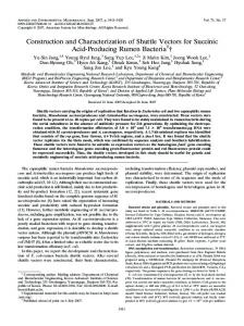
1,5kb Hind III fragment carrying the 19 lactamose gene lacking Its signal sequence
*\
wN
HP14 4.2 kb
P
Inserted in pHP14 digested with Hind III and Pst I
=-
CS (mp 13)
,H\
B
Bcl I
o
pGPA11 (14) 5,8kb CIa
ori
pBR 322
pTA186\
l p GPB11(14) \ 5,7kb
Hind m
~.lactamase MCS
l I
-npC194 pE194 -
Inserted in Hind III site of pHP14