Osborne, R. S., and T. J. Silhavy. 1993. PrlA suppressor mutations .... Rev. Biochem. 60:101â124. 62. Yuan, J., R. Henry, M. McCaffery, and K. Cline. 1994.
JOURNAL OF BACTERIOLOGY, June 1995, p. 3518–3526 0021-9193/95/$04.0010 Copyright q 1995, American Society for Microbiology
Vol. 177, No. 12
Suppression of Signal Sequence Defects and Azide Resistance in Escherichia coli Commonly Result from the Same Mutations in secA JANET L. HUIE
AND
THOMAS J. SILHAVY*
Department of Molecular Biology, Princeton University, Princeton, New Jersey 08544 Received 15 November 1994/Accepted 5 April 1995
The SecA protein of Escherichia coli is required for protein translocation from the cytoplasm. The complexity of SecA function is reflected by missense mutations in the secA gene that confer several different phenotypes: (i) conditional-lethal alleles cause a generalized block in protein secretion, resulting in the cytoplasmic accumulation of the precursor forms of secreted proteins; (ii) azi alleles confer resistance to azide at concentrations up to 4 mM; and (iii) prlD alleles suppress a number of signal sequence mutations in several different genes. To gain further insights into the role of SecA in protein secretion, we have isolated and characterized a large number of prlD mutations, reasoning that these mutations alter a normal function of wild-type SecA. Our results reveal a striking coincidence of signal sequence suppression and azide resistance: the majority of prlD alleles also confer azide resistance, and all azi alleles tested are suppressors. We suggest that this correlation reflects the mechanism(s) of signal sequence suppression. There are two particularly interesting subclasses of prlD and azi alleles. First, four of the prlD and azi alleles exhibit special properties: (i) as suppressors they are potent enough to allow PrlD (SecA) inactivation by a toxic LacZ fusion protein marked with a signal sequence mutation (suppressor-directed inactivation), (ii) they confer azide resistance, and (iii) they cause modest defects in the secretion of wild-type proteins. Sequence analysis reveals that all four of these alleles alter Tyr-134 in SecA, changing it to Ser, Cys, or Asn. The second subclass consists of seven prlD alleles that confer azide supersensitivity, and sequence analysis reveals that six of these alleles are changes of Ala-507 to Val. Both of the affected amino acids are located within different putative ATP-binding regions of SecA and thus may affect ATPase activities of SecA. We suggest that the four azide-resistant mutations slow an ATPase activity of SecA, thus allowing successful translocation of increased amounts of mutant precursor proteins. Proteins such as LamB (maltoporin, l receptor) and MalE (maltose-binding protein), which are destined for secretion to the outer membrane and the periplasm of Escherichia coli, respectively, are synthesized in precursor form with a cleavable signal sequence at the amino terminus. This sequence targets proteins to the secretion pathway for translocation out of the cytoplasm. Mutations that alter the signal sequence prevent proper targeting and result in accumulation of the precursor form of the molecule in the cytoplasm. A variety of genetic and biochemical methods have been employed to determine the role of the signal sequence and to understand the mechanism by which proteins are translocated across the cytoplasmic membrane (for reviews, see references 6, 48, and 61). One approach that has been successfully employed in E. coli to identify components of the cellular secretion machinery involves the isolation and characterization of suppressors of signal sequence mutations. These suppressors, which are termed prl for protein localization, appear to broaden the specificity of the secretion machinery to allow recognition of precursor molecules with defective signal sequences. Three genes, prlA, prlG, and prlD, have been identified repeatedly in a variety of suppressor selections. Subsequent analysis revealed that these suppressors are alleles of secY, secE, and secA, respectively. Recessive, loss-of-function mutations in each of these three genes block the secretion pathway and cause a generalized precursor accumulation in the cytoplasm. Biochemical analysis supports the view that these proteins play a critical role in the translocation reaction; indeed, successful
reconstitution of translocation in vitro with only these three proteins has been reported (reference 1, but also see references 10, 11, and 60). SecY/PrlA and SecE/PrlG are integral cytoplasmic membrane proteins. Recently, homologs of these two proteins have been identified in other bacteria, archaebacteria, yeasts, and mammals, and it has been suggested that these proteins may form a channel through which secreted proteins pass as they leave the cytoplasm (16, 24, 26). This discovery, together with the universal presence of signal sequences in secreted proteins, argues that the basic mechanism of protein secretion has been conserved throughout nature. While homologs of SecA have been found in other bacteria, in plastids of algae, and in chloroplasts of plants (5, 14, 34, 35, 39, 46, 47, 59, 62), none has been discovered in yeasts or mammals. Whether this reflects an important difference between prokaryotes and eukaryotes remains to be determined. The SecA protein is large (102 kDa) and complex. It is found both in soluble form in the cytoplasm and embedded in the cytoplasmic membrane (12). This unusual behavior in fractionation studies may reflect a role for SecA in the targeting of precursor proteins from the cytoplasm to the translocation sites (SecY/E) in the membrane (25). SecA functions as a dimer (3, 17), and it is known to exhibit ATPase activity (30). This hydrolytic activity, which is required for translocation, is stimulated by precursor proteins, acidic phospholipids, and SecY/E (31). Recently, ATP hydrolysis was shown to drive SecA insertion and deinsertion from inner membrane vesicles (18, 27), and this likely reflects a key role for SecA in the energetics of translocation. Several years ago, mutations conferring resistance to azide
* Corresponding author. Phone: (609) 258-5899. Fax: (609) 2586175. 3518
prlD SUPPRESSORS CONFER AZIDE RESISTANCE
VOL. 177, 1995
were found to be located in secA (23, 37). Thus, as it turns out, the first secA mutations were actually isolated by Lederberg (29), who termed them azi. Azide is known to inhibit certain types of ATPases, consistent with the known activities of SecA. Apparently, SecA is the most azide-sensitive, essential ATPase in E. coli under the selection conditions employed. This resistance is low level (up to 4 mM azide); higher levels kill Azr cells presumably by other mechanisms. Additionally, it is known that azide inhibits the ATPase activity required for both in vivo and in vitro translocation (37), although the nature of the interactions between azide and SecA remains to be elucidated. The prlA and prlG suppressor alleles provide useful tools for analyzing the translocation reaction. For example, these alleles form the basis for a genetic technique termed suppressordirected inactivation (SDI). This technique has been used to block the secretion pathway at various stages and works in the following manner: LamB-LacZ hybrid proteins that contain a functional signal sequence can cause a lethal jamming of the cellular secretion machinery. Signal sequence mutations in the hybrid gene prevent jamming. Certain prlA and prlG suppressors restore lethality, and under these conditions, it can be demonstrated with diploid analysis that the suppressor gene product is inactivated by the toxic fusion protein. Apparently the LamB-LacZ fusion protein tagged with a defective signal sequence can inactivate the suppressor protein that is trying to secrete it. Further analysis of these jammed complexes suggested temporal order in the secretion pathway: SecA targets precursor proteins to the membrane at SecE, and SecY is recruited into the complex at a later step (7–9). It seems likely that suppressor mutations alter a normal activity of the wild-type protein rather than creating an entirely new activity. Accordingly, an understanding of Prl suppressor action could reveal insights into the functions of these critical Sec proteins. It has long been assumed that these suppressors restore recognition of mutant signal sequences. However, recent work suggests an alternative model (15, 22, 38). This view posits a signal sequence proofreading activity for the wild-type secretion machinery, which normally causes rejection of precursor molecules with a defective signal. The prlA and prlG suppressors may act by bypassing this proofreading step, thus allowing defective molecules to proceed through the secretory pathway. In order to gain insight into the complex nature of SecA function, we have undertaken an analysis of the prlD suppressor alleles. The results presented here show a striking correlation between signal sequence suppression and azide resistance. On the basis of these results, we propose that at least a subset of the prlD suppressors affect an activity of SecA related to ATP hydrolysis. MATERIALS AND METHODS Media and chemicals. Solid and liquid media have been described previously (52). Maltodextrins were obtained from Pfanstiel Laboratories, Inc. The purification of maltodextrins by dialysis has been described previously (58). For selecting suppressors, a concentration of the purified maltodextrin solution in M63 minimal agar was chosen such that MC4100 (lamB1) grows and KB81 (lamB14D leu::Tn10) does not grow. Sodium azide was obtained from Sigma Chemical Co. [35S]methionine was obtained from DuPont, NEN Research Products. Bacterial strains. E. coli K-12 strain MC4100 [F2 araD139 D(argF-lac)U169 rpsL150 relA1 flbB5301 deoC1 ptsF25 rbsR] (13) is the isogenic parent strain for pop3186 [lamB-lacZ(42–1)] (53), SE1073 [lamB17D-lacZ(42–1)] (20), and all prlD and azi derivatives of these strains constructed in this study. STA1000 is isogenic to MC4100 (57) and is the isogenic parent strain for STA14D (lamB14D), KB81 (lamB14D leu::Tn10) (K. Bieker), STA1013 (lamBD78), NT178 [secA51(Ts)] (N. Trun), and all prlD and azi derivatives of these strains constructed in this study. W208 (CGSC 1867; azi-4), x148 (CGSC 6492; azi-6), Hfr Hayes (CGSC 5492; azi-7), and SS320 (CGSC 6414; azi-9), sources of the Lederberg (29) azi mutations, were obtained from the E. coli Genetic Stock
3519
Center, Department of Biology, Yale University (B. Bachmann). Mapping markers zac-3093::Tn10kan and zad-3094::Tn10kan were obtained from the collection of Singer et al. (54). LP68 [MC4100 nadA::Tn10 D(gal-att-bio)] was provided by L. Pratt. Strains were constructed by standard genetic techniques (32, 52). prlD, azi, and secA alleles were moved via P1 transduction with linkage to a Tn10 insertion in leuB (1.7 min). The presence or absence of the alleles in each strain background was verified by screening for the azide phenotype and/or by moving the allele via P1 transduction into a lamB14D strain and checking for the suppressor phenotype (marker rescue). Phages and plasmids. P1 transductions and preparation of P1vir and lvir lysates were carried out as previously described (52). pMF8, a pBR322-derived plasmid carrying the geneX-secA operon, has been described previously (50). pJH1 was constructed by the removal of a 9geneX-secA fragment from pMF8 by an EcoRI digest, gel purification of the vector, religation, and transformation into the appropriate strains. pJH1 was used as a negative control in all experiments with pMF8. lPR9 is a limm21 phage that carries the secA1 and envA1 genes (41), and it was provided by D. Oliver. Strains with lPR9 were constructed by plating the phage on the desired strain and purifying lysogens by screening for immunity to the l phage B500 (l h80 imm21 cI) and screening for sensitivity to phage f80vir to ensure that the cells are still phage sensitive (52). To check whether lPR9 integrated at the chromosomal attB l integration site and not into the secA locus, nadA::Tn10 linked to a gal-att-bio deletion was transduced into these strains from LP68. The resulting Gal2 transductants had lost immunity to the phage B500 and also lost the lPR9-mediated phenotypes, thus verifying the correct integration of lPR9. Localized spontaneous mutagenesis. Localized spontaneous mutagenesis was previously described by Trun and Silhavy (58). prlD mutants were selected in the strain KB81 (lamB14D leu::Tn10). On two separate occasions, KB81 was streaked on Luria-Bertani agar containing tetracycline and incubated at 378C overnight. Each time, 10 tubes containing 5 ml of Luria-Bertani agar each were then inoculated from 10 individual colonies and grown overnight at 378C. The independent cultures were pelleted and resuspended in one-half volume of 10 mM MgSO4–5 mM CaCl2, and 0.1 ml from each was spread on maltodextrin M63 minimal agar and incubated at 378C. Dex1 suppressors arose at a frequency of 1026. The Dex1 colonies were then pooled for each plate, and a P1 phage lysate was prepared from each pool. The lysates were used to transduce STA14D (lamB14D) to tetracycline resistance (Tetr) on dextrin-tetracycline MacConkey plates at 378C (thus selecting for P1 phage carrying regions of the chromosome containing leu::Tn10). Tetr Dex1 transductants (red or pink colonies on dextrintetracycline MacConkey agar) were purified for further characterization. These strains were screened for l sensitivity by cross-streaking against lvir on glucose (LamB repressed), glycerol (LamB not induced), and maltose (LamB induced) minimal agar plates. Strength of suppression was determined with these phenotypic assays. Isolation of azide resistant mutants. Azide resistant mutants (azi) were selected by plating independent cultures on Luria-Bertani agar plates containing 3.5 mM sodium azide at 378C. The strain used for selection was the same as that used for the procedure described above (KB81), and the azide resistance and suppression phenotypes were reassessed after moving these mutations into STA14D and MC4100 via P1 transduction; mapping was also performed with these strains. Maltose sensitivity and azide sensitivity disk assays. Cells of the appropriate strains were grown in Luria-Bertani media (with antibiotics when necessary) to saturation, pelleted, and resuspended in one-half volume of 10 mM MgSO4–5 mM CaCl2. A total of 0.1 ml of the cell suspension and any supplemental amino acids or antibiotics was then mixed with 3 ml of molten F-top agar and plated on glycerol M63 minimal agar for the maltose sensitivity assay or Luria-Bertani agar for the azide sensitivity assay. Seven-millimeter-diameter paper filter disks (Schleicher and Schuell) were then placed on top of the hardened F-top agar. For the maltose sensitivity assay, 10 ml of 2.5% maltose was added to the disks, and for the azide sensitivity assay, 10 ml of 1 M azide was added to the disks. The plates were incubated overnight at 308C (maltose assay) or 378C (azide assay). The diameter of the zone of sensitivity was measured, subtracting the diameter of the filter disk (7 mm). Pulse-chase assay for MalE processing in LacZ fusion strains. lamB-lacZ (with or without the 17D signal sequence mutation) strains were grown to saturation in glycerol M63 liquid medium at 378C and subcultured (1:20) in fresh glycerol M63 liquid medium at 308C to an optical density at 600 nm of ;0.25. LamB-LacZ fusion protein and MalE synthesis were then induced with 0.2% maltose for 40 min, after which the cells were labeled with [35S]methionine for 20 s, followed by chase time points of 10 s through 4 min. LamB-LacZ and MalE were immunoprecipitated with anti-LacZ and MalE antibodies, respectively. LacZ antibodies (rabbit anti-b-galactosidase) were obtained from 5 prime33 prime, Inc. Samples were subjected to electrophoresis with sodium dodecyl sulfate (SDS)–10% polyacrylamide gels, followed by autoradiography and imaging with a PhosphorImager (Molecular Dynamics). Pulse-chase assay for MalE and LamB processing. Cells were grown to saturation in glycerol M63 liquid medium at 378C and subcultured (1:20) in fresh glycerol M63 liquid medium at 308C, with 0.4% maltose to induce synthesis of MalE and LamB, to an optical density at 600 nm of approximately 0.30. This procedure has been described previously (55). [35S]methionine labeling was performed with a 20-s pulse followed by chase time points of 10 s to 4 min, and
3520
HUIE AND SILHAVY
J. BACTERIOL. TABLE 1. Phenotypic characterization of the prlD suppressors Phage sensitivity of lvir ona:
Allele(s) lamB
lamB1 lamB14D lamB14D lamB14D lamB14D lamB14D lamB14D
prl
prlD1 prlD1 prlD2, -4, -5, -20–24, -26, -27 and azi-17 prlD28–33 prlD25, -34–49 and azi-4, -6, -7, -9, -11–16 prlA4 prlG1
Glu
Gly
Mal
S R S/R R R S S/R
S R S/R R/S R S S/R
S R S S S/R S S
Use of maltodextrinsb
Rd Wh Rd DPk Pk or Wh/Rd centers Rd Rd
a Phage sensitivity was determined by cross-streaking of each strain across lvir on M63 minimal agar supplemented with glucose (Glu), glycerol (Gly), or maltose (Mal). R, resistant; S, sensitive; R/S, more resistant than sensitive; S/R, more sensitive than resistant. b Ability to utilize maltodextrins was scored on dextrin MacConkey agar. Rd, red (LamB1); Wh, white (LamB2); DPk, dark pink; Pk, pink; Wh/Rd centers, white with red centers (LamB6).
samples were immunoprecipitated with anti-MalE, anti-LamB, and/or anti-SecA polyclonal antibodies, as described in reference 58. Anti-SecA antibody was provided by D. Oliver. Samples were subjected to electrophoresis with SDS–10% polyacrylamide gels, followed by autoradiography and imaging with a PhosphorImager. Quantitation of the percentage of mMalE. Densitometry was performed with the ImageQuant system (Molecular Dynamics) to analyze images obtained from the PhosphorImager. The percentage of mature MalE (mMalE) as a function of total MalE protein (including precursor [pMalE]) was obtained by the following equation: mMalE 2 bkgd/[(mMalE 1 pMalE) 2 2(bkgd)], where bkgd is the background for each lane in the gel. The amounts of background, mMalE, and pMalE in each time point lane in the gel were determined by placing rectangular boxes (identical in size) around each band and subsequent integration of the boxes with ImageQuant. mMalE has lost three of its original nine methionines by signal sequence cleavage; to correct for the corresponding loss in radioactivity of mMalE from pMalE, the amount of mMalE from each time point was multiplied by 9/6 (40). PCR amplification and DNA sequencing of prlD and azi alleles. The secA gene was amplified by PCR with the single colony method as described previously (43), with four primers in order to amplify overlapping halves of the gene: SecA1 (nucleotides [nt] 635 to 654, 59GGCACGCCGTCTGAAAAGGG39) and SecA6 (nt 2396 to 2377, 59CGCGGCAACTTCTGCCTGCC39) and SecA10 (nt 2088 to 2105, 59CTGCCGGACCTGGTCTAC39) and SecA4 (nt 3668 to 3648 59GCG CATCTGCTGCGCGACGCG39), with nt designating the nucleotide position within the E. coli geneX-secA DNA sequence (51). Standard DNA sequencing of the PCR products was then accomplished with primers spaced approximately every 200 to 300 bp within secA. The prlD20, -21, -22, -23, -28, and -43 mutations are the only alterations that were found within the entire secA coding sequence, including the Shine-Dalgarno site. Because prlD24, -26, -27, and -31 are identical to prlD20 and -28, the complete DNA sequence was not determined.
RESULTS Previously characterized prlD suppressors. The first prlD alleles, prlD1, -2, -3, -4, and -5, were isolated as suppressors of mutations that alter the hydrophobic core of the signal sequence of MalE (4, 44, 45). DNA sequence analysis verified that prlD2, -4, and -5 are unique alleles of secA and are located in three widely spaced regions of SecA (21). prlD3 is the same mutation as prlD2. The prlD1 allele appeared to lie outside of secA (4); however, cells carrying this mutation grew poorly, and the mutation has been lost. Isolation of additional prlD suppressors. LamB functions in E. coli as the maltoporin and also as the phage l receptor. Cells carrying lamB signal sequence mutations cannot grow with maltodextrins as a sole carbon source (Dex2), are resistant to l phage (lr), and accumulate the precursor form of LamB in the cytoplasm. Suppressors that restore export of the mutant precursor protein are easily selected by demanding growth on maltodextrin minimal agar (Dex1 phenotype). Recently, in a large screen for prl suppressors of the lamB14D allele (a change of valine to aspartate at amino acid position 14 in the signal sequence), our laboratory isolated 17 extragenic suppressors at or near the secA locus. Linkage was shown by
transductional mapping with leu::Tn10, which is approximately 50% linked to secA (56). In this study, we employed a different method, localized spontaneous mutagenesis (described in Materials and Methods), to obtain 13 additional prlD suppressors of lamB14D. In transductional crosses, the 13 new alleles and the previously isolated 17 alleles are tightly linked to secA51(Ts) and similarly linked to outside markers: 48% linkage to zac-3093::Tn10kan (2.0 min), 42% to leu::Tn10 (1.7 min), 13% to araC (1.4 min), and 4% to zad-3094::Tn10kan (3.5 min). The 30 prlD alleles were numbered on the basis of the strength of their suppressor phenotypes (Dex1 and ls) in the lamB14D strain. These data are summarized in Table 1. For comparison, the three unique prlD mutations isolated by the Bassford group are also included. Thus, the total number of prlD suppressors examined in this study is 33. As can be seen in Table 1, 10 of the prlD suppressors are comparable in strength to prlG suppressors, while the rest are weaker. In general, prlA suppressors are more potent than either prlG or prlD suppressors. Suppressor strength correlates directly with the amount of LamB in the outer membrane. Suppression and azide resistance are highly coincident. Azide resistance, which maps to the secA locus (23, 37), was used for fine structure mapping to more firmly establish the linkage observed between the new alleles and secA51(Ts). Surprisingly, we found that most of the suppressors already confer azide resistance (Azr). Not only are 20 of 33 of the prlD alleles resistant to 3 mM azide on L agar, but all 3 of the original prlD alleles also confer resistance. To quantitate the level of Azr, we used a disk assay as described in Materials and Methods. The results revealed that almost one-fourth (7 of 33) of all the prlD suppressors cause another azide phenotype, supersensitivity to azide (Azss), compared with the same strain containing secA1. Almost an additional quarter of the total number of prlD alleles (6 of 33) confer similar azide sensitivity (Azs) to secA1 strains. To further investigate the correlation between suppression and Azr, we obtained (from B. Bachmann) Azr mutations (azi-4, -6, -7, and -9) that were isolated by Lederberg (29). These secA alleles also turn out to be suppressors of lamB14D, as judged by the Dex1 and ls phenotypes of these strains (Table 1). Suppression is similar to that of the other prlD alleles. Thus, the first prlD suppressors were actually isolated in 1950 as azi mutations. In addition, we isolated seven new azi mutations (azi-11, -12, -13, -14, -15, -16, and -17) by selecting for resistance to 3.5 mM azide. All contain mutations that map to secA. These mutations are also suppressors of lamB14D; they confer similar Dex1 and
prlD SUPPRESSORS CONFER AZIDE RESISTANCE
VOL. 177, 1995
3521
TABLE 2. Azide resistance is highly coincident with suppression Allele(s)
secA1 prlD2, -4, -5, -21– 23, -29, -30, -34, -36– 42, -45– 48 prlD20, -24, -26–28, -31, -43 prlD25, -32, -33, -35, -44, -49 azi-4, -6, -7, -9, -11– 17
Suppressiona
Azide phenotype (disk assay)b
1
S R
1 1 1
SS S R
FIG. 1. Suppression of lamBD78 by prlD21 and prlD2. Cells were pulselabeled with [35S]methionine, and immunoprecipitations were performed as described in Materials and Methods. Shown is the processing of LamBD78 in the following strains: STA1013 (a), STA1013 containing prlD21 (b), and STA1013 containing prlD2 (c). p represents the precursor form of LamB, and m represents the mature form of this protein. Time points indicate the time of the trichloroacetic acid precipitation after the addition of chase.
a
See Table 1. Resistance was scored with a disk assay as described in Materials and Methods. R, resistance (0- to 5-mm diameter of the zone of sensitivity at 378C to 10 ml of 1 M azide after subtraction of the disk diameter [7 mm]); S, sensitive (15 to 17 mm); SS, supersensitive (20 to 28 mm). b
ls phenotypes to the prlD alleles (Table 1). One of these, azi-17, is comparable to the strongest subset of prlD suppressors. As summarized in Table 2, the majority (82%) of prlD suppressors exhibit altered azide sensitivity. In addition, all azi alleles, both new and previously isolated, suppress lamB14D with the efficiency of prlD alleles. We think it improbable that the high degree of correlation of these two phenotypes is a coincidence, and we believe it provides insight into the mechanism of prlD suppression. Diploid analysis: the azide-resistant suppressors are allelic to secA. Fikes and Bassford (21) found that suppression of malE14-1 by prlD2 is dominant, similar to the dominance observed with prlA and prlG (6, 56). This is as expected, because prl alleles are gain-of-function mutations. All of the new prlD suppressors are dominant in diploid analysis with lPR9 (lsecA1). We were concerned that dominance could be attributed to a simple increase in the levels of SecA because slight suppression of malE14-1 could be effected by a high-copy-number plasmid carrying secA1 (21). We observed that pMF8, a similar plasmid carrying secA1, allows weak suppression of lamB14D. Nevertheless, lPR9 lysogens, which are true secA diploids, do not exhibit detectable levels of suppression, confirming that suppression by the new prlD alleles is indeed dominant. Azr, however, is recessive (23, 37), and this allows mapping by complementation. The Azr phenotype of all of the prlD and azi suppressors is recessive to secA1 in trans (lPR9), proving that these mutants are allelic to secA. Thus, the new Azr prlD suppressors and the seven new azi alleles are all secA alleles that merit the name prlD. The Azss phenotype conferred by prlD alleles is dominant, and the Azs alleles also cannot be used in complementation tests. However, because their linkage to secA is indistinguishable from that of the Azr alleles, we think it likely that these are secA alleles as well. Biochemical characterization of suppression. To quantitate suppression of signal sequence mutations, we utilized a pulsechase assay to monitor precursor maturation over time. This assay, while useful for quantitation, is not as sensitive an indicator of suppression as the phenotypic plate assays. For example, prlA suppressors are so potent that increased processing of mutant precursors can be observed in the pulse-chase assay even with strongly defective signal sequence mutations; processing cannot be observed with the same mutations in strains containing prlG (19, 55, 56). Suppression by prlG alleles can be observed in the pulse-chase assay with a signal sequence mutation that causes a less-severe secretion defect, such as lamBD78. We were unable to detect suppression of lamB14D by the
prlD alleles in the pulse-chase assay. However, all of the suppressors checked, including prlD2, -4, -5, -21, -22, and -23 and azi-7 and -17, show suppression of lamBD78 (Fig. 1 [data shown for prlD2 and -21]). Suppression of lamBD78 is relatively weak compared with that of prlA suppressors, coincident with the weak Dex1 and ls phenotypes exhibited by these strains. We were unable to reliably detect differences in strength of suppression between different prlD and azi alleles with the pulse-chase assay. However, the prlD21, -22, and -23 and azi-17 strains appear to produce mature LamB only by 2 min, while the prlD2, -4, and -5 and azi-7 strains show significant suppression by 10 s (Fig. 1, compare prlD2 and -21). The difference in kinetics may reflect a difference in the mechanism of suppression between these sets of alleles (described below). We also used the pulse-chase assay to measure suppression of lamBD78 in lPR9 lysogens and found that suppression by these representative alleles is dominant (data not shown). As noted above, wild-type lPR9 lysogens do not exhibit a suppressor phenotype. Overexpression of SecA, with pMF8, allows suppression of lamBD78 (data not shown). However, the level of suppression is even less than that observed with prlD2, -4, -5, -21, -22, and -23 and azi-7 and -17. As a whole, biochemical assays support our phenotypic observations. Suppression by the representative alleles tested, prlD2, -4, -5, -21, -22, and -23 and azi-7 and -17, is similar to that observed with the prlG suppressors, and it can only be detected in the pulse-chase assay with the leaky signal sequence mutation lamBD78. Four of the prlD and azi alleles allow SDI. An additional test of suppression measures the ability of a prl allele to restore proper targeting of a mutant fusion protein, LamB17D-LacZ, into the secretion pathway (7–9). LamB-LacZ is lethal when induced to high levels with maltose (maltose sensitivity [Mals]) (53). However, the signal sequence mutation 17D relieves this toxicity (maltose resistance [Malr]) (20). A certain number of the prlA and prlG suppressors redirect the fusion protein into the secretion pathway and restore Mals, while other suppressor alleles fail this test. We found that 4 of the 44 suppressor alleles (9%) are able
TABLE 3. Suppression of lamB17D-lacZ by prlD21 and prlD2 Strain
prlD1 lamB-lacZ.................................................................. prlD1 lamB17D-lacZ........................................................... prlA4 lamB17D-lacZ............................................................ prlG1 lamB17D-lacZ ........................................................... prlD21 lamB17D-lacZ ......................................................... prlD2 lamB17D-lacZ ...........................................................
Diam (mm) of zone of maltose sensitivitya
22 0 15 14 14 0
a Values were obtained with a disk assay as described in Materials and Methods and indicate the diameter of the zone of sensitivity at 308C to 10 ml of 2.5% maltose after subtraction of the disk diameter (7 mm).
3522
HUIE AND SILHAVY
J. BACTERIOL.
FIG. 2. prlD21 causes a severe, recessive secretion defect in a lamB17D-lacZ fusion strain. Cells were pulse-labeled and immunoprecipitated with antibody to MalE and otherwise prepared as described in Materials and Methods. Shown is the processing of MalE in the following strains: SE1073 (a), SE1073 containing prlD21 (b), and SE1073 containing prlD21 and lPR9 (secA1) (c). p represents the precursor form of MalE, and m represents the mature form of this protein. Time points indicate the time of the trichloroacetic acid precipitation after the addition of chase.
to restore proper targeting of LamB17D-LacZ into the secretion pathway (Mals): prlD21, -22, and -23 and azi-17 (Table 3 [data shown for prlD21; prlD22 and -23 and azi-17 are similar]). These alleles are all Azr and are among the strongest prlD suppressors of lamB14D (Table 1). However, the three original prlD alleles, prlD2, -4, and -5, are also Azr and equally strong suppressors but do not restore Mals to the lamB17D-lacZ strain. In addition, Azr does not correlate with strength of suppression; for example, some Azss suppressors are stronger than some Azr suppressors. Therefore, we conclude that although all of the alleles capable of restoring Mals confer Azr and are strong suppressors, Azr and strength of suppression are not generally correlated with the ability to restore Mals. In diploid analysis with lPR9 lysogens, the Mals phenotype generated by the four alleles is recessive, shown as follows. The diameters (millimeters) of the zones of maltose sensitivity (as described in Materials and Methods) were 0 for prlD1 lamB17D-lacZ, lPR9 (secA1); 14 for prlD21 lamB17D-lacZ; and 0 for prlD21 lamB17D-lacZ, lPR9. (Data for prlD22 and -23 and azi-17 were similar.) This implies that the LamB17DLacZ fusion protein is specifically inactivating the PrlD21, -22, and -23 and Azi-17 proteins (SDI). If this is true, we would expect the Mals phenotype to result from a secretion defect caused by the loss of PrlD (SecA) function. Pulse-chase experiments support this view (Fig. 2 [data shown for prlD21; prlD22 and -23 and azi-17 are similar]). The prlD21 allele causes a pronounced secretion defect for MalE in the lamB17D-lacZ strain (35% mMalE produced at the 10-s time point compared with 84% with secA1). This defect is relieved by the presence of secA1 in trans (57% mMalE at 10 s). Thus, despite weak suppression of lamB14D and lamBD78 by the prlD and azi alleles, four of these alleles are able to restore Mals and a secretion defect to the lamB17D-lacZ strain. The results are similar to those obtained with certain prlA and prlG suppressors. In all cases, it appears that the toxic fusion protein is inactivating the Prl suppressor protein that is trying to secrete it. A secretion defect is associated with the four special alleles. While performing the pulse-chase assays described above, we noticed a modest defect in the secretion of wild-type MalE in strains containing prlD21 and lamBD78 but not in isogenic strains containing prlD2, -4, or -5 or azi-7. This secretion defect is due solely to the presence of the prlD21 allele. When the suppressor is introduced into a wild-type strain, a decrease in the rate of signal sequence processing is seen for both LamB and MalE, compared with strains containing wild-type SecA or SecA overexpressed (pMF8) (Fig. 3). The test was repeated with 11 suppressors representative of each of the phenotypic classes found except Azs. This list includes prlD21, -22, and -23 and azi-17 (allowing SDI and conferring Azr); prlD2, -4, -5, and -37 and azi-7 (conferring Azr); and prlD20 and -28 (conferring Azss). Only prlD21, -22, and -23 and azi-17, those that allow SDI, cause a secretion defect. The percentage of MalE pro-
FIG. 3. Effect of prlD21 on the secretion of wild-type LamB and MalE precursors. Cells were pulse-labeled and immunoprecipitated with antibodies to LamB, MalE, and SecA and otherwise prepared as described in Materials and Methods. Strains are as follows: MC4100 containing prlD21 (a), MC4100 (b), and MC4100 carrying the secA1 plasmid pMF8 (c). p represents the precursor forms of LamB and MalE, and m represents the mature forms of these proteins. Time points indicate the time of the trichloroacetic acid precipitation after the addition of chase.
cessed after 10 s ranges from 69 to 77% for prlD21, -22, and -23 and azi-17, compared with 92 to 100% for the wild type and the other seven prlD and azi strains. The secretion defects are recessive to a single extra copy of secA1 on the chromosome (data not shown). Thus, these four mutant SecA proteins do not actively interfere with secretion. Rather, they appear to have reduced function. It is known that SecA synthesis is regulated according to the secretion needs of the cell (42, 50). Consistent with the secretion defect conferred by prlD21, -22, and -23 and azi-17 is derepression of secA (Fig. 3, for example, as seen for prlD21). The other Azr and Azss alleles that were checked do not cause derepression of secA. Thus, prlD21, -22, and -23 and azi-17, which allow SDI, are the only alleles that confer significant secretion defects. We suggest that the secretion defect is closely correlated and perhaps integral to the ability of these alleles to restore proper targeting of LamB17D-LacZ. The fact that only a subset of the suppressor alleles confer both a secretion defect and SDI argues that these four mutations cause distinctive changes in SecA function. DNA sequence analysis of a subset of the prlD and azi alleles. We performed DNA sequence analysis of the four special alleles as well as of the Azss class of prlD alleles with the purpose of identifying a specific region of SecA that mediates these phenotypes. First, sequence analysis of prlD21, -22, and -23 and azi-17 revealed that these four alleles all alter a single amino acid in SecA: Tyr-134. prlD21 causes a change of Tyr-134 to Ser and, additionally, Glu-148 to Lys; the codon changes (underlined) are from TAC to TCA and GAA to AAA, respectively. prlD23 also causes a change of Tyr-134 to Ser (TAC to TCT), but without the second mutation. prlD22 and azi-17 change Tyr134 to Cys (TAC to TGC) and Asn (TAC to AAC), respectively. prlD21 and prlD23 were obtained by UV mutagenesis, so the multiple mutations observed are not unusual. Second, sequence analysis showed that six of the seven Azss prlD alleles (prlD20, -24, -26, -27, -28, and -31) are alterations of Ala-507 to Val (GCG to GTG). prlD43 is an alteration of His-484 to Gln (CAC to CAG). In sum, the four special alleles prlD21, -22, and -23 and azi-17 all alter the same amino acid in the N terminus of secA; six of the seven Azss alleles alter a particular amino acid in the C terminus of secA. Thus, genetic analysis revealed significant clustering of mutations in secA with distinct and identical phenotypes.
prlD SUPPRESSORS CONFER AZIDE RESISTANCE
VOL. 177, 1995
3523
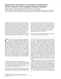
FIG. 4. Compilation of the known prlD and azi mutational alterations of the E. coli SecA protein. The horizontal line represents the linear amino acid (aa) sequence of the protein, and numbers indicate specific residues. Changes caused by previously identified mutations are indicated above the line; changes caused by mutations identified in this study are indicated below the line. Azide phenotypes are indicated for prlD alleles. Boxes indicate functional regions of the molecule (see text for details): µ, ABC I (high-affinity ATP-binding site); ■, ABC II (low-affinity ATP-binding site); h, putative signal sequence-binding site.
DISCUSSION The SecA protein is an essential component of the cellular secretion machinery. It binds the signal sequence and mature regions of precursor proteins and, in an ATP-dependent fashion, catalyzes their membrane insertion (26, 31, 49, 61). In an effort to better understand the function(s) of SecA, we undertook a detailed study of the prlD suppressor alleles. One possible mechanism of PrlD-mediated suppression posits a broadened specificity of signal sequence recognition. Viewed in this manner, the prlD mutations might be expected to alter the signal sequence recognition domain of SecA, allowing it to recognize mutant sequences it would normally ignore. However, our results demonstrate a high correlation between suppression and azide resistance: the majority of the prlD suppressors confer azide resistance (Azr), and all of the azi alleles are suppressors. Another class of prlD alleles confers azide supersensitivity (Azss). Because the broadened specificity model does not clearly predict or easily explain this correlation, we suggest that the mechanism of suppression may be more complex. Available DNA sequence data, shown in Fig. 4, support this view. A putative ATP-binding and hydrolysis region consisting of two subsites is located in the N terminus of SecA (Fig. 4) (33). This region is called the ATP-binding cassette (ABC I) and corresponds to a high-affinity site identified by Mitchell and Oliver (33). Intriguingly, ATP binding at this site appears to cause a conformational change that drives membrane insertion of SecA (18, 27) (Fig. 5, step iii). All prlD and azi mutations in this region confer Azr and are suppressors of signal sequence mutations, including prlD4 (T111N) (21) and azi-7 (N179Y) (29, 37). Particularly interesting is the clustering of prlD21 (Y134S and E148K), -22 (Y134C), and -23 (Y134S) and azi-17 (Y134N), which have been shown in this work to confer unique phenotypes. These mutations all alter Tyr-134. As will be discussed further below, we believe that these special alleles alter the ATPase activity of ABC I. Consistent with this view, Nakane et al. (36) have shown that alterations of Thr-128 in Bacillus subtilis SecA confer Azr and cause increased ATPase activity. None of the prlD or azi mutations are located within the actual subsites of ABC I (amino acids 102 to 109 and 198 to 210); perhaps, as Mitchell and Oliver (33) suggested, alterations of amino acids in these sites abolish ATP binding and/or hydrolysis entirely and are therefore lethal. The second ABC (ABC II) is located in the C-terminal half of SecA and was identified as a low-affinity site (33). It has been proposed that ATP hydrolysis at this second site allows
SecA to remove itself (deinsert) from the membrane (18, 27). All azide supersensitive (Azss) alleles alter amino acids near or within the first subsite of ABC II (amino acids 503 to 511): prlD20, -24, -26, -27, -28, and -31 (all A507V) and -43 (H484Q). It is tempting to speculate that Azss may be related to a defect in the membrane deinsertion reaction. An azide-induced block at this step would leave SecA embedded in the membrane, where it may cause lethality by titrating other essential components of the secretion machinery. This model could explain the dominance of Azs and Azss in diploid analysis. In addition to all of the Azss alleles, Azr is also conferred by suppressor mutations within ABC II. These Azr mutations are located within or near either subsite of the cassette, rather than in the intervening sequence, as is the case for mutations in ABC I. prlD2 (A288V) (21) is located near the first subsite. azi-4 (L645Q) (29, 37), azi-201 (A630V), and azi-248 (R656C) (23) are all alterations of amino acids near or within the second subsite (amino acids 631 to 653), but the suppressor phenotype of azi-4 is particularly weak and those of azi-201 and -248 are unknown. An additional suppressor, prlD5 (A373V), is located near a region defined by Kimura et al. (28) as the signal sequence binding site (amino acids 267 to 340). Evidence for such a site came from cross-linking of proteolytic fragments of SecA with proOmpF-Lpp, a model precursor protein. Perhaps prlD5 alters the interaction of SecA with signal sequences (2). Note, however, that prlD5 confers Azr. This result, coupled with the fact that Azr mutations are also found in both ABC I and II, demonstrates the complexity of the azide resistance phenotype. Clearly, mutational alterations in several different functional domains of SecA can confer Azr, and the mechanism of signal sequence suppression for the different alterations is probably not the same. We have identified four alleles (out of the total of 44) that exhibit special properties. These alleles, prlD21, -22, and -23 and azi-17, confer azide resistance, are potent enough with LamB17D-LacZ to allow SDI, and cause defects in the secretion of wild-type proteins. Strikingly, all alter Tyr-134, a residue conserved in all known SecA proteins (5, 14, 34, 35, 39, 46, 47, 59, 62). We suspect that these four mutations may slow an ATPase activity of SecA. Our model for suppression (Fig. 5) is based on recent studies with prlA and prlG (15, 22, 38) and biochemical studies performed by Schiebel et al. (49). The prlA and prlG suppressors appear to act by reducing a signal sequence proofreading function. In wild-type strains, defective signal sequences are re-
3524
HUIE AND SILHAVY
J. BACTERIOL.
FIG. 5. Model for the mechanism of action of the four prlD suppressors. (i) SecB and SecA are recruited to maintain the precursor protein in an export-competent state and to target it to the translocator, composed of SecY and SecE. (ii) After docking, the precursor protein, SecA, binds ATP. (iii) Binding of ATP provides the energy for a conformational change that drives the precursor into the translocator, provided that the signal sequence is read correctly. (iv.a and v) ATP hydrolysis allows translocation to proceed. (iv.b) If the signal sequence is defective (ss*), the mutant precursor is rejected from the pathway. We propose that the prlD suppressors act to slow the step of ATP hydrolysis, the proofreading step, thus providing the translocator with additional time to engage. This allows successful translocation of increased amounts of mutant precursor.
jected from the secretion pathway. In suppressor strains, compromised proofreading allows mutant precursor secretion. The biochemical studies indicate that ATP binding by SecA causes a dramatic conformational change that drives precursor translocation to the extent that signal sequence processing occurs (18, 27, 49) (Fig. 5, iii). We suggest that signal sequence proofreading occurs at this ATP-dependent step. If SecY recognizes the signal sequence, the conformational change is allowed, and a functional translocase forms; ATP hydrolysis then drives further translocation (Fig. 5, iv.a) (49). However, if SecY rejects the signal sequence, ATP hydrolysis instead releases the defective precursor and translocation is aborted (Fig. 5, iv.b). All signal sequence mutations, even deletions, are leaky. For example, a complete signal sequence deletion in phoA still allows secretion of 1% of PhoA protein (15). If these special prlD suppressors slow the ATPase activity, the extra time allotted by the increased half-life of the SecA-ATP complex should permit the successful translocation of increased amounts of mutant precursor. This putative decreased ATPase activity can explain the other properties of these prlD suppressors. First, azide is known to affect certain types of ATPases, and it seems likely that mutations which confer azide resistance will alter this activity. Second, if these mutations slow the ATPase activity, the mutant SecA protein might remain complexed with the toxic LamB17D-LacZ fusion protein for longer periods of time, and this would explain suppressor-directed inactivation of PrlD. Third, if the ATPase activity is slowed, normal function of the protein is then slowed, and this would explain the defects observed for the secretion of wild-type precursor proteins. Our model predicts that mutant signal sequences can still be recognized by wild-type SecA. As noted above, the coincidence
of suppression and azide resistance argues that few, if any, of the Azr prlD suppressors act by restoring mutant signal sequence recognition. This is understandable if SecA can recognize defective signal sequences already, and it also explains why prlA suppressors are so potent. In prlA strains, suppression is SecA dependent (15). Thus, SecA must still be able to bind and properly direct both wild-type and mutant precursors into the secretion machinery efficiently. A second prediction of the model relates to the proposed increased half-life of the precursor-SecA-ATP complex. If this is true, then it should be possible to effect suppression by simply increasing the concentration of this complex. Thus, we would predict that an increase in the amount of SecA should promote mutant precursor secretion by mass action. Indeed, increased levels of SecA do cause suppression of signal sequence mutations, albeit at low levels (reference 21 and this study). Additionally, we found that increased levels of SecA restore a modest level of Mals in the lamB17D-lacZ fusion strain. This suggests increased targeting of the toxic fusion protein as well. The secretion defects caused by the four special prlD alleles result in derepression of PrlD synthesis. Accordingly, we have considered the possibility that the mechanism of action of these suppressors could be simply attributed to increased levels of PrlD (SecA). We think this unlikely for the following reasons. First, neither the suppressor nor the SDI phenotypes conferred by SecA overexpression are as strong as those conferred by these four alleles, despite the fact that SecA levels in the overexpression strain are two- to threefold higher than those in the suppressor strains (Fig. 3 [similar results obtained with prlD22 and -23 and azi-17]). Second, increased levels of SecA do not cause a secretion defect or azide resistance, as do
prlD SUPPRESSORS CONFER AZIDE RESISTANCE
VOL. 177, 1995
the suppressors. Third, the Mals conferred by the suppressor alleles is recessive to a single extra copy of secA (lPR9). In this case, increasing the gene dosage of secA relieves SDI. The model is based on and offers explanation for only the four special prlD alleles. The remaining 40 are more difficult to explain. We think that the complexity of PrlD phenotypes reflects the multiple functions of SecA, and certain of the other alleles may provide further insights into the role of SecA and its ATPase activities in protein translocation. ACKNOWLEDGMENTS We especially thank Liya Shi for invaluable work and help with DNA sequence analysis of the prlD and azi alleles. We also acknowledge John Carlson, who noted the secretion defect caused by prlD21. Special thanks are given to Kristina Bieker-Brady, under whose expert guidance the initial part of this work was accomplished. We thank D. Oliver for providing the plasmid pMF8, the phage lPR9, and antiSecA antibody; B. Bachmann for azi strains; and Leslie Pratt for strain LP68. Weihong Hsing, Craig Parker, Tony Greenberg, Paul Danese, Bill Snyder, Chris! Harris, Deborah Allen, and Susan DiRenzo were especially helpful in critical reading of the manuscript. This work was supported by Public Health Services grants GM07388 (J.L.H.) and GM34821 (T.J.S.). REFERENCES 1. Akimura, J., S. Matsuyama, H. Tokuda, and S. Mizushima. 1991. Reconstitution of a protein translocation system containing purified SecY, SecE, and SecA from Escherichia coli. Proc. Natl. Acad. Sci. USA 88:6545–6549. 2. Akita, M., S. Sasaki, S. Matsuyama, and S. Mizushima. 1990. SecA interacts with secretory proteins by recognizing the positive charge at the amino terminus of the signal peptide in Escherichia coli. J. Biol. Chem. 265:8164– 8169. 3. Akita, M., A. Shinkai, S. Matsuyama, and S. Mizushima. 1991. SecA, an essential component of the secretory machinery of Escherichia coli, exists as homodimer. Biochem. Biophys. Res. Commun. 174:211–216. 4. Bankaitis, V. A., and P. J. Bassford, Jr. 1985. Proper interaction between at least two components is required for efficient export of proteins to the Escherichia coli cell envelope. J. Bacteriol. 161:169–178. 5. Barbrook, A. C., J. C. Packer, and C. J. Howe. 1993. Components of the protein translocation machinery in the thermophilic cyanobacterium Phormidium laminosum. Biochem. Biophys. Res. Commun. 197:874–877. 6. Bieker, K. L., G. J. Phillips, and T. J. Silhavy. 1990. The sec and prl genes of Escherichia coli. J. Bioenerg. Biomembr. 22:291–310. 7. Bieker, K. L., and T. J. Silhavy. 1989. PrlA is important for the translocation of exported proteins across the cytoplasmic membrane of Escherichia coli. Proc. Natl. Acad. Sci. USA 86:968–972. 8. Bieker, K. L., and T. J. Silhavy. 1990. PrlA (SecY) and PrlG (SecE) interact directly and function sequentially during protein translocation in E. coli. Cell 61:833–842. 9. Bieker-Brady, K. L., and T. J. Silhavy. 1992. Suppressor analysis suggests a multistep, cyclic mechanism for protein secretion in Escherichia coli. EMBO J. 11:3165–3174. 10. Brundage, L., C. J. Fimmel, S. Mizushima, and W. Wickner. 1992. SecY, SecE, and Band1 form the membrane-embedded domain of Escherichia coli preprotein translocase. J. Biol. Chem. 267:4166–4170. 11. Brundage, L., J. P. Hendrick, E. Schiebel, A. J. M. Driessen, and W. Wickner. 1990. The purified E. coli integral membrane protein SecY/E is sufficient for reconstitution of SecA-dependent precursor translocation. Cell 62:649– 657. 12. Cabelli, R. J., K. M. Dolan, L. Qian, and D. B. Oliver. 1991. Characterization of membrane-associated and soluble states of SecA protein from wild-type and secA51(ts) mutant strains of Escherichia coli. J. Biol. Chem. 266:24420– 24427. 13. Casadaban, M. J. 1976. Transposition and fusion of the lac genes to selected promoters in Escherichia coli using bacteriophage lambda and mu. J. Mol. Biol. 104:541–555. 14. de Cock, H., and J. Tommassen. 1991. Conservation of components of the Escherichia coli export machinery in prokaryotes. FEMS Microbiol. Lett. 64:195–199. 15. Derman, A. I., J. W. Puziss, P. J. Bassford, Jr., and J. Beckwith. 1993. A signal sequence is not required for protein export in prlA mutants of Escherichia coli. EMBO J. 12:879–888. 16. Dobberstein, B. 1994. Protein transport: on the beaten pathway. Nature (London) 367:599–600. 17. Driessen, A. J. 1993. secA, the peripheral subunit of the Escherichia coli precursor protein translocase, is functional as a dimer. Biochemistry 32: 13190–13197.
3525
18. Economou, A., and W. Wickner. 1994. SecA promotes preprotein translocation by undergoing ATP-driven cycles of membrane insertion and deinsertion. Cell 78:835–843. 19. Emr, S. D., and P. J. Bassford, Jr. 1982. Localization and processing of outer membrane and periplasmic proteins in Escherichia coli strains harboring export-specific suppressor mutations. J. Biol. Chem. 257:5852–5860. 20. Emr, S. D., and T. J. Silhavy. 1980. Mutations affecting localization of an Escherichia coli outer membrane protein, the bacteriophage lambda receptor. J. Mol. Biol. 141:63–90. 21. Fikes, J. D., and P. J. Bassford, Jr. 1989. Novel secA alleles improve export of maltose-binding protein synthesized with a defective signal peptide. J. Bacteriol. 171:402–409. 22. Flower, A. M., R. C. Doebele, and T. J. Silhavy. 1994. prlA and prlG suppressors reduce the requirement for signal sequence recognition. J. Bacteriol. 176:5607–5614. 23. Fortin, Y., P. Phoenix, and G. R. Drapeau. 1990. Mutations conferring resistance to azide in Escherichia coli occur primarily in the secA gene. J. Bacteriol. 172:6607–6610. 24. Gorlich, D., S. Prehn, E. Hartmann, K. Kalies, and T. A. Rapoport. 1992. A mammalian homolog of SEC61p and SecYp is associated with ribosomes and nascent polypeptides during translocation. Cell 71:489–503. 25. Hartl, F.-U., S. Lecker, E. Schiebel, J. P. Hendrick, and W. Wickner. 1990. The binding cascade of SecB to SecA to SecY/E mediates preprotein targeting to the E. coli plasma membrane. Cell 63:269–279. 26. Hartmann, E., T. Sommer, S. Prehn, D. Gorlich, S. Jentsch, and T. A. Rapoport. 1994. Evolutionary conservation of components of the protein translocation complex. Nature (London) 367:654–657. 27. Kim, Y. J., T. Rajapandi, and D. Oliver. 1994. SecA protein is exposed to the periplasmic surface of the E. coli inner membrane in its active state. Cell 78:845–853. 28. Kimura, E., M. Akita, S. Matsuyama, and S. Mizushima. 1991. Determination of a region in SecA that interacts with presecretory proteins in Escherichia coli. J. Biol. Chem. 266:6600–6606. 29. Lederberg, J. 1950. The selection of genetic recombinations with bacterial growth inhibitors. J. Bacteriol. 59:211–215. 30. Lill, R., K. Cunningham, L. Brundage, K. Ito, D. Oliver, and W. Wickner. 1989. The SecA protein hydrolyzes ATP and is an essential component of the protein translocation ATPase of E. coli. EMBO J. 8:961–966. 31. Lill, R., W. Dowhan, and W. Wickner. 1990. The ATPase activity of SecA is regulated by acidic phospholipids, SecY, and the leader and mature domains of precursor proteins. Cell 60:271–280. 32. Miller, J. H. 1972. Experiments in molecular genetics. Cold Spring Harbor Laboratory Press, Cold Spring Harbor, N.Y. 33. Mitchell, C., and D. Oliver. 1993. Two distinct ATP-binding domains are needed to promote protein export by Escherichia coli SecA ATPase. Mol. Microbiol. 10:483–497. 34. Nakai, M., A. Goto, T. Nohara, D. Sugita, and T. Endo. 1994. Identification of the SecA protein homolog in pea chloroplasts and its possible involvement in thylakoid protein transport. J. Biol. Chem. 269:31338–31341. 35. Nakai, M., T. Nohara, D. Sugita, and T. Endo. 1994. Identification and characterization of the SecA protein homologue in the cyanobacterium Synechococcus PCC7942. Biochem. Biophys. Res. Commun. 200:844–851. 36. Nakane, A., H. Takamatsu, A. Oguro, Y. Sadaie, K. Nakamura, and K. Yamane. 1995. Acquisition of azide-resistance by elevated SecA ATPase activity confers azide-resistance upon cell growth and protein translocation in B. subtilis. Microbiology 141:113–121. 37. Oliver, D. B., R. J. Cabelli, K. M. Dolan, and G. P. Jaorsik. 1990. Azideresistant mutants of Escherichia coli alter the SecA protein, an azide-sensitive component of the protein export machinery. Proc. Natl. Acad. Sci. USA 87:8227–8231. 38. Osborne, R. S., and T. J. Silhavy. 1993. PrlA suppressor mutations cluster in regions corresponding to three distinct topological domains. EMBO J. 12: 3391–3398. 39. Overhoff, B., M. Klein, M. Spies, and R. Freudl. 1991. Identification of a gene fragment which codes for the 364 amino-terminal amino acid residues of a SecA homologue from Bacillus subtilis: further evidence for the conservation of the protein export apparatus in gram-positive and gram-negative bacteria. Mol. Gen. Genet. 228:417–423. 40. Pogliano, K. J., and J. Beckwith. 1993. The Cs sec mutants of Escherichia coli reflect the cold sensitivity of protein export itself. Genetics 133:763–773. 41. Riggs, P. D., A. I. Derman, and J. Beckwith. 1988. A mutation affecting the regulation of a secA-lacZ fusion defines a new sec gene. Genetics 118:571– 579. 42. Rollo, E. E., and D. B. Oliver. 1988. Regulation of the Escherichia coli secA gene by protein secretion defects: analysis of secA, secB, secD, and secY mutants. J. Bacteriol. 170:3281–3282. 43. Russo, F. D., J. M. Slauch, and T. J. Silhavy. 1993. Mutations that affect separate functions of OmpR, the phosphorylated regulator of porin transcription in Escherichia coli. J. Mol. Biol. 231:261–273. 44. Ryan, J. P. 1985. Ph.D. thesis. University of North Carolina, Chapel Hill. 45. Ryan, J. P., and P. J. Bassford, Jr. 1985. Post-translational export of maltosebinding protein in Escherichia coli strains harboring malE signal sequence
3526
46. 47. 48. 49. 50. 51. 52. 53. 54.
HUIE AND SILHAVY
mutations and either prl1 or prl suppressor alleles. J. Biol. Chem. 260:14832– 14837. Sadaie, Y., H. Takamatsu, K. Nakamura, and K. Yamane. 1991. Sequencing reveals similarity of the wild-type div1 gene of Bacillus subtilis to the Escherichia coli secA gene. Gene 98:101–105. Scaramuzzi, C. D., R. G. Hiller, and H. W. Stokes. 1992. Identification of a chloroplast-encoded secA gene homologue in a chromophytic alga: possible role in chloroplast protein translocation. Curr. Genet. 22:421–427. Schatz, P. J., and J. Beckwith. 1990. Genetic analysis of protein export in Escherichia coli. Annu. Rev. Genet. 24:215–248. Schiebel, E., A. J. M. Driessen, F.-U. Hartl, and W. Wickner. 1991. DmH1 and ATP function at different steps of the catalytic cycle of preprotein translocase. Cell 64:927–939. Schmidt, M. G., and D. B. Oliver. 1989. SecA protein autogenously represses its own translation during normal protein secretion in Escherichia coli. J. Bacteriol. 171:643–649. Schmidt, M. G., E. E. Rollo, J. Grodberg, and D. B. Oliver. 1988. Nucleotide sequence of the secA gene and secA(Ts) mutations preventing protein export in Escherichia coli. J. Bacteriol. 170:3404–3414. Silhavy, T. J., M. Berman, and L. Enquist. 1984. Experiments with gene fusions. Cold Spring Harbor Laboratory Press, Cold Spring Harbor, N.Y. Silhavy, T. J., H. A. Shuman, J. Beckwith, and M. Schwartz. 1977. Use of gene fusions to study outer membrane protein localization in Escherichia coli. Proc. Natl. Acad. Sci. USA 74:5411–5415. Singer, M., T. A. Baker, G. Schnitzler, S. M. Deischel, M. Goel, W. Dove, K. J. Jaacks, A. D. Grossman, J. W. Erickson, and C. A. Gross. 1989. A
J. BACTERIOL.
55. 56. 57. 58. 59. 60. 61. 62.
collection of strains containing genetically linked alternating antibiotic resistance elements for genetic mapping of Escherichia coli. Microbiol. Rev. 53:1–24. Stader, J., S. A. Benson, and T. J. Silhavy. 1986. Kinetic analysis of lamB mutants suggests the signal sequence plays multiple roles in protein export. J. Biol. Chem. 261:15075–15080. Stader, J., L. J. Gansheroff, and T. J. Silhavy. 1989. New suppressors of signal-sequence mutations, prlG, are linked tightly to the secE gene of Escherichia coli. Genes Dev. 3:1045–1052. Stader, J., and T. J. Silhavy. 1988. A progenitor of the outer membrane LamB trimer. J. Bacteriol. 170:1973–1974. Trun, N. J., and T. J. Silhavy. 1987. Characterization and in vivo cloning of prlC, a suppressor of signal sequence mutations in Escherichia coli K12. Genetics 116:513–521. Valentin, K. 1993. secA is plastid-encoded in a red alga: implications for the evolution of plastid genomes and the thylakoid protein import apparatus. Mol. Gen. Genet. 236:245–250. Watanabe, M., and G. Blobel. 1993. SecA protein is required for translocation of a model precursor protein into inverted vesicles of Escherichia coli plasma membrane. Proc. Natl. Acad. Sci. USA 90:9011–9015. Wickner, W., A. J. M. Driessen, and F.-U. Hartl. 1991. The enzymology of protein translocation across the Escherichia coli plasma membrane. Annu. Rev. Biochem. 60:101–124. Yuan, J., R. Henry, M. McCaffery, and K. Cline. 1994. SecA homolog in protein transport within chloroplasts: evidence for endosymbiont-derived sorting. Science 266:796–798.