GRONEt, AND J. L. RUTH: *School of Biological Sciences, University ... chemical method, devised by Maxam and Gilbert (1), selec- tively cleaves DNA strands ...
Proc. Natl. Acad. Sci. USA Vol. 85, pp. 5610-5614, August 1988
Genetics
Continuous, on-line DNA sequencing using oligodeoxynucleotide primers with multiple fluorophores (lasers/dideoxy sequencing/automated sequencing)
JOHN A. BRUMBAUGH*, LYLE R. MIDDENDORFt, DANIEL L. GRONEt, AND J. L. RUTH: *School of Biological Sciences, University of Nebraska, Lincoln, NB 68588-0118; tLi-Cor, Inc., Lincoln, NB 68504; and tMolecular Biosystems, San Diego, CA 92121
Communicated by Myron K. Brakke, April 21, 1988 (received for review February 29, 1988)
ABSTRACT A method for sequencing DNA by using a difluoresceinated primer and laser excitation is described. Dideoxy protocols have been determined that provide sequences for 600 bases starting with base 1 with 350 samples have been sequenced by using this method. In addition to the test specimen from pBR325, DNA segments from maize, a chlorella virus, Xenopus, mouse, Drosophila, tobacco, and man have been sequenced. The method appears very reliable and gives consistent sequences to between 500 and 600 bases starting with the first base past the primer. Table 1 shows the average cumulative reading errors for the test specimen. The average percent error for runs between 550 and 600 bases is 0.83%. Table 1 summarizes data obtained over an 11-month period. These error rates are based upon a comparison of our sequence data to the published sequence data for the cloned fragment of pBR325 (21). Our sample did have a point mutation at base 5696 where a thymine replaced the cytosine of the published sequence. Since this was consistent in all runs, we used the thymine as the normal sequence for our sample. It is more difficult to obtain an estimation of sequencing accuracy when the sequences being analyzed have not been determined. Table 2 shows a comparison of sequence data obtained from separate isolations of maize alcohol dehydrogenase fragments. Comparisons were made within a given sample but derived from separate DNA isolations and separate polymerase reactions. It can be seen from Table 2 that only seven single reading differences to 500 bases were found in 11 samples, about 0.13% average variation. The errors were of two types. Some errors were due to conditions involving the enzymatic and chemical reactions, such as bands appearing in more than one lane at a given position or bands missing at a given position. Such errors were not due to the detection system and would be present when manual methods are used. The second type of error occurs near the end of a sequence when the band separations become less distinct and difficult to interpret. Sequencing Traits of Fluorescently Labeled Primers. Standard autoradiographic techniques showed that the linker arms did not interfere with biological activity and allowed sequence data to be generated in a conventional manner (Fig. 3). The bands containing the difluoresceinated primer miTable 1. Average cumulative reading errors using test specimen (fragment of pBR325) Bases 1-300 To 400 To 450 To 500 To 550 To 600 Average number 5.25 0.34 0.84 1.74 3.15 4.62 of errors* 28 58 86 n 120 116 107 Average % 0.83 0.84 0.63 0.39 errors 0.11 0.25 Gel concentrations of 6% acrylamide/8 M urea were used at 50TC. Samples were read using the semiautomated software described in the text. n, Number of trials. *Average number of errors per sample analyzed to base number indicated.
Table 2. Comparison of sequence data obtained from separate isolations of maize alcohol dehydrogenase fragments read to 500 bases Total no. of differences No. of Clone clones between clones designation 0 3 18N 3 3 18Q 3 2 19D 1 3 19N
grated more slowly in the gel than the bands produced using the "linker arm only" primer but yielded a normal pattern (Fig. 3). The difluoresceinated primer, therefore, served as a good substrate for Klenow primer extension and thus was suitable for fluorescent DNA sequencing. Reactions for Automated Sequencing. To prime 5 ,ug of template DNA (=2 pmol), 50 ng of primer (':7.2 pmol) was used, a molar ratio of 1:3.6. This provides enough product for three to six fluorescent sequencing runs (100-250 ng of template per well or 400-1000 ng per sample). The concentrations of the deoxy and dideoxy nucleotide pools were adjusted such that bands had nearly even brightness over the entire run. The concentrations described in Materials and Methods provided broad coverage for moderately (A + T)- to moderately (G + C)-rich samples in the range from 1 to 600 bases. Gel Electrophoresis. Eight percent gels gave excellent well morphology and well-defined bands but produced sequence data rather slowly. Four percent gels, on the other hand, produced sequence data very fast but gave poor well morphology, which caused interpretation problems. Six percent gels gave good well morphology and resolution, producing data at about 1.0 base per min per four lanes and were used routinely. Electrophoresis was done by using constant power. Tests were conducted in the range between 16 and 22 W. There was a slight resolution advantage in interpreting data produced at 16 W over that of higher wattages, but this did not compensate for the slower speed at which data were produced. We chose to use 20-22 W. Detection. Several factors enhanced fluorescent detection that enabled 500-600 bases to be sequenced in a single load. The long time constant of background fluorescence was discriminated from signal fluorescence by chopping the laser beam at 10 kHz. This was especially needed when soda lime glass was used. Focusing the laser beam allowed for the resolution of closer band-to-band intervals, which decrease as a function of electrophoresis time (unpublished observa~no
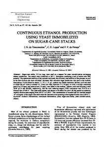
es
-*"
gram of
Sequence autoradioMl3mpl8 with a pBR32S
~~~segment
inserted at the HindIll
FIG.
3.
site. The left four lanes show the
produced by the difluoresprimer; the right four lanes were produced by using unfluoresceinated primer. The arrow indicates the region of the HindIll pattern
ceinated
site (AAGCTT). The difluorescemated
bands than
C
G
primer produced discrete more
that
migrated
slowly
unfluoresceinated primer due
to the molecular weight of the T
A A
T
C
fluoresceins.
Genetics:
Brumbaugh et al.
Proc. Natl. Acad. Sci. USA 85 (1988)
tions). Background fluorescence at 585 nm due to the Raman radiation of water in the gel was a significant part of the collected signal that was filtered out. The lower limits of detection were determined by directly loading and electrophoresing a dilution series of difluoresceinated primer (54, 27, 5.4, 2.7, 0.54, 0.27, 0.054, and 0.027 fmol). The lowest detectable band was 0.054 fmol (-3 X 10⁷ molecules), with the 0.54-fmol band intensity most like that of an "average" band. Imaging. Thirty to 35 screens were filled per run. The images were designed to resemble standard autoradiograms. Klenow polymerase produces certain band patterns that are apparent by autoradiographic and fluorescent sequencing. For example, a double cytosine has a light first band compared to the second band. Thus, the guidelines for interpreting the results are the same as those used in autoradiography (25). There is, however, a significant difference between the fluorescent display and autoradiograms, since band separations with the fluorescent method are in the time domain rather than the spatial domain. As the DNA fragments increase in length, they pass the detector more slowly causing the bands to become increasingly wider, but the band center-to-band center distances remain relatively constant. This is unlike autoradiograms, where large fragment bands become sharper but closer together. Well morphology becomes important as fragment length increases because small aberrations become accentuated in the time domain, particularly with the slower fragments. Data Analysis. The semiautomated software programs described in Materials and Methods were used to analyze the data. As each screen is displayed from memory, the cursor is used to write the sequence directly to the disk. Fig. 4 shows a picture of a screen with cursor bars displayed on the right center sample of the 16-lane, four-sample format. A programable "jump" is employed to advance the cursor to the next band. An optional software routine for band alignment was used for screen-by-screen analysis. This alignment program was used when well aberrations caused band distortions. Humpbacked or U-shaped bands could be straightened by this program so that their appropriate position between other bands could be accurately determined. This program was used 40% of the time. Sequencing the video data took about 30 min for 500-600 bases.
A
I
( X
A T
G
C
A
T G
C A
T
G
C
FIG. 4. A typical screen of four samples (16 lanes) at 130 bases. The right center sample has cursor lines on the bands that have been written to the computer disk (6% acrylamide/8 M urea, 500C, 20 W).
5613
DISCUSSION The primary reason for scaling up the amount of DNA produced and reacted was to eliminate the necessity of repeating isolations and reactions. Clearly less DNA can be used since three to six runs can be made per reaction. The lower limits have not yet been determined but would appear by calculation to be in the 1-,pg range (see Results). Use of the 7-deazaguanine analog eliminated most compressions, although it was still a problem with poly(CG) linkers (26, 27). Smith et al. (13) found that primers with attached dyes migrated at different rates than those that did not have attached dyes; in this respect their results were similar to ours. Smith et al. (16) also used four different dyes and varied the linkers to compensate for differing mobility effects (28), but the migration of the four primers still varied approximately one-fourth of a base due to the effects of the different fluorophores. The four-dye single-lane format (16, 28) can be compared with our single-dye four-lane format. The four-dye single-lane format requires four independent hybridizations followed by four independent elongation reactions. The single-dye four-lane format requires just a single hybridization followed by four independent reactions. Prober et al. (17) use fluorescently tagged dideoxy analogs and do reactions in one tube. Ansorge et al. (14, 15), Smith et al. (16), Prober et al. (17), and Connell et al. (28) have only single reporter groups per DNA fragment. This limits the amount of signal. The difluoresceinated primer increases the amount of signal, which increases sensitivity. If needed, additional fluoresceins could be added to further increase sensitivity; we have tested up to three fluoresceins per primer but did not need the added sensitivity. Ansorge and Barker (29) demonstrated that for longer fragments 4% acrylamide gave better resolution than 6%. If 4% or 5% gels can be made with good well morphology, the number of readable bases in a single load may be extended even further. A comparison of the various detection systems is interesting. Chopping the laser signal to discriminate between background and sample fluorescence as described here is unique. Ansorge et al. (14, 15), Prober et al. (17), and our method filter out the Raman shift. The limits of detection determined by Smith et al. (16) were 0.1-1 fmol. Our method detects