Journal of
Mechanics of Materials and Structures
CONTINUUM-BASED COMPUTATIONAL MODELS FOR CELL AND NUCLEAR MECHANICS Ashkan Vaziri, Arvind Gopinath and Vikram S. Deshpande
Volume 2, Nº 6
June 2007 mathematical sciences publishers
JOURNAL OF MECHANICS OF MATERIALS AND STRUCTURES Vol. 2, No. 6, 2007
CONTINUUM-BASED COMPUTATIONAL MODELS FOR CELL AND NUCLEAR MECHANICS A SHKAN VAZIRI , A RVIND G OPINATH
AND
V IKRAM S. D ESHPANDE
Deciphering the relationship between cellular processes and the structure of living cells is a key step toward understanding and predicting cell functions with direct implications for understanding human health and disease. The active nature of these cellular processes, which span several decades of spatial and temporal scales, pose significant challenges to unraveling this complex structure-function paradigm. Complementing novel experimental techniques with robust computational approaches capable of modeling mechanical response at varying scales provides new avenues to resolving this paradigm. We provide an overview of continuum-based computational approaches used in studying and interpreting responses of individual cells and nuclei, we outline techniques used for measuring the mechanical characteristics of living cells, and we discuss some of the key insights provided by these approaches.
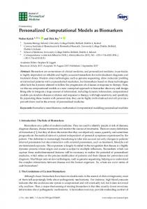
1. Introduction Recent developments in the field of nanotechnology have delivered sophisticated experimental tools for measuring forces as small as piconewtons and deformations as small as microns or less. These developments have enabled accurate measurement and monitoring of the mechanical properties and function of living cells, subcellular structures and even single biomolecules [Bao and Suresh 2003; Van Vilet et al. 2003; Huang et al. 2004; Yu et al. 2006]. Specifically, work done over the last two decades at cellular scales has led to the formulation of experimental frameworks for measuring the mechanical response of cells in vitro. Quantifying the mechanical characteristics of subcellular structures based on these frameworks, a key step towards understanding the structure-function paradigm of living cells, is a fundamental challenge facing the biomechanics, biomaterials, and biophysics communities. This challenge arises due to the intricate nature of the cell, a delicate assembly of numerous subcellular components with vastly different geometrical, material, and biochemical characteristics; see, for example, Figure 1. These subcellular components evolve during various cell functions at temporal scales that extend over several decades. Figure 2a illustrates the mapping between length scales and time scales via selected functionality of living cells and various organelles. While most short-term cellular responses are locally mediated, long-term events generally involve alterations in gene expression [Kamm and KaazempurMofrad 2004]. Even what is seemingly a single cell function can be governed by completely different mechanisms at different time scales, as illustrated recently for cell adhesion to substrates [Curvelier et al. 2007]. Keywords: cell mechanics, nuclear mechanics, computational mechanics. This work has been supported by the Division of Engineering and Applied Sciences, Harvard University. 1169
1170
ASHKAN VAZIRI, ARVIND GOPINATH
A
microtubule
AND
VIKRAM S. DESHPANDE
B
endoplasmic reticulum
outer membrane nuclear lamina
cell membrane nucleus nuclear envelope nuclear envelope
cytoplasm
heterochromatin nuclear matrix network
nucleolus
cytoplasmic network
golgi apparatus mitochondrion focal adhesions
(a)
(b)
Figure 1. Schematics of (a) an eukaryotic cell and (b) its nucleus.
In the process of modeling the deformation and mechanics of cells, subcellular structures, and constituent biomolecules, the method of choice depends on the length and time scales of interest. Computational approaches can be classified into two main categories: continuum-based approaches and micro/nanostructural approaches [Vaziri and Gopinath 2007]. Integrated models that combine aspects of these two approaches and are useful in predicting the response of living cells at physiologically relevant temporal and spatial scales are still in their infancy. This article reviews continuum-based computational models in cell and nuclear mechanics. Our aim is to provide an overview of the current state of the art in such approaches and comment on their applicability in interpreting as well as predicting the deformation and mechanics of living cells and organelles. The advantages and deficiencies of the more commonly used models are emphasized. Insights provided by these approaches, specifically in understanding the connection to human health and disease, are discussed. This article complements a recent review by Vaziri and Gopinath [2007] that focuses on the insights provided by micro/nanostructural and continuum computational approaches in cell and biomolecular mechanics. We will highlight computational studies on both whole cell response as well as studies on isolated nuclei, which have been the subject of much research in recent years. Continuum methods are applicable when the length scales of interest are much larger than microscopically relevant structural features that act as small perturbations to global variations in properties. To illustrate this, consider, for instance, steady AFM indentation of the individual cells at micron level in which the length scale characterizing the deformation field is typically larger than the spatial dimensions of individual cellular elements. Consequently, a continuum description is appropriate when studying the nature of this deformation field. The same picture holds for studies on cell migration and flow and deformation of red blood cells in capillaries. Even though microscopic features affect the cell shape and behavior at small length scales, the overall response of a blood cell to flow or stress as it moves in and out of capillaries is amenable to a macroscopic analysis. On the other hand, when studying the
A
Time
days Diffusion time of Cell migration CONTINUUM-BASED COMPUTATIONAL protein across cellMODELS FOR CELL AND NUCLEAR MECHANICS Cell differentiation Gene A Cell proliferation Binding of protein expression to chromatin hour days Diffusion time of protein across cell
Nuclear envelope Bindingbreakdown of protein
sec
Time
Gene expression
10
Cell migration Cell differentiation Cell proliferation
to chromatin
hour
Transition betwen protein Nuclear envelope states (active/inactive) breakdown + + Na / K Transition transport betwen protein
sec
msec
1171
-9msec
10 10
-9
states (active/inactive) -7 + + -6 Na / K transport 10 10
-8 10
-8
Length, m -7 -6
10
10
Electric signal transfer through a cell
10
-5
10
-5
Electric signal transfer through a cell
10
-4
10
10
-4
10
0
0
Length, m Biomolecules Cytoplasmic filaments
Cell and Nucleus
Biomolecules Cytoplasmic filaments
B
Cell and Nucleus
Tissues Tissues
(a) B CONTINUUM-BASED MODELS FOR CELL AND NUCLEAR MECHANICS
CONTINUUM-BASED MODELS FOR CELL AND NUCLEAR MECHANICS
Elastic continua Viscoelastic Elastic continua Viscoelastic continuacontinua • Linear models
Biphasic continua Biphasic continua
• Maxwell and Modified Maxwell models
• Maxwell and Modified Maxwell models • Linear models • Power-law structural damping models • Non-linear models • Power-law structural damping models • Non-linear models
• Poroelastic models
• Poroelastic models • Poro-viscoelastic models • Poro-viscoelastic models
Active Activecontinua continua • Bio-Chemo-Mechanical models
• Bio-Chemo-Mechanical models
• Active poroelastic gels
• Active poroelastic gels
(b) Figure 2. (a) Mapping between length and time scales in cell and biomolecular mechanics via selected functions. Shown is the vast difference in length scales from biomolecules to tissues as well as concomitant time scales for some processes and function at biomolecular/cellular level. (b) Computational toolbox encapsulating current continuum-based computational approaches for living cells and isolated nuclei.
mechanics of individual biomolecules, as in protein folding and fracture, the length scales of interest, for example, spatial scales characterizing the deformation field, are comparable to the molecular size. In this case, continuum-based models are inappropriate, and atomistic and molecular simulations should be employed. Most of the atomistic and molecular simulations in biomechanics are based on Monte Carlo or Molecular Dynamics algorithms reviewed in a recent article by Vaziri and Gopinath [2007]. Most continuum-based computational models in cell mechanics, and more generally in biomechanics, are based on the finite element method. Significant reasons for its popularity are its efficiency, ease of computation, and speed of implementation. In addition, material as well as geometric nonlinearities can be easily incorporated. Numerical schemes associated with this method are also well-developed and have been implemented in various commercially available software. It must be pointed out, however, that alternative computational approaches have also been used when appropriate for particular applications. For instance, the boundary element method, which is also based on fundamental solutions of the governing partial differential equations and requires discretization on only the domain boundary, has the
1172
ASHKAN VAZIRI, ARVIND GOPINATH
AND
VIKRAM S. DESHPANDE
potential to reduce significantly the computation time specifically for complex geometries. Such alternate computational approaches offer advantages in certain specific problems in cell mechanics [Discher et al. 1998; Haidar and Guilak 2002; Cristini and Kassab 2005], but the finite element method seems to be the algorithm of choice in most applications. The perspectives and overview presented here are necessarily brief and are intended to convey only broad concepts. The readers are encouraged to consult the papers cited in this review for details on the topics addressed here including the list of key references. 2. Cell and nuclear mechanics: insight from computational approaches Advances and development of new techniques in numerical methods over the last decade have provided reliable tools to model materials with complex characteristics. A key challenge in developing appropriate computational models in cell mechanics is the selection of material laws capable of representing the multiaxial and highly nonlinear stress-strain relationships as well as their alteration due to mechanical, biochemical, or electrical stimuli with high fidelity. Figure 2b displays material laws ranging from simple linear elasticity to more complex biphasic models and active continua that have been employed in various computational models in cell and nuclear mechanics. Most of these models, with the exception of those in the active continua category, assume that the cell and its subcellular structures respond passively to the stimulus. The material constants associated with these models are usually obtained by measuring the overall response of a cell (or isolated nuclei) using experimental techniques such as micropipette aspiration and atomic force microscopy (AFM) indentation experiments, and comparing these experimental results with computational predictions. Table 1 shows the available computational models developed for common experimental techniques that are typically performed on whole cells. A companion list of the computational methodologies developed for modeling the deformation and response of an isolated nucleus is provided in Table 2. An illustration of the method by which material constants are obtained is shown in Figure 3 for microplate compression tests on a round endothelial cell and an isolated nucleus extracted from cell cultures by chemical treatment [Caille et al. 2002]. The corresponding computational simulations at each stage of deformation are also shown and performed by assuming that the cell and the nucleus are separate homogeneous isotropic materials and calculating the response to compression. Interpreting the experimental measurements using simulations shows that the nucleus of an endothelial cell is about 10 times stiffer than its cytoplasm (Figures 3c and 3d). Characteristic properties of cells have been experimentally obtained by considering responses over a limited range of excitation frequencies. Recent studies have indicated that the cytoskeleton response is governed by a ubiquitous mechanical behavior called power law rheology over a broad frequency spectrum [Fabry et al. 2001; 2003; Alcaraz et al. 2003; Lenormand et al. 2004; Desprat et al. 2005; Bursac et al. 2005; Hoffman et al. 2006]. Investigations of the rheology of actin gels containing a single actin crosslinking protein indicate that that the power law behavior might be an intrinsic feature of actin systems with only one or two binding proteins present [Gardel et al. 2006]. The emergence of this relatively simple power law behavior for complex structures such as the cytoskeleton and actin gels has motivated both theoretical and computational efforts to interpret these experimental observations. In Section 2.1, we provide an overview of such a computational model developed by Vaziri et al. [2007].
CONTINUUM-BASED COMPUTATIONAL MODELS FOR CELL AND NUCLEAR MECHANICS
Experiment AFM Indentation
Material model
References
Linear elastic
[Costa and Yin 1999] [Ohashi et al. 2002; Ng et al. 2007]
Nonlinear elastic
[Costa and Yin 1999]
Nonlinear elastic
[McElfresh et al. 2002]
Linear elastic
[Shin and Athanasiou 1999]
Poroelastic
[Shin and Athanasiou 1999]
Cytoindentation
Nonlinear elastic
[Mijailovich et al. 2002] [Charras and Horton 2002] [Ohayon et al. 2005]
Maxwell viscoelastic
[Karcher et al. 2003]
Power law structural damping
[Vaziri and Gopinath 2007]
Linear elastic
[Charras and Horton 2002] § [Cao et al. 2007; Ferko et al. 2007] §
Nonlinear elastic
[Jadhav et al. 2005]
Linear elastic
[Nelson et al. 2005]
Modified Maxwell viscoelastic
[McGarry et al. 2005] §
Biochemomechanical
[Deshpande et al. 2006; 2007a; 2007b] [Wei et al. 2007]
Nonlinear elastic
[Caille et al. 2002]
Linear elastic Nonlinear elastic Maxwell viscoelastic Modified Maxwell viscoelastic Poroelastic Poroviscoelastic
[Haidar and Guilak 2002] [Zhou et al. 2005; Baaijens et al. 2005] [Baaijens et al. 2005; Trickey et al. 2006] [Haidar and Guilak 2000] [Baaijens et al. 2005] [Baaijens et al. 2005; Trickey et al. 2006]
Nonlinear elastic
[Dao et al. 2003; Mills et al. 2004] [Suresh et al. 2005]
Modified Maxwell viscoelastic
[Mills et al. 2004]
Linear elastic Magnetic Twisting Cytometry
Shear Flow
Cell Contraction (Microarrays/substrate)
Microplate Compression
Micropipette Aspiration
Optical Tweezers
Table 1. Computational models for whole cell deformation in common experimental techniques in cell mechanics. Most of the current models are based on the finite element method. Alternate approaches include studies based on boundary integral methods as in [Haidar and Guilak 2000; 2002]. (§ The model of the cell in [Caille et al. 2002; McGarry et al. 2005; Ferko et al. 2007] includes a separate component representing the nucleus.)
1173
1174
ASHKAN VAZIRI, ARVIND GOPINATH
Experiment
AND
VIKRAM S. DESHPANDE
Material model
Reference
Maxwell viscoelastic
[Vaziri et al. 2006]
Nonlinear elastic
[Caille et al. 2002]
Maxwell viscoelastic
[Vaziri et al. 2006]
Modified Maxwell viscoelastic
[Vaziri et al. 2007]
AFM Indentation
Microplate Compression
Micropipette Aspiration
Table 2. Available computational models for the deformation of an isolated nucleus.
Most material models presented in Figure 2b assume a passive nature for the response of cells and subcellular components to mechanical stimuli. In a living cell, however, the cytoskeletal structure undergoes active changes in response to mechanical stimuli and chemical signals [Wang et al. 2000; Bao and Suresh 2003; Kaunas et al. 2005; Chien 2006; Kumar et al. 2006]. Recent efforts by several groups have led to the development of biochemomechanical models for cells in an effort to capture this active mechanical response [Deshpande et al. 2006; 2007a; Deshpande et al. 2007b; Pathak et al. 2007; Yang and Saif 2007]. These new approaches can help immensely in understanding the interplay of mechanics and biochemistry during the active cellular response and cell function as explained in Section 2.2. Recent theoretical efforts have also focused on developing a general framework for describing the largescale dynamics of an active-filament network as is found in the cytoskeleton [Kruse and Julicher 2006]. Using pairwise interactions between active filaments and general conservation principles, effective medium expressions are developed for the stresses generated in such a filamentous structure. This description may be linked to continuum theories of active polar gels and provides a useful starting point for the numerical modeling of such systems. An additional property of the deformation response to mechanical forces, present even in the passive limit, is the inherent inhomogeneity in cell structure. Computational models that treat the cell and its nucleus as a homogeneous isotropic material have proven successful in modeling certain aspects of the cell behavior. This approximate treatment however fails to predict some major physiological observations such as transmission of the forces applied via focal adhesions to long distances in the cytoskeleton [Maniotis et al. 1997; Hu et al. 2003]. Developing computational biomechanical models which account for inhomogeneity of the cell and nuclear structure constitutes another important step towards understanding the behavior and functions of living cells and their relationship with mechanics. In Section 2.3, we
F (n
200 100
5µ m 0 0
10
20
30
Height reductio
A
C
CONTINUUM-BASED COMPUTATIONAL MODELS FOR CELL AND NUCLEAR MECHANICS
D 1175
C
1500
B500
500 400
isolated nucleus
15 µ m 400 300
F (nN)
15 µ m
round cell
200 100
500
5µ m 100
0
10
0
B C 500
200
5µ m 1005 µ m
round cell
30
40
30
40
Height reduction (%)
(c)
50
5 kPa
isolated nucleus 1000 model isolated nucleus 1000 model
5 kPa
500 500
1 kPa
0 0
0.5 kPa 1 kPa 0.5 kPa0.1 kPa 0.1 kPa 50 75
25 25
25
50
Height reductio
10 kPa
0 20
0
50
10 kPa
0
10
50
(b) (%) Height reduction
0 0
40
F (nN)
F (nN)
F (nN)
20
1500
isolated nucleus
300
D
30
Height reduction (%)
1500
D
15 µ m 400 15 µ m 15 µ m
10
D
B
20
0
0
(a)
round cell
5µ m
0
5µ m
isolated nucleus model
1000
300 200
F (nN)
15 µ m
isolated nucleus
F (nN)
A
50
75
Height reduction (%)
Height reduction (%)
(d) 10 kPa
F (nN)
1500 Figure 3. (a) Top panels show digitized images of a round endothelial cell compressed by flexible microplates. Left: cell at rest; middle: 30% compression of cell height; 5 kPa Bottom panels show the finite element model right: 50%isolated compression nucleus of cell height. 1000 model of the compression test at same stages; the nucleus within the cell is distinguished by the bold line and was modeled as a separate element with different mechanical proper500 ties. (b) Same as (a) for an isolated nucleus extracted from endothelial cell by chemical 1 kPa treatment. (c) Average force-height reduction of round endothelial cells (n = 17) and 0.5 kPa 0.1 kPa isolated0 nuclei (n = 21) measured experimentally. (d) Numerical simulation of force0 25 50 75 height reduction of an isolated nucleus for various values of the nuclear elastic modulus. Height reduction (%) Fitting to experimental data indicates that the nucleus is about 10 times stiffer than the cytoplasm in endothelial cells. In numerical simulations both cytoplasm and nucleus are modeled as homogeneous, incompressible, isotropic, hyperelastic materials. (Figures and data are modified from [Caille et al. 2002]. Copyright © 2002, Elsevier Science. Reprinted with permission.)
discuss the details of a new computational model proposed for studying the deformation and mechanics of isolated nuclei. Complete characterization of the biomechanics of the cell and its subcellular structures cannot be achieved by a single experiment and requires combinations of several experimental techniques and detailed computational simulations. For example, Baaijens et al. [2005] show that computational models using a viscoelastic material model or a biphasic viscoelastic material model are capable of accurately
1176
ASHKAN VAZIRI, ARVIND GOPINATH
AND
VIKRAM S. DESHPANDE
simulating the experimentally measured creep response of chondrocytes aspired into a micropipette. A similar discussion is provided by Vaziri and Kaazempur-Mofrad [2007] for the deformation of an isolated nucleus in micropipette aspiration experiment. 2.1. Power law rheology based models. The power law rheology exhibited by emulsions, pastes, foams, and colloids is an intrinsic feature of materials composed of numerous discrete elements that experience weak interactions with inherently disordered and metastable microstructural geometry. Surprisingly, recent experiments have indicated that under certain conditions this rheological law is also valid for the cytoplasm as described in Section 2. A computational model of the magnetic twisting cytometry experiment has been recently developed in which the material law associated with this power law rheology is incorporated. In Figure 4, results from the computational simulations based on this coarse-grained model are compared with experimental observations on HASM cells from Fabry et al. [2003]. Interpreting these observations using the computational model yields an accurate assessment of the material constants associated with the power law rheology. It should be noted that this computational model is phenomenological, and fails to provide any physical insight into the mechanisms responsible for the emergence of this peculiar behavior of the cytoskeleton. When the simple law described above proves inadequate, the framework may be extended to incorporate more complex frequency dependent viscoelastic behavior. As an example, the intranuclear region of Swiss 3T3 fibroblasts is shown to have a frequency dependent viscoelastic characteristic that in general does not follow the power law rheology over a wider range of excitation frequencies [Tseng et al. 2004]. Another example is the additional Newtonian viscosity term suggested by Fabry et al. [2003] to account for the complex behavior of the cytoplasm at relatively high frequencies of excitation. Alternatively, mechanical prestress in the active cytoskeleton, generated in part due to the activity of molecular motors, also significantly influences its rheology and behavior of the cell [Stamenovic et al. 2002; Wang and Suo 2005]. This prestress regulates the transition between solid-like and fluid-like behavior in cells, and is indicated in the power law rheology as a decrease in the power law exponent with increasing prestress [Stamenovic et al. 2002; Stamenovic et al. 2004]. The developed computational model is capable of incorporating the mechanical prestressing and can provide insight into the role of this mechanical factor. 2.2. Active continua computational models. The cytoskeleton is a protein scaffold found inside most eukaryotic cells. It consists of three major classes of filaments: actin, microtubules, and intermediate filaments along with accessory proteins that crosslink the filaments to the plasma membrane. In mammalian cells with compliant cell membranes (no cell walls), the dynamic structure and active nature of the cytoskeleton largely determine the morphology of a cell and its mechanics [Howard 2001]. Here we briefly review the main features of computational approaches that attempt to account for the active nature of the cytoskeleton and its effect on the mechano-sensitivity of cell adhesion. 2.2.1. Cytoskeletal contractility. The tension generated inside the contractile microfilaments in the cytoskeleton plays a critical role in control of cell morphology and function [Ingber 1997]. Contractile bundles composed of actin and myosin proteins form and dissociate in response to external mechanical or chemical stimuli. Typically, these contractile bundles are found in three forms: (i) circumferential belts (in epithelial cells), (ii) stress fibers along the ventral surfaces, and (iii) contractile rings at the equator that form during cytokinesis. A suitable model must incorporate the following basic biomechanochemical
CONTINUUM-BASED COMPUTATIONAL MODELS FOR CELL AND NUCLEAR MECHANICS
1177
Figure 4. Power law rheology of the cytoskeleton. (a) Scanning electron microscopy of a magnetic microbead bound to surface of a human airway smooth muscle cell (HASMc) in magnetic twisting cytometry (MTC) experiments (see Table 1 for the schematic of the experiment). (b) Ferromagnetic beads bind to the actin cytoskeleton (stained with fluorescently labeled phalloidin) of HASM cells via cell adhesion molecules (integrins). (c) Microbead displacement versus specific torque (mechanical torque/bead volume) of the cytoskeleton at different frequencies of excitation of the microbead in MTCE for HASMc. (d) Geometry of computational model of MTC experiment. (e) Numerical results for response in MTC experiment at various excitation frequencies of rigid microbead. Both average radius of ferromagnetic microbeads in experiment and of rigid spheres in numerical simulations is 2.25 µm. See Table 1 for alternative computational models developed for understanding cytoskeletal response in MTC experiment. (A, B and C are taken from [Fabry et al. 2003]. Copyright © 2003, American Physical Society. Reprinted with permission. D and E are modified from [Vaziri et al. 2007]. Copyright © 2007, Elsevier Science (D and E). Reprinted with permission.) processes [Deshpande et al. 2006; 2007a]: (i) the formation of stress fibers induced by an activation signal, (ii) polymerization of actin filaments and the phosphorylation of myosin II that promotes the assembly of myosin II into bipolar filaments triggered by the activation signals. These myosin filaments enter into the α actinin bound actin filament bundles, resulting in the formation of stress fibers, and (iii) tension generated in stress fibers by crossbridge cycling between the actin and myosin filaments. When the tension is allowed to relax, the actin filaments are no longer held in place by the bipolar myosin filaments and the stress fibers disassemble. Motivated by these processes, Deshpande et al. [2006; 2007a] have proposed a model for the contractility of the cytoskeleton that supersedes oversimplified models in which the cytoskeleton is regarded as a passive mechanical structure augmented only by a thermal contraction induced by a deus ex machina
1178
ASHKAN VAZIRI, ARVIND GOPINATH
A
CELSSL
A A
AND
VIKRAM S. DESHPANDE
B B k =3.88 k =3.88k =3.88
B
CELSSL CELSSL
CELL CELL
CELL
k CELL
kk
(b)
CELL CELL
k =0.2 k =0.2 k =0.2
(a)
η 0
0.1
η η
0 0.1 0.2 00.2 0.10.3 0.2
0.3 σp 0.3
σp 0.25σ p
0.25 0.25
(c)
Figure 5. (a) Schematic of the cell on 4 posts. Effect of reducing stiffness of top right support (denoted by k) is illustrated in (b) and (c), wherein contours of stress fiber concentration η¯ are plotted along with line segments indicating direction of maximum principal stress. (The figures are modified from [Deshpande et al. 2006]. Copyright © 2006, National Academy of Sciences, USA.) [Satcher and Dewey 1996; Nelson et al. 2005; Mohrdieck et al. 2005]. In the model, the mechanical response of the stress fibers comprises three coupled phenomena: (i) an activation signal that triggers the formation of stress fibers; (ii) a fiber formation rate dependent on the activation signal, coupled with a dissociation rate dependent on the tension; and (iii) a contraction rate (contractility) for the stress fiber that depends on the tension through the crossbridge dynamics. Simple phenomenological relations are used to model these coupled phenomena for a single stress fiber, and thereafter the relations are generalized to two and three dimensional cytoskeletal networks by homogenizing over all possible fiber orientations at each point in the cell. Simulation of the cell response is carried out by solving the equations of force equilibrium as the cytoskeleton forms to create a contractile network consistent with the tensegrity hypothesis [Ingber 1997]. By considering a square cell fixed between four support posts (see Figure 5a), the model predicts reconstruction of the cytoskeleton in a manner dependent on the external mechanical constraints. Predictions of the distributions of the stress fibers (Figures 5b and 5c) are in line with experimental findings that the stress fiber network is dependent on the spring constants of the supports with a higher concentration of fibers located adjacent to stiffer supports. Moreover, the model captures the many key features observed in experimental studies including: (a) the decrease of the forces generated by the cell with increasing substrate compliance, and (b) the influence of cell shape and boundary conditions on the development of structural anisotropy. It is apparent from these studies that
A CONTINUUM-BASED COMPUTATIONAL MODELS FOR CELL AND NUCLEAR MECHANICS A prediction prediction
1179
increasing increasing vinclulin vinclulindensity density
experiment experiment
experiment experiment
(a)
prediction prediction
increasing increasing actin actindensity density
B B
(b) Figure 6. Observations (left) and predictions (right) of the vinculin (a) and actin (b) distributions in infinity telomerase-immortalized retinal pigment epithelial human cells placed on a V-shaped fibronectin pattern. The edge of the V is 46 µm. Observations are reproduced from [Théry et al. 2006], while the predictions are from [Pathak et al. 2007]. mechanics and the conditions of force equilibrium play a central role in the processes of cytoskeleton formation and contractility. 2.2.2. Cooperativity between cell adhesion and cytoskeletal contractility. Cell adhesion to the extracellular matrix (ECM) is critical to functions ranging from migration and proliferation to apoptosis. The primary subcellular structures that mediate the regulatory effects of ECM adhesion are the focal adhesions (FAs). These macromolecular complexes not only mediate cell anchorage by physically coupling integrins to the contractile actin cytoskeleton but also orient and concentrate numerous signaling proteins at sites of integrin binding and clustering. Because FAs function not only as the mechanical linkages but also as biochemical signaling hubs for many regulatory pathways, regulation of their assembly and disassembly is a critical mechanism for controlling cell function. FA formation is itself controlled mechanically with the tractions generated at the contact sites increasing the rate of assembly of the focal adhesions [Balaban et al. 2001]. Moreover, FA formation can be increased or decreased by respectively raising or lowering cytoskeletal tension using pharmacological agents such as BDM that curtail actomyosin activity [Chen et al. 2003]. The global shape of cells and their internal cytoskeletal structure also play an important role in constraining the magnitude, distribution, and direction of such tractions across the cell surface [Parker et al. 2002; Chen et al. 2003], and thereby control local FA assembly
1180
ASHKAN VAZIRI, ARVIND GOPINATH
AND
VIKRAM S. DESHPANDE
and organization. The fact that many proteins perform both structural and signaling functions [Burridge and Chrzanowska-Wodnicka 1996] hinders the ability of traditional approaches to parse the mechanisms that regulate FA dynamics: for example, knocking out an adhesion protein by genetic manipulation may also eliminate an essential structural component. Consequently, a combination of critical experiments guided by computational models that include the salient features of the mechanics and biochemistry is needed in order to understand the observed behaviors. Th´ery et al. [2006] have investigated the effect of cell shape on the distribution of actin and vinculin at fixed cell area, as exemplified in the upper part of Figure 6, which visualizes a human endothelial cell placed on a V-shaped fibronectin ligand pattern. Such studies indicate a complex interaction among cell shape, ligand patch geometry, and cytoskeletal response with strong stress fibers forming along the nonadhesive edges and high focal adhesion densities (as exemplified by the vinculin density) along the edges of the ligand patch. Deshpande et al. [2007b] have developed a model that accounts for the cooperativity between cytoskeletal contractility and focal adhesion formation and growth. This thermodynamically-motivated computational approach has three essential features: (i) coexistence of the low and high affinity integrins in thermodynamic equilibrium, (ii) mobility of the low affinity integrins within the plasma membrane, and (iii) mechanical equilibrium of the contractile forces generated by the stress fibers; these forces affect the free energies of the integrins giving rise to a coupled thermomechanical response. An initial prediction based on the model of Deshpande et al. [2007b] is included in bottom part of Figure 6. The predictions show distributions of the normalized focal adhesion densities (as parameterized by the high affinity integrin concentration) and contours of the stress fiber density. Also included with the predicted stress fiber concentration distributions are line segments showing the effective direction of the stress fiber. The predictions are remarkably consistent with trends observed in the experiment: (i) since the cell exerts maximum tractions on the ECM near the edges of the ligand patch, high focal adhesion densities are predicted in this region, and (ii) aligned stress fibers form along the longest nonadhesive edge; see [Pathak et al. 2007] for details and interpretation of the analysis. 2.3. Computational model of an isolated nucleus. The nucleus of eukaryotic cells, containing most of the cell’s genetic material, is an assembly of numerous subnuclear components which work cooperatively to maintain the structural integrity and proper function of the nucleus, as shown in Figure 1b. The nuclear envelope is one of the major elements of the nucleus, and is comprised of inner and outer membranes, nuclear lamina, nuclear pore complexes, and the perinuclear region. The nuclear lamina is a polymeric network composed of coiled-coil proteins called lamins [Aebi et al. 1986; McKeon et al. 1986] and is known to play a critical role in maintaining the structural integrity of the nucleus. It is built of two types of proteins, lamin A/C and lamin B, and comprises the majority of the protein composition within the nucleus. Within the nucleoplasm are one or several nucleoli, responsible for the production of ribosomes that are then transported to the rough endoplasmic reticulum where protein synthesis occurs. Continuum-based computations have also been used in studying the mechanical properties of isolated and intact nuclei. These studies can further our understanding of the mechanisms by which mechanical stresses are transmitted to biochemical reactions and ultimately alterations in gene expression and cell function. A continuum-based computational model of an isolated nucleus that incorporates the nuclear inner and outer membranes, nuclear lamina and nucleoplasm as separate components with different material characteristics has been developed recently by Vaziri et al. [2006]; see Figures 7a and 7b. Using
CONTINUUM-BASED COMPUTATIONAL MODELS FOR B CELL AND NUCLEAR MECHANICS
A
1181
outer membrane, 7.5 nm inner membrane 7.5 nm
25 nm
nuclear lamina
nucleoplasm
rigid indenter
C 0.5 Experiment
Indentation force, nN
0.4
0.3
-19
K =13 10
nucleoplasm
N
-19
0.2
K =24 10 N
-19
6.0 µm
K =0.13 10
0.1
rigid substrate
N
axis of symmetry no nuclear envelope
0 0
200
400
600
800
1000
Indentation depth, nm
D Figure 7. From top left clockwise: (a) three-dimensional images of isolated nuclei on a glass substrate (black in the image) via two-photon microscopy; (b) schematic von Mises stress, Pa Displacement, nm 1000computational model of an isolated nucleus in 7.85 of the AFM indentation, consisting of 6.50 nucleoplasm, inner and outer membranes, and nuclear lamina, with different material 830 5.47 characteristics; and (c) dependence of force-indentation response of isolated nucleus on 660 bending stiffness of nuclear lamina, K N . Nuclear elements 4.44 are modeled as linear elastic materials with following characteristics: Poisson ratio υ = 0.485, nucleoplasm Young’s 500 3.41 modulus =25 Pa, and bending stiffness of nuclear membrane 1.8 × 10⁻¹⁹ Nm. Exper230 imental result gives the average response of 23 nuclei2.38 from mouse embryo fibroblasts. Error of indentation. (The figures are 160bars give the standard error at specific values 1.35 modified from [Vaziri et al. 2006]. Copyright © 2006, Materials Research Society.) 0
0.33
this approach, the contributions of individual nuclear elements to the overall response of an isolated nucleus can be calculated; this is shown for the case of AFM indentation in Figure 7c and discussed for the case of micropipette aspiration in [Vaziri and Kaazempur-Mofrad 2007]. Numerical simulation of the indentation of an isolated nucleus reveals that under localized indentation, the nuclear envelope transfers the force applied locally to a large portion of the nucleoplasm as the nuclear envelope undergoes
6.0 µm
I
K =0.13 10
0.1
substrate
N
axis of symmetry no nuclear envelope
0 0
200
400
600
800
1000
Indentation depth, nm 1182
ASHKAN VAZIRI, ARVIND GOPINATH
D
Displacement, nm 1000 830 660
AND
VIKRAM S. DESHPANDE
von Mises stress, Pa 7.85 6.50 5.47 4.44
500
3.41
230
2.38
160
1.35
0
0.33
Figure 8. Distribution of nodal displacement and von Mises stress in the nucleoplasm at probe displacement of 1000 nm from the isolated nucleus model in Figure 7 for nuclear lamina bending stiffness of K N L = 13.6×10⁻¹⁹ N.m. In computational simulations sharp probe has a half angle of 35◦ , consistent with experiments. (The figures are modified from [Vaziri et al. 2006]. Copyright © 2006, Materials Research Society.) bending and stretching; see Figure 8. The overall response of the nucleus to point indentation is predicted to be a complex function of the material characteristics of its individual elements. Similar theoretical and computational studies, carried out to study the mechanisms of response of living cells under point indentation, indicated the necessity of accounting for the mechanics of biomembrane in the response [McElfresh et al. 2002; Baesu et al. 2004]. These results clearly reveal the inadequacy of the commonly employed Hertzian contact model used in interpreting results from indentation experiments; see [Vaziri et al. 2006] for further discussion. Recent experimental observations indicate that the nuclei of endothelial cells undergo permanent structural remodeling when shear stress is applied to the cell. This cytoskeleton-mediated deformation of the nucleus is hypothesized as a possible pathway through which shear stresses are transduced to gene regulating signals [Deguchi et al. 2005]. The underlying mechanisms of this process and the details of the mechanical connection between the cell membrane and its nucleus are still far from being understood, but it is known that subcellular structures play an important role in this process. Computational models capable of distinguishing and incorporating the structural role of different subcellular structures and nuclear elements can help immensely in understanding these mechanisms. Moreover, these computational models are robust tools that can help in resolving the apparent discrepancy of the current data on mechanical properties of cells and subcellular structures [Vaziri et al. 2006; Vaziri and KaazempurMofrad 2007]. 3. Implications in understanding the state of human health and disease Initiation and progression of many diseases are associated with alterations in the structure and function of subcellular and subnuclear components which in turn modify the function of the cell [Cornhill and Roach 1974; Sinzinger et al. 1980; Shieh and Athanasiou 2002; Trickey et al. 2000; Suresh et al. 2005; Wilson 2005; Mattout et al. 2006; Suresh 2006; Cao et al. 2007]. For instance, mutations in gene
CONTINUUM-BASED COMPUTATIONAL MODELS FOR CELL AND NUCLEAR MECHANICS
1183
encoding lamin A and its binding partners have been associated with a variety of human diseases including Emery–Dreifuss muscular dystrophy, dilated cardiomyopathy, and Hutchinson–Gilford progeria syndrome [Lammerding et al. 2004; Wilson 2005; Mattout et al. 2006]. As another example, various cancer cells have altered nuclear structure and irregularity in shapes and spatial organization of subnuclear structures [Zink et al. 2004]. The exact role of mechanics in the progression of these diseases is still unclear and computational models can play a key role in unraveling this relationship. An example of such studies is the recent work on biomechanics of red blood cells infected by the parasite P. falciparum [Dao et al. 2003; Mills et al. 2004; Suresh et al. 2005]. This work, which combines novel experimental techniques and continuumbased computational models, has significantly enhanced our fundamental knowledge of the underlying mechanisms of disease progression at the cellular level and provides a template for extensions of this approach to other applications. Moreover, insights obtained from detailed computations that distinguish between the effects of subcellular structures can serve to guide further experiments for characterizing the mechanics of altered subcellular structures. For example, modeling the response of an isolated nucleus in micropipette aspiration and indentation experiments by employing the computational model of an isolated nucleus discussed in Section 2.3 indicated that the overall response of the nucleus in micropipette aspiration is highly sensitive to the stiffness of the nuclear envelope [Vaziri and Kaazempur-Mofrad 2007], while in the indentation experiment the overall response is more sensitive to the nucleoplasm material properties. This indicates that micropipette aspiration is robust enough to examine the influence of various alterations in the nuclear lamina such as mutations in gene encoding lamin A and its bindings. Another example in which mechanics might play a role involves the development of atherosclerotic lesions. These lesions develop in the vicinity of branch points in the arterial vasculature, while straight unbranched regions tend to be protected [Cornhill and Roach 1974; Sinzinger et al. 1980]. Namely, the tubular geometry of the straight segments ensures that the cyclical rise and fall of blood pressure results in uniaxial stretch. Conversely, the complex geometry of artery branches promotes stretching of the blood vessel along more than one axis. These differences result in endothelial cells having nonaligned stress fibers at branch points, while those in the straight segments are aligned in the longitudinal direction [Davies 1995]. Isotropic or random cytoskeletal arrangements give rise to continued activation of Jun Kinase (JNK) [Kaunas et al. 2006] that triggers a cascade of events that eventually induce structural changes at the branch points including the increased deposition of ECM collagen and elastin. These changes lead to the atherosclerotic narrowing and hardening of coronary arteries at vessel branches. Wei et al. [2007] have shown that the biochemomechanical model discussed in Section 2.2 accurately predicts the cytoskeletal structures that evolve in endothelial cells due to a range of hemodynamic and mechanical stimuli. It is suggested that this model could be employed to investigate the onset of atherosclerotic lesions. Applications are also seen in the field of viral studies and especially in studies on infectivity and its connection to structure. An excellent example is described in recent work by Kol et al. [2007]. This work deals with the internal morphological reorganization that HIV and some other retrovirus particles undergo following budding from a cell. During this maturation process, the immature HIV particle changes from being roughly spherically symmetric with a thick protein shell to a state characterized by a thin protein shell and a conical core. Nanoindentation experiments via AFM indicate that immature HIV particles are 14 times stiffer than mature particles and this difference is primarily due to the HIV
1184
ASHKAN VAZIRI, ARVIND GOPINATH
AND
VIKRAM S. DESHPANDE
envelope cytoplasmic tail domain. Finite element simulations used to elucidate the effects of deleting the cytoplasmic tail domain offer strong evidence that changes in mechanical properties, in this case the softening of the viral particle, might be a crucial step in the infection process. Such studies offer immense scope in using computations to anticipate effects of changes in structure arising due to mutations or as a course of natural morphological change during the progression of disease. Once an understanding of this mechanical change is gained, attempts to block or change this process may be used as a means to circumvent this intrinsic morphological switch and retard infection. Finally, the function of the cell and its nucleus is ultimately governed by the characteristics of appropriate biomolecules and their alteration by biochemical reactions. Therefore, understanding the mechanisms of the diseases associated with altered cell and nuclear function entails resolving the behavior at much smaller spatial scales. Computational approaches based on micro/nano approaches can help immensely in this regard. Examples are recent works on the effects of hyperglycemia on collagenolysis [Stultz and Edelman 2003; Stultz 2006]. 4. Multiscale approaches: from cell mechanics to collective tissue behavior The complexity of individual subcellular structures and the range of spatial and temporal scales associated with their interactions make it almost impossible to analyze these structures using a single theoretical or computational model. Complete characterization of the cell entails interconnecting several models each valid in a certain range of scales and forming a composite picture that reflects accurately mechanical and biochemical behavior at all scales. A major challenge in this process is to appropriately couple macroscale dynamics and cell kinetics to microscale deformation and mechanics; for example, protein mechanics, filament dynamics and active motor behavior. A significant complication arises due to the strongly nonlinear interactions as macroscopic mechanical effects provoke signal pathways that may give rise to both local effects and nonlocal cell-wide changes. In a living cell, the continuous consumption of fuel results in a constant state of nonequilibrium at all length scales. Even when considering properties and responses at macroscopic length scales it is imperative to take into account the cumulative effects of active microscopic processes. Multiscale approaches developed in the field of cell mechanics can be classified into two categories. In the first category, micro/nano simulations at subcellular, microscopic, or single molecule level are used as input into macroscopic computations. A specific example would be using network simulations of erythrocytes to obtain effective elastic constants of the membrane and the underlying filamentous structure [Li et al. 2005; Dao et al. 2006]. This is discussed further in [Vaziri and Gopinath 2007]. In this article, we provide a selected set of examples regarding a second complementary multiscale approach wherein results from continuum computations at the single cell or subcellular level are used as input to study behavior and response at the tissue or organ level, as illustrated in Figure 9. Multiscale computational approaches have been employed in understanding various aspects of the circulatory system from the mechanical properties of the heart muscle due to aging and plaque to cardiovascular circulatory mechanics [Shim et al. 2006]. We expect that continuum based computations will be of use in coupling the dynamical characteristics of the circulation to the activation-induced mechanical contraction of the heart muscle. Another physiologically relevant example is studying the process by which air exchange occurs in the lung [Fredberg and Kamm 2006]. Evidence strongly suggests that
CONTINUUM-BASED COMPUTATIONAL MODELS FOR CELL AND NUCLEAR MECHANICS
1185
Figure 9. Multiscale approaches in biomechanics connecting microscopic characteristics of biomolecules to collective tissue behavior. multiscale phenomena span the length scales that exist and govern integrative lung behavior. Another example of application of multiscale approaches is in understanding the underlying mechanisms of morphogenesis, a complex developmental phenomenon that involves cell growth, division, rearrangement, as well as flow in a well defined manner [Chaturvedi et al. 2005]. In tissue engineering, alginate hydrogels are increasingly used as biomaterials to develop new cell delivery methods as well as support matrices [Augst et al. 2006; Ling et al. 2006; Khademhosseini et al. 2006; Rivest et al. 2007]. It is expected that these biomaterials play the role of an artificial extracellular matrix that will surround living tissues and provide space for repair, regeneration, or growth of new tissue as well as offering mechanical support and traction. These composite biomaterials are living analogues of inanimate composites that have been so intensively studied until now in materials science and engineering. The possibility of drug delivery from such materials is also of great interest [Khademhosseini and Langer 2006]. In all these applications, an accurate mechanical characterization is essential, and multiscale continuum methods offer a good route to such understanding. Numerical investigations of such composite biomaterials employ solutions to basic unit cell geometries coupled with homogenization techniques to probe macroscopic variations. Typically, the cells are treated as deformable inclusions responding to the macroscopically imposed external stress. The stress results in a macroscopic deformation field, the magnitude of which has to be self-consistently determined by solving a microscopic unit cell problem. Computations can then be used to quantify effects of scaffold stiffness and inclusion interactions on cell deformation. Such multiscale studies are critical in evaluating the effects of mechanical processing on the viability of tissue constructs. For example, Guilak and Mow [2000] used a biphasic FEM model to study biomechanical interactions between the chondrocyte and the extracellular matrix. In a similar vein, Breuls et al. [2002] introduced a large strain model to study the local deformation fields in tissue composites when subject to macroscopic external loads. A similar approach is possible in the analysis of naturally occurring composites like bone tissues [Kowalczyk 2006]. 5. Concluding remarks Mechanical characteristics of biological materials, although far more complex than the behavior of manmade materials, are still amenable to studies based on computational frameworks. Indeed, the steps toward a coherent synthesis of computations and experiments, with an aim to understanding cell and
1186
ASHKAN VAZIRI, ARVIND GOPINATH
AND
VIKRAM S. DESHPANDE
subcellular mechanical characteristics, have already begun in earnest. Considering recent advances in computational efficiency and algorithms and the inclination of many researchers to enter interdisciplinary fields, fast progress in the utilization of computational approaches in the field of cell and nuclear mechanics is anticipated. This in turn can lead to the emergence of new applications in various fields such as tissue engineering, medicine, and nanobiotechnology, as well as novel avenues for treatment and diagnosis of diseases. Acknowledgements AV would like to thank Mohammad R. K. Mofrad, John W. Hutchinson, Roger D. Kamm, Bashir A. Tafti and Amir H. Sadrzadeh for many insightful discussions. References [Aebi et al. 1986] U. Aebi, J. Cohn, L. Buhle, and L. Gerace, “The nuclear lamina is a meshwork of intermediate-type filaments”, Nature 323 (1986), 560–564. [Alcaraz et al. 2003] J. Alcaraz, L. Buscemi, M. Grabulosa, X. Trepat, B. Fabry, R. Farré, and D. Navajas, “Microrheology of human lung epithelial cells measured by atomic force microscopy”, Biophys. J. 84 (2003), 2071–2079. [Augst et al. 2006] A. D. Augst, H. J. Kong, and D. J. Mooney, “Alginate hydrogels as bio-materials”, Macromol. Biosci. 6 (2006), 623–633. [Baaijens et al. 2005] F. P. Baaijens, W. R. Trickey, T. A. Laursen, and F. Guilak, “Large deformation finite element analysis of micropipette aspiration to determine the mechanical properties of the chondrocyte”, Ann. Biomed. Eng. 33 (2005), 494–501. [Baesu et al. 2004] E. Baesu, R. E. Rudd, J. Belak, and M. McElfresh, “Continuum modeling of cell membranes”, Internat. J. Non-Linear Mech. 39 (2004), 369–377. [Balaban et al. 2001] N. Q. Balaban, U. S. Schwarz, D. Riveline, P. Goichberg, G. Tzur, I. Sabanay, D. Mahalu, S. Safran, A. Bershadskay, L. Addadiand, and B. Geiger, “Force and focal adhesion assembly: a close relationship studied using elastic micropatterned substrates”, Nat. Mater. 3 (2001), 466–472. [Bao and Suresh 2003] G. Bao and S. Suresh, “Cell and molecular mechanics of biological materials”, Nat. Mater. 2 (2003), 715–725. [Breuls et al. 2002] R. G. M. Breuls, B. G. Sengers, C. W. J. Oomens, C. V. C. Bouten, and F. P. T. Baaijens, “Predicting local cell deformation in engineering tissue constructs, A multilevel finite element approach”, J. Biomech. Eng. 124 (2002), 177–187. [Burridge and Chrzanowska-Wodnicka 1996] K. Burridge and M. Chrzanowska-Wodnicka, “Focal adhesions, contractility and signaling”, Ann. Rev. Cell Dev. Bio. 12 (1996), 463–469. [Bursac et al. 2005] P. Bursac, G. Lenormand, B. Fabry, M. Oliver, D. Weitz, V. Viasnoff, J. Butler, and J. J. Fredberg, “Cytoskeletal remodeling and slow dynamics in the living cells”, Nat. Mater. 4 (2005), 557–561. [Caille et al. 2002] N. Caille, O. Thoumine, Y. Tardy, and J. J. Meister, “Contribution of the nucleus to the mechanical properties of endothelial cells”, J. Biomech. 35 (2002), 177–187. [Cao et al. 2007] Y. Cao, R. Bly, W. Moore, Z. Gao, A. M. Cuitino, and W. Soboyejo, “On the measurement of human osteosarcoma cell elastic modulus using shear assay experiment”, J. Mat. Sci.: Materials in Medicine 18 (2007), 103–109. [Charras and Horton 2002] G. T. Charras and M. A. Horton, “Determination of cellular strains by combined atomic force microscopy and finite element modeling”, Biophys. J. 83 (2002), 858–879. [Chaturvedi et al. 2005] R. Chaturvedi, C. Huang, B. Kazmierczak, T. Schneider, J. A. Izaguirre, T. Glimm, H. G. Hentschel, J. A. Glazier, S. A. Newman, and M. S. Alber, “On multiscale approaches to three dimensional modeling of morphogenesis”, J. Royal Society Interface 2 (2005), 237–253.
CONTINUUM-BASED COMPUTATIONAL MODELS FOR CELL AND NUCLEAR MECHANICS
1187
[Chen et al. 2003] C. S. Chen, J. L. Alonso, E. Ostuni, G. M. Whitesides, and D. E. Ingber, “Cell shape provides global control of focal adhesion assembly”, Biochem. Biophys. Res. Comm. 307 (2003), 355–361. [Chien 2006] S. Chien, “Molecular basis of rheological modulation of endothelial functions: Importance of stress direction”, Biorheology 43 (2006), 95–116. [Cornhill and Roach 1974] J. F. Cornhill and M. R. Roach, “Quantitative method for evaluation of atherosclerotic lesions”, Atherosclerosis 20 (1974), 131–136. [Costa and Yin 1999] K. D. Costa and F. C. Yin, “Analysis of indentation: implications for measuring mechanical properties with atmic force microscopy”, J. Biomech. 121 (1999), 462–471. [Cristini and Kassab 2005] V. Cristini and G. S. Kassab, “Computer modeling of red blood cell rheology in the microcirculation: A brief overview”, Ann. Biomed. Eng. 33 (2005), 1724–1727. [Curvelier et al. 2007] D. Curvelier, M. Théry, Y. S. Chu, S. Dufour, J. P. Thiéry, M. Bornens, P. Nassoy, and L. Mahadevan, “The universal dynamics of cell spreasing”, 2007. [Dao et al. 2003] M. Dao, J. Li, and S. Suresh, “Mechanics of the human red blood cell deformed by optical tweezers”, J. Mech. Phys. Solids 51 (2003), 2259–2280. [Dao et al. 2006] M. Dao, J. Li, and S. Suresh, “Molecularly based analysis of deformation of spectrin network and human erythrocyte”, Mater. Sci. Eng. C: Biomimetic Mater., Sens. Syst. 26 (2006), 1232–1244. [Davies 1995] P. F. Davies, “Flow-mediated endothelial mechanotransduction”, Phys. Rev. 75 (1995), 519–560. [Deguchi et al. 2005] S. Deguchi, K. Maeda, T. Ohashi, and M. Sato, “Flow-induced hardening of endothelial nucleus as an intracellular stress-bearing organelle”, J. Biomech. 38 (2005), 1751–1759. [Deshpande et al. 2006] V. S. Deshpande, R. M. McMeeking, and G. A. Evans, “A bio-chemo-mechanical model for cell contractility”, Proc. Nat. Acad. Sci. 103 (2006), 14015–14020. [Deshpande et al. 2007a] V. S. Deshpande, R. M. McMeeking, and G. A. Evans, “A model for the contractility of the cytoskeleton including the effects of stress fiber formation and dissociation”, 2007. [Deshpande et al. 2007b] V. S. Deshpande, M. Mrksich, R. M. McMeeking, and G. A. Evans, “A bio-mechanical model for coupling cell contractility with focal adhesion formation”, 2007. submitted (2007b). [Desprat et al. 2005] N. Desprat, A. Richert, J. Simeon, and A. Asnacios, “Creep function of a single living cell”, Biophys. J. 88 (2005), 2224–2233. [Discher et al. 1998] D. E. Discher, D. H. Boal, and S. K. Boey, “Simulations of the erythrocyte cytoskeleton at large deformation II Micropipette aspiration”, Biophys. J. 75 (1998), 1584–1597. [Fabry et al. 2001] B. Fabry, G. N. Maksym, J. P. Butler, M. Glogauer, D. Navajas, and J. J. Fredberg, “Scaling the microrheology of living cells”, Phys. Rev. Lett. 87 (2001), 148102. [Fabry et al. 2003] B. Fabry, G. N. Maksym, J. P. Butler, M. Glogauer, D. Navajas, N. A. Taback, E. J. Millet, and J. J. Fredberg, “Time scale and other invariants of integrative mechanical behavior in living cell”, Phys. Rev. E 68 (2003), 041914. [Ferko et al. 2007] M. C. Ferko, A. Bhatnagar, M. B. Garcia, and P. J. Butler, “Finite-element stress analysis of a multicomponent model of sheared and focally-adhered endothelial cells”, 2007. [Fredberg and Kamm 2006] J. J. Fredberg and R. D. Kamm, “Stress transmission in the lung: pathways from organ to molecule”, Annual Rev. Physiol. 68 (2006), 507–541. [Gardel et al. 2006] M. L. Gardel, F. Nakamura, J. H. Hartwig, J. C. Crocker, T. P. Stossel, and D. A. Weitz, “Prestressed F-actin networks cross-linked by hinged filamins replicate mechanical properties of cells”, Proc. Nat. Acad. Sci. 103 (2006), 1762–1767. [Guilak and Mow 2000] F. Guilak and V. C. Mow, “The mechanical environment of the chondrocyte: a biphasic finite element model of cell-matrix interactions in articular cartilage”, J. Biomech. 33 (2000), 1663–1673. [Haidar and Guilak 2000] M. A. Haidar and F. Guilak, “An axisymmetric boundary integral model for incompressible linear viscoelasticity: application to the micropipette aspiration contact problem”, J. Biomech. Eng. 122 (2000), 236–44.
1188
ASHKAN VAZIRI, ARVIND GOPINATH
AND
VIKRAM S. DESHPANDE
[Haidar and Guilak 2002] M. A. Haidar and F. Guilak, “An axisymmetric boundary integral model for assessing elastic cell properties in the micropipette aspiration contact problem”, J. Biomech. Eng. 124 (2002), 586–595. [Hoffman et al. 2006] B. D. Hoffman, G. Massiera, K. M. Van Citters, and J. C. Crocker, “The consensus mechanics of cultured mammalian cells”, Proc. Nat. Acad. Sci. 103 (2006), 10259–10264. [Howard 2001] J. Howard, Mechanics of motor proteins and the cytoskeleton, Sinauer Associates, Inc., Massachusetts, USA, 2001. [Hu et al. 2003] S. Hu, J. Chen, B. Fabry, Y. Numaguchi, A. Gouldstone, D. E. Ingber, J. J. Fredberg, J. P. Butler, and N. Wang, “Intracellular stress tomography reveals stress focusing and structural anisotropy in cytoskeleton of living cells”, Am. J. Physiol. Cell Physiol. 285 (2003), C1082–C1090. [Huang et al. 2004] H. Huang, R. D. Kamm, and R. T. Lee, “Cell mechanics and mechanotransduction: pathways, probes, and physiology”, Am. J. Physiol. Cell Physiol. 287 (2004), C1–11. [Ingber 1997] D. E. Ingber, “Tensegrity: The architectural basis of cellular mechanotransduction”, Annual Rev. Physiol. 57 (1997), 575–599. [Jadhav et al. 2005] S. Jadhav, C. D. Eggleton, and K. Konstantopoulos, “A 3-D computational model predicts that cell deformation affects selectin-mediated leukocyte rolling”, Biophys. J. 88 (2005), 96–104. [Kamm and Kaazempur-Mofrad 2004] R. D. Kamm and M. R. Kaazempur-Mofrad, “On the molecular basis for mechanotransduction”, Mech. Chem. Biosyst. 1 (2004), 201–210. [Karcher et al. 2003] H. Karcher, J. Lammerding, H. Huang, R. T. Lee, R. D. Kamm, and M. R. Kaazempur-Mofrad, “A three-dimensional viscoelastic model for cell deformation with experimental verification”, Biophys. J. 85 (2003), 3336–3349. [Kaunas et al. 2005] R. Kaunas, P. Nguyen, S. Usami, and S. Chien, “Cooperative effects of Rho and mechanical stretch on stress fiber organization”, Proc. Nat. Acad. Sci. 102 (2005), 15895–15900. [Kaunas et al. 2006] R. Kaunas, S. Usami, , and S. Chien, “Regulation of stretch-induced JNK activation by stress fiber orientation”, Cellular Signalling 18 (2006), 1924–1931. [Khademhosseini and Langer 2006] A. Khademhosseini and R. Langer, “Nanobiotechnolgy for tissue engineering and drug delivery”, Chem. Eng. Prog. 102 (2006), 38–42. [Khademhosseini et al. 2006] A. Khademhosseini, R. Langer, J. Borenstein, and J. Vacanti, “Microscale technologies for tissue engineering and biology”, Proc. Nat. Acad. Sci. 103 (2006), 2480–2487. [Kol et al. 2007] N. Kol, Y. Shi, M. Tsitov, D. Barlam, R. Z. Shneck, M. S. Kay, and I. Rousso, “Stiffness Switch in Human Immunodeficiency Virus”, 2007. [Kowalczyk 2006] P. Kowalczyk, “Orthotropic properties of cancellous bone modeled as parametrized cellular material”, Computer Meth. Biomech. Biomed. Eng. 9 (2006), 135–147. [Kruse and Julicher 2006] K. Kruse and F. Julicher, “Dynamics and mechanics of motor-filament systems”, Eur. Phys. J. E 20 (2006), 459–465. [Kumar et al. 2006] S. Kumar, I. Z. Maxwell, A. Heisterkamp, T. R. Polte, T. P. Lele, M. Salanga, E. Mazur, and D. E. Ingber, “Viscoelastic retraction of single living stress fibers and its impact on cell shape, cytoskeletal organization, and extracellular matrix mechanics”, Biophys. J. 90 (2006), 3762–3773. [Lammerding et al. 2004] J. Lammerding, C. Schulze, T. Takahashi, S. Kozlov, T. Sullivan, R. D. Kamm, C. Stewart, and R. T. Lee, “Lamin A/C deficiency causes defective nuclear mechanics and mechanotransduction”, J. Clin. Invest. 113 (2004), 370–378. [Lenormand et al. 2004] G. Lenormand, E. Millet, B. Fabry, J. P. Butler, and J. J. Fredberg, “Linearity and time-scale invariance of the creep function in living cells”, J. Royal Society Interface 1 (2004), 91–97. [Li et al. 2005] J. Li, M. Dao, C. T. Lim, and S. Suresh, “Spectrin-level modeling of the cytoskeleton and optical tweezers stretching of the erythrocyte”, Biophys. J. 88 (2005), 3707–3719.
CONTINUUM-BASED COMPUTATIONAL MODELS FOR CELL AND NUCLEAR MECHANICS
1189
[Ling et al. 2006] Y. Ling, J. Yeh, J. Gantz, J. Yeh, G. Eng, J. Fukuda, R. Langer, and A. Khademhosseini, “Micromolding of photocrosslinkable hydrogels for cell encapsulation”, Biomaterials 27 (2006), 5391–5398. [Maniotis et al. 1997] A. J. Maniotis, C. S. Chen, and D. E. Ingber, “Demonstration of mechanical connections between integrins, cytoskeletal filaments, and nucleoplasm that stabilize nuclear structure”, Proc. Nat. Acad. Sci. 94 (1997), 849–854. [Mattout et al. 2006] A. Mattout, T. Dechat, S. A. Adam, R. D. Goldman, and Y. Gruenbaum, “Nuclear lamins, diseases and aging”, Curr. Opin. Cell Biol. 18 (2006), 335–341. [McElfresh et al. 2002] M. McElfresh, E. Baesu, R. Balhorn, J. Belak, M. J. Allen, and R. E. Rudd, “Combining constitutive materials modeling with atomic force microscopy to understand the mechanical properties of living cells”, Proc. Nat. Acad. Sci. 99 (2002), 6493–6497. [McGarry et al. 2005] J. P. McGarry, B. P. Murphy, and P. E. McHugh, “Computational mechanics modeling of cell–substrate contact during cyclic substrate deformation”, J. Mech. Phys. Solids 53 (2005), 2597–2637. [McKeon et al. 1986] F. D. McKeon, M. W. Kirschner, and D. Caput, “Homologies in both primary and secondary structure between nuclear envelope and intermediate filament proteins”, Nature 319 (1986), 463–468. [Mijailovich et al. 2002] S. M. Mijailovich, M. Kojic, M. Zivkovic, B. Fabry, and J. J. Fredberg, “A finite element model of cell deformation during magnetic bead twisting”, J. Appl. Phys. 93 (2002), 1429–1436. [Mills et al. 2004] J. P. Mills, L. Qie, M. Dao, C. T. Lim, and S. Suresh, “Nonlinear elastic and viscoelastic deformation of the human red blood cell with optical tweezers”, Mech. Chem. Biosyst. 1 (2004), 169–180. [Mohrdieck et al. 2005] C. Mohrdieck, A. Wanner, W. Roos, A. Roth, E. Sackmann, J. P. Spatz, and E. Arzt, “A theoretical description of elastic pillar substrates n biophysical experiments”, ChemPhysChem 6 (2005), 1492–1498. [Nelson et al. 2005] C. M. Nelson, R. P. Jean, J. L. Tan, W. F. Liu, N. J. Sniadecki, A. A. Spector, and C. S. Chen, “Emerging patterns of growth controlled by multicellular form and mechanics”, Proc. Nat. Acad. Sci. 102 (2005), 11594–11599. [Ng et al. 2007] L. Ng, H. H. Hung, A. Sprunt, S. Chubinskaya, C. Ortiz, and A. Grodzinsky, “Nanomechanical properties of individual chondrocytes and their developing growth factor-stimulated pericellular matrix”, 2007. [Ohashi et al. 2002] T. Ohashi, Y. Ishii, Y. Ishikawa, T. Matsumoto, and M. Sato, “Experimental and numerical analyses of local mechanical properties measured by atomic force microscopy for sheared endothelial cells”, Biomed. Mater. Eng. 12 (2002), 319–327. [Ohayon et al. 2005] J. Ohayon, P. Tracqui, R. Foldi, S. Fereol, V. M. Laurent, E. Planus, and D. Isabey, “Analysis of nonlinear responses of adherent epithelial cells probed by magnetic bead twisting: A finite element model based on homogenization approach”, J. Biomech. Eng. 126 (2005), 685–698. [Parker et al. 2002] K. K. Parker, A. L. Brock, C. Brangwynne, R. J. Mannix, N. Wang, E. Ostuni, N. A. Geisse, J. C. Adams, G. M. Whitesides, and D. E. Ingber, “Directional control of lamellipodia extension by constraining cell shape and orienting cell tractional forces”, FASEB J. 16 (2002), 1195–1204. [Pathak et al. 2007] A. Pathak, V. S. Deshpande, R. M. McMeeking, and A. G. Evans, “Analysis of stress fiber and focal adhesion distributions of cells on micro-patterned substrates”, 2007. submitted (2007). [Rivest et al. 2007] C. Rivest, D. W. G. Morrison, B. Ni, J. Rubin, V. Yadav, A. Mahdavi, J. M. Karp, and A. Khademhosseini, “Microscale hydrogels for medicine and biology: Synthesis, characteristics and applications”, 2007. J. Mech. Mater. Solids (2007), in press. [Satcher and Dewey 1996] R. L. J. Satcher and C. F. J. Dewey, “Theoretical estimates of mechanical properties of the endothelial cell cytoskeleton”, Biophys. J. 71 (1996), 109–118. [Shieh and Athanasiou 2002] A. C. Shieh and K. A. Athanasiou, “Biomechanics of single chondrocytes and osteoarthritis”, Critical rev. biomed. eng. 30 (2002), 307–343. [Shim et al. 2006] E. B. Shim, C. H. Leem, Y. Abe, and A. Noma, “A new multi-scale simulation model of the circulation: from cells to systems”, Philos. trans. Series A Math. phys. eng. sci. 364 (2006), 1483–1500. [Shin and Athanasiou 1999] D. Shin and K. R. Athanasiou, “Cytoindentation for obtaining cell biomechanical properties”, J. Orthopaedic Res. 17 (1999), 880–890.
1190
ASHKAN VAZIRI, ARVIND GOPINATH
AND
VIKRAM S. DESHPANDE
[Sinzinger et al. 1980] H. Sinzinger, K. Silberbauer, and W. Auerswald, “Quantitative investigation of sudanophilic lesions around the aortic ostia of human fetuses, newborn and children”, Blood Vessels 17 (1980), 44–52. [Stamenovic et al. 2002] D. Stamenovic, Z. Liang, J. Chen, and N. Wang, “Effect of the cytoskeletal prestress on the mechanical impedance of cultured airway smooth muscle”, J. Appl. Phys. 92 (2002), 1443–1450. [Stamenovic et al. 2004] D. Stamenovic, B. Suki, B. Fabry, N. Wang, J. J. Fredberg, and J. E. Buy, “Rheology of airway smooth muscle cells is associated with cytoskeletal contractile stress”, J. Appl. Phys. 96 (2004), 1600–1605. [Stultz 2006] C. M. Stultz, “The folding mechanism of collagen-like model peptides explored through detailed simulations”, Protein Science 15 (2006), 2166–2177. [Stultz and Edelman 2003] C. M. Stultz and E. R. Edelman, “A structural model that explains the effects of hyperglycemia on collagenolysis”, Biophys. J. 85 (2003), 2198–2204. [Suresh 2006] S. Suresh, “Mechanical response of human red blood cells in health and disease: Some structure-propertyfunction relationships”, J. Mater. Res. 21 (2006), 1871–1877. [Suresh et al. 2005] S. Suresh, J. Spatz, J. P. Mills, A. Micoulet, M. Dao, C. T. Lim, M. Beil, and T. Sefferlein, “Connections between single-cell biomechanics and human disease states: gastrointestinal cancer and malaria”, Acta Biomaterialia 1 (2005), 16–30. [Théry et al. 2006] M. Théry, A. Pépin, E. Dressaire, Y. Chen, and M. Bornens, “Cell distribution of stress fibres in response to the geometry of the adhesive environment”, Cell Motil. Cytoskeleton 63 (2006), 341–355. [Trickey et al. 2000] W. R. Trickey, G. M. Lee, and F. Guilak, “Viscoelastic properties of chondrocytes from normal and osteoarthritic human cartilage”, J. Orthopaedic Res. 18 (2000), 891–898. [Trickey et al. 2006] W. R. Trickey, F. P. Baaijens, T. A. Laursen, L. G. Alexopoulos, and F. Guilak, “Determination of the Poisson’s ratio of the cell: recovery properties of chondrocytes after release from complete micropipette aspiration”, J. Biomech. 39 (2006), 78–87. [Tseng et al. 2004] Y. Tseng, J. S. Lee, T. P. Kole, I. Jiang, and D. Wirtz, “Micro-organization and visco-elasticity of the interphase nucleus revealed by particle nanotracking”, J. Cell Sci. 117 (2004), 2159–2167. [Van Vilet et al. 2003] K. J. Van Vilet, G. Bao, and S. Suresh, “The biomechanics toolbox: experimental approaches for living cells and biomolecules”, Acta Materialia 51 (2003), 5881–5905. [Vaziri and Gopinath 2007] A. Vaziri and A. Gopinath, “Computational approaches in cell and biomolecular mechanics”, 2007. in press. [Vaziri and Kaazempur-Mofrad 2007] A. Vaziri and M. R. Kaazempur-Mofrad, “Mechanics and deformation of the nucleus in micropipette aspiration experiment”, 2007. J. Biomech., in press (2007). [Vaziri et al. 2006] A. Vaziri, H. Lee, and M. R. Kaazempur-Mofrad, “Deformation of the cell nucleus under indentation: Mechanics and mechanisms”, J. Mater. Res. 21 (2006), 2126–2135. [Vaziri et al. 2007] A. Vaziri, Z. Xue, R. D. Kamm, and M. R. Kaazempur-Mofrad, “A computational study on cell mechanics based on power-law rheology”, 2007. [Wang and Suo 2005] N. Wang and Z. Suo, “Long-distance propagation of forces in a cell”, Biochem. Biophys. Res. Commun. 328 (2005), 1133–1138. [Wang et al. 2000] J. C. Wang, P. Goldschmidt-Clermont, and F. C. P. Yin, “Contractility affects stress fiber remodeling and reorientation of endothelial cells subjected to cyclic mechanical stretching”, Ann. Biomed. Eng. 28 (2000), 1165–1171. [Wei et al. 2007] Z. Wei, V. S. Deshpande, R. M. McMeeking, and A. G. Evans, “Analysis and interpretation of stress fiber organization in cells subjected to cyclic stretch”, 2007. Submitted (2007). [Wilson 2005] K. Wilson, “Integrity matters: linking nuclear architecture to lifespan”, Proc. Nat. Acad. Sci. 102 (2005), 18767– 18768. [Yang and Saif 2007] S. Yang and M. T. A. Saif, “Force response and actin remodeling (agglomeration) in fibroblasts due to lateral indentation”, Acta Biomaterialia 3 (2007), 77–87.
CONTINUUM-BASED COMPUTATIONAL MODELS FOR CELL AND NUCLEAR MECHANICS
1191
[Yu et al. 2006] J. Yu, J. Xiao, X. Ren, K. Lao, and X. S. Xie, “Probing gene expression in live cells, one protein molecule at a time”, Science 311 (2006), 1600–1603. [Zhou et al. 2005] E. H. Zhou, C. T. Lim, and S. T. Quek, “Finite element simulation of the micropipette aspiration of a living cell undergoing large viscoelastic deformation”, Mech. Adv. Mater. Struct. 12 (2005), 510–512. [Zink et al. 2004] D. Zink, A. H. Fischer, and J. A. Nickerson, “Nuclear structure in cancer cells”, Nat. Rev. Cancer 4 (2004), 677–687. Received 16 Feb 2007. Accepted 24 Feb 2007. A SHKAN VAZIRI :
[email protected] Division of Engineering and Applied Sciences, Harvard University, Cambridge, MA 02138, United States www.deas.harvard.edu/~avaziri A RVIND G OPINATH :
[email protected] Division of Engineering and Applied Sciences, Harvard University, Cambridge, MA 02138, United States V IKRAM S. D ESHPANDE :
[email protected] Engineering Department, Cambridge University, Trumpington Street, Cambridge, CB2 1PZ, United Kingdom