Larsen, P. R., Harney, J. W. & Moore, D. D. (1986) Proc. Natl. Acad. Sci. USA 83, 8283-8287. 43. Goodbourn, S., Burstein, H. & Maniatis, T. (1986) Cell 45, 601-.
Proc. Natl. Acad. Sci. USA Vol. 85, pp. 7302-7306, October 1988 Genetics
Coordinate regulation of two genes encoding gluconeogenic enzymes by the trans-dominant locus Tse-1 (tissue specificity/microceli hybrids/extinction)
JANIS LEM, ALLISON C. CHIN, MATHEW J. THAYER, ROBIN J. LEACH, AND R. E. K. FOURNIER* Department of Microbiology and the Comprehensive Cancer Center, University of Southern California School of Medicine, Los Angeles, CA 90033
Communicated by Salome G. Waelsch, June 17, 1988
complete rat hepatoma-mouse fibroblast hybrids, but reexpression occurs upon loss of relatively few fibroblast chromosomes (12). Second, extinction of particular liver genes is mediated by discrete genetic loci that map to single murine fibroblast chromosomes (13, 14). These tissue-specific extinguisher (Tse) loci affect expression of unlinked structural genes in trans. The studies described in this report were designed to investigate the possibility that individual Tse loci might affect expression of multiple liver-specific genes. We report that the genes encoding two gluconeogenic enzymes, tyrosine aminotransferase (TAT) and phosphoenolpyruvate carboxykinase (PEPCK), are coordinately regulated in trans by the previously defined locus, Tse-1 (13).
Tissue-specific extinguisher-i (Tse-1) is a ABSTRACT mouse genetic locus that can repress liver-specific tyrosine aminotransferase gene expression in trans. To search for other Tse-l-responsive genes, hepatoma microcell hybrids retaining mouse chromosome 11 or human chromosome 17, containing murine Tse-1 and human TSEI, respectively, were screened for expression of liver-specific mRNAs. While most liver gene activity was unaffected in such hybrids, phosphoenolpyruvate carboxykinase and tyrosine aminotransferase gene expression was coordinately repressed in these clones. Extinction of both genes was apparently mediated by a single genetic locus that resides on human chromosome 17.
Cellular differentiation is generally viewed as an orchestrated process in which specific gene sets are activated or repressed at defined developmental times. These events culminate in the establishment of lineage-dependent patterns of transcription that are the basis of cell specialization. The molecular mechanisms that control these processes are poorly understood. Tissue-specific genes are primarily regulated at the level of transcription (1, 2), and discrete sequence elements are required in cis for proper developmental control (3). These observations support the widely-held view that trans-acting factors play key roles in regulating eukaryotic gene activity. Further analysis of this mechanism of gene control will require the characterization of specific regulatory factors in both genetic and biochemical terms. The first clear evidence for trans-regulation of differentiated functions in mammalian cells was reported by Davidson et al. in 1966 (4). They observed that melanoma-fibroblast hybrid cells failed to produce the melanin pigment characteristic of their differentiated melanoma parent. This "extinction" phenomenon proved to be both general and bidirectional: most stable hybrids formed by fusing distinctly different cell types fail to express the tissue-specific products of either parent (5, 6). However, extinguished traits can be reexpressed in hybrid segregants that have eliminated chromosomes of one of the parental cells (7-9). In reexpressing segregants, heterologous gene activation may be observed (10, 11). Thus, expression of tissue-specific genes can be manipulated experimentally in intertypic hybrids, and this provides a system with the potential to define genetic factors that regulate gene activity in trans. Liver-specific gene expression in intertypic hepatoma hybrids has been studied for many years, and, thanks largely to the work of Weiss and coworkers (7-11), this remains the most comprehensively analyzed hybrid cell system to date. Two important facts about tissue-specific gene expression in this system have recently been established. First, virtually all liver-specific gene activity is repressed in genotypically
MATERIALS AND METHODS Cell Lines and Culture Conditions. The rat hepatoma lines
FAO-1 (13) and FTO-2B (15) are derivatives of H4IIEC3 (16). Mouse embryo fibroblast (MEF) cultures were prepared by standard techniques (17). The isolation and characterization of rat hepatoma-mouse fibroblast hybrid clones FF5-1 and FF3-3 have been described (13). Virtually all liver-specific gene activity is repressed in these karyotypically complete hybrid clones (12). Rat hepatoma microcell hybrids were also used in these studies (13). F(11)J, F(11)U, F(11)Y, and F(11)G are microcell hybrids that selectively retain mouse chromosome 11, while FB(11)J, FB(11)U, FB(11)Y, and FB(11)G are their respective back-selectants from which chromosome 11 has been removed. Similarly, rat hepatoma microcell hybrids HF(17)E and HF(17)I selectively retain human chromosome 17, while their back-selectants [HFB(17)E and HFB(17)I] have segregated that single human chromosome. The 7A-series clones are a set of deletion hybrids that retain fragments of human chromosome 17. These lines were constructed by microcell fusion using donor cells [L(17n)C] in which the retroviral vector ZIPneoSV(X)1 had integrated into human chromosome 17 (18). Fragments of that human chromosome were transferred and fixed in hepatoma recipients by selecting for the G418-resistant phenotype encoded by the neo gene of the integrated viral vector. The isolation and characterization of this set of deletion hybrids will be described in detail elsewhere (R.J.L., M.J.T., and R.E.K.F., unpublished observations). All cells were cultured in 1:1 (vol/vol) Ham's F12 medium/Dulbecco's modified Eagle's medium with 10% (vol/vol) fetal bovine serum and without antibiotics as described (13). FF-, F(11)-, and HF(17)-series hybrids were propagated in medium supplemented with hypoxanthine/ Abbreviations: TAT, tyrosine aminotransferase; PEPCK, phosphoenolpyruvate carboxykinase. *Present address: Department of Molecular Medicine, Fred Hutchinson Cancer Research Center, 1124 Columbia Street, Seattle, WA 98104.
The publication costs of this article were defrayed in part by page charge payment. This article must therefore be hereby marked "advertisement" in accordance with 18 U.S.C. §1734 solely to indicate this fact.
7302
aminopterin/thymidine (HAT), whereas the 7A-series clones was grown in medium containing 500 pmg of G418 per ml. RNA Blotting Analysis. Cytoplasmic RNA was isolated from the various cell lines by sequential extraction with
phenol and chloroform extraction (19). Blots were prepared by fractionation of the RNA samples on 1.2% agarose/ formaldehyde gels followed by capillary transfer onto Zetabind (AMF-Cuno, Meriden, CT). Slot blots were also prepared on Zetabind membranes with serial dilutions of the RNA samples in 7.5 x standard sodium citrate solution (SSC; 1 x SSC is 0.15 M sodium chloride/0.015 M sodium citrate)/4.3 M formaldehyde. All blots were UV-crosslinked to immobilize the RNA and were prehybridized for several hours at 420C in 1% bovine serum albumin/2 mM disodium EDTA/500 mM sodium phosphate, pH 7.2/5% sodium dodecyl sulfate (SDS). Hybridizations were performed at 420C in fresh buffer containing probe labeled with 32P by nicktranslation (specific activity, 2-4 x 10⁸ cpm/,4g) or by random hexamer-primed labeling (specific activity, 109 cpm/,ug). Blots were washed sequentially in 2 x SSC/0.1% SDS (15 min at room temperature), 0.2 x SSC/0.1% SDS (15 min at room temperature), and 0.2 x SSC/0.1% SDS (30-60 min at 550C). Autoradiography was for 2 hr to several days with Kodak XAR or XRP film with a single intensifying screen at - 70'C. Densitometry was accomplished by using a Hoefer scientific scanning densitometer (model GS300) with a Hewlett-Packard integrator (model 3392A). DNA Marker Analysis. High molecular weight cellular DNA (20) was digested to completion with either EcoRI or HindIII (New England Biolabs), electrophoresed on 0.5% agarose gels, and transferred to Zetabind membranes by the method of Southern (21). Plasmids pUC8TK (22), pTHH59 (23), pMO4-6 (24), p10-3 (25), Hf677 (26), and phPKC-a7 (27), containing sequences from the human TK, THH59, HOX2, MYH, COLIA1, and PKCA loci (see Table 1), respectively, were oligolabeled by using random hexamer primers and were hybridized to membrane-bound DNA as described (28). The filters were washed in 2 x SSC/0.1% SDS for 15 min at room temperature and then in two changes of 0.1 x SSC/ 0.1% SDS for 30 min at 65°C. Autoradiography was for 2472 hr at - 70°C with Kodak XRP film and a single screen. Under these conditions, the probes containing TK, THH59, and MYH sequences hybridized specifically to human DNA; no cross-hybridization to homologous rodent sequences was observed. For the HOX2 and COLIAI probes, hybridization to both human and rat DNA occurred, but human-specific restriction fragments could be obtained after digestion with HindIII or EcoRI, respectively. Human-specific PCKA restriction fragments were obtained with either enzyme.
RESULTS Hepatoma microcell hybrids that retain only mouse fibroblast chromosome 11 fail to express hepatic TAT activity or to accumulate TAT mRNA, but removal of chromosome 11 from the cells by back-selection results in reexpression of the Tat-i gene product to full parental levels (13). In contrast, hepatoma hybrids containing a variety of other fibroblast chromosomes continue to express the Tat-i gene at levels comparable to those of parental hepatoma cells (29). These data indicate that extinction of Tat-i expression in hepatomafibroblast hybrids is a specific genetic effect mediated by a
on Zetabind, and the blots were probed with labeled cDNA
clones. mRNAs encoding liver-specific TAT, PEPCK, transferrin, serum albumin, alcohol dehydrogenase, and the prod-
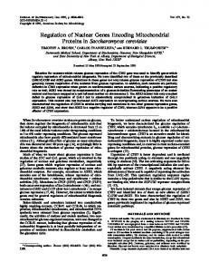
uct of pliv10 were all expressed in hepatoma cells (FTO-2B), but none of these sequences was detected in RNA prepared from mouse embryo fibroblasts (Fig. 1). Although all of these tissue-specific mRNAs were extinguished in genotypically complete hepatoma-fibroblast hybrids (12), most continued to be expressed in monochromosomal clones that retained only fibroblast chromosome 11 (Fig. 1). For example, transferrin, serum albumin, alcohol dehydrogenase, and pliv10 mRNAs were expressed at similar levels in parental hepatoma cells (FTO-2B), in monochromosomal hybrids [F(11)J and F(11)U], and in hybrid back-selectants [FB(11)J and FB(11)U]. Other liver genes, including those encoding alanine aminotransferase, aldehyde dehydrogenase, sorbital dehydrogenase, aldolase B, pyruvate kinase, a1-antitrypsin, and the products of pliv7, -9, and -10 showed a similar pattern of continued expression in the F(11) clones (ref. 13; also data not shown). Thus, extinction of these genes in intertypic hepatoma hybrids (12) is mediated by genetic loci distinct from Tse-i. In contrast to the continued expression of most liver genes in the F(11) hybrids, Tat-i and Pck-i (the gene encoding PEPCK) expression was specifically repressed in these clones. For example, scanning densitometry indicated that TAT mRNA levels in F(11)U and F(11)J were only 5% and 11% of parental FTO-2B levels, but the respective backselectants [FB(11)U and FB(11)J] reexpressed TAT mRNA to full hepatoma levels (Fig. 1). These results are in accord with previously published observations (13, 29). Significantly, PEPCK mRNA levels were also depressed by factors of 10-20 in the F(11) hybrids but were restored to parental hepatoma levels upon segregation of fibroblast chromosome 11 (Fig. 1). Thus, the genes encoding TAT and PEPCK were coordinately extinguished and reexpressed in F(11) hybrids and their back-selectants. To verify that accumulation of TAT and PEPCK mRNAs was specifically affected in the F(11) hybrids, blot-hybridization experiments were performed (Fig. 2). RNA samples were fractionated on agarose/formaldehyde gels, transferred
hybrids retaining that single fibroblast chromosome were screened for expression of liver-specific mRNAs. Cytoplasmic RNAs from parental and hybrid cells were immobilized
I
l
TAT j
TRF
I
P I|
PEPCKg
l111111I11| II 111,111 i1 II I
IIlI
||
RSA egg ADH
Ig
plivlO
- ...____ II ....... I._ _
ciTU
discrete locus on mouse fibroblast chromosome 11. This
locus, which can repress Tat-i activity in trans, has been designated tissue-specific extinguisher-i (Tse-i) (13). To identify other liver genes whose expression was regulated by loci on mouse chromosome 11, hepatoma microcell
7303
Proc. Natl. Acad. Sci. USA 85 (1988)
Genetics: Lem et al.
I
I
I
pg RNA:
u?
LO
I
I
I
r1 --I