â Magnetic Resonance Centre, University of Nottingham, University Park, Nottingham, NG7 ... Queen's Medical Centre require ADC maps as part of their MRI.
Correction of Distortion in ADC maps using the Reversed Gradient Method † Paul S.Morgan, Alan R. Moody, Steve J. Allder*, and Richard W.Bowtell
Academic Radiology, and *Clinical Neurology, University Hospital, Queen’s Medical Centre, Nottingham, NG7 2UH, England † Magnetic Resonance Centre, University of Nottingham, University Park, Nottingham, NG7 2RD, England
Introduction The measurement of apparent diffusion coefficients (ADC) shows promise in the diagnosis of many disease processes, especially in the fields of stroke and multiple sclerosis. While diffusion weighted images can be produced with conventional MRI scanning techniques, it is preferable to use a fast imaging sequence, of which echo planar imaging (EPI) offers the shortest acquisition and highest signal to noise. With EPI becoming an increasingly common option on clinical scanners, the interest in the routine production of ADC maps has increased in recent years. A number of trials currently being performed at the Queen’s Medical Centre require ADC maps as part of their MRI protocols. In order to apply diffusion weighting to a spin-echo EP image, large magnetic gradients are applied either side of the refocusing RF pulse. These have the potential to induce eddy currents, which in turn affect the homogeneity of the magnetic fields during image acquisition. Due to its inherently low bandwidth per point, EPI is highly susceptible to the effects of magnetic field inhomogeneities, leading to spatial distortion in the EP image. The production of ADC maps depends upon acquiring images with varying amounts of diffusion weighting. These images may therefore have differing amounts of spatial distortion. Identification of the same pixel in the images with differing amounts of spatial distortion is difficult; if no attempt to correct for this effect is made, then incorrect values of ADC may be calculated. This also will cause errors in properties calculated from the ADC maps, such as the diffusion tensor. A method to correct this distortion was required prior to introducing the production of ADC map into routine use.
3
gradient of reversed polarity . All images were corrected for distortion, and ADC maps calculated, assuming monoexponential diffusion processes within each voxel.
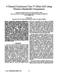
Results The technique was verified by use of a water filled phantom containing Balsa wood to give directional diffusion contrast. The figures presented here show the results from an acute stroke patient. Figure 1 shows a raw image (b=0); Figure 2 a raw diffusion weighted image; Figure 3 is the average ADC map produced directly from the raw images; Figure 4 is the average ADC map produced from images corrected for spatial distortion using the reversed gradient method.
1. T2 weighted
2. Diffusion weighted
3. Uncorrected ADC
4. Corrected ADC
Method Previous work has concentrated on reducing the effects of the diffusion gradients at the time of image acquisition, or to correct the distortion post-acquisition using phase map methods, to 5 ensure that all images are in alignment with one another . This work attempts to correct for the distortion after image acquisition with the application of the reversed gradient distortion correction 2 3 method applied to EPI . This correction method only corrects distortion caused by static magnetic field inhomogeneities; eddy currents would be expected to cause time varying inhomogeneities. The time courses of the inhomogeneities on the MR scanner used for this work (Siemens 1·5 T Vision) have been measured throughout the diffusion weighted image acquisition used. By inserting an increasing time delay after the last diffusion gradient, the evolution of phase in the image may 4 be measured . This has the advantage of allowing the phase evolution with time to be measured at various points throughout the image, allowing any spatial variation to be detected. The non-linear contribution was found to be less than 10° of phase at all points within the image throughout the acquisition. Hence, the assumption that the field inhomogeneities remain constant throughout the EPI acquisition was felt to be valid. Fat saturated images were acquired using a SE EP diffusion -2 weighted sequence (b=1000 s mm , 128×128 matrix, 2×2×5 mm voxel size, TE=103 ms, 0·8 ms per line). The sequence was surrounded by dummy diffusion gradients of opposite polarity to 1 reduce the effect of eddy currents during the acquisition . Three diffusion weighted images were obtained per slice, with the diffusion gradient applied along each primary axis in turn. An image with no diffusion weighted gradients was also obtained. These acquisitions were repeated with a phase encoding
This method has been introduced into routine use and has successfully produced ADC maps for over 75 patients (24 stroke, 27 MS, 25 NIA, 3 others).
Discussion The correction works well, allowing undistorted ADC maps to be produced. Misregistration artefacts, seen in Figure 3 as bright edges and heterogeneity, are eliminated in Figure 4. Disadvantages of this method include the inability to correct for any time varying magnetic field inhomogeneities, or through-slice distortion; it also relies on good edge detection of the object in the image. Advantages include correction of the spatial distortion inherent in EPI as well as the additional distortion caused by the diffusion gradients, and improved signal to noise in the corrected image compared to a single acquisition.
References 1. C.Boesch et al. Magnetic Resonance in Medicine 20 p.268-284. (1991) 2. H.Chang & J.M.Fitzpatrick. IEEE Transactions in Medical Imaging 11 (3) p.319-329. (1992) nd 3. R.W.Bowtell et al. Proc. SMR 2 Meeting p.411. (1994) 4. P.Jezzard et al. Magnetic Resonance in Medicine 34 p.65-73. (1995) 5. F.Calamante et al. Magnetic Resonance in Medicine 41 p.95 (1999)