Heywang S, Wolf A. PrussE, Hilbertz T, Eier mann W, Permanetter W. MR imaging of the breast with Gd-DTPA: use and limitations. Radi ologv1989:171:95ââ¬â ...
Correlation of Dynamic Contrast Enhanced MR Imaging with HistologicTumor Grade: Comparisonof Macromolecularand Small-MolecularContrast Media OBJECTIVE. Theendothelial integrityof microvessels is disrupted in malignanttumors.
HeikeDaldrup1'2 DavidM.Shames1 MichaelWendland1 Yoshitaka Okuhata1 ThomasM.Link1 WernerRosenau1 YingLu1 RobertC.Brasch1
Quantitative assays of tumor microvascular characteristics based on dynamic MR imaging were correlated with histopathologic grade in mammary soft-tissue tumors.
MATERIALS AND METHODS. A spectrum of tumors,benignthroughhighlymalig nant, was induced in 33 female rats by administration of N-ethyl-N-nitrosourea, a potent car cinogen. Dynamic contrast-enhanced MR imaging was performed using a small-molecular contrast medium (gadopentetate, molecular weight = 0.5 kDa) and a macromolecular contrast medium (albumin-(Gd-DTPA)30, molecular weight = 92 kDa) at an interval of 1—2 days. Per meability surface area product (PS), as estimated by the corresponding endothelial transfer coefficient (K@5), and fractional plasma volume (JPV) were calculated for each tumor and each contrast agent using a two-compartment bidirectional kinetic model. MR imaging mi crovascular characteristics were correlated with histopathologic tumor grade.
RESULTS. Tumorpermeabilityto macromolecular contrastmedium,characterized by K@5, showed a highly positive correlation with tumor grade (r2 = .76, p < lO_10). K@5 values were zero for all benign and some low-grade carcinomas, greater than zero in all other carci nomas, and increased in magnitude with higher tumor grade. A considerably smaller but sig nificantly positive correlation was found betweenfPV and tumor grade using macromolecular contrast medium (r2 = .25, p < .003). No correlation between K@5orfPV values and tumor grade was found using gadopentetate (r2 = .01, p > .95 and r2 = .03, p > .15, respectively).
CONCLUSION. Quantitative tumormicrovascular permeabilityassaysgenerated with macromolecular MR imaging contrast medium correlate closely with histologic tumor grade. No significant correlation is found using small-molecular gadopentetate.
U
se of contrast enhancement in con junction
with
MR
imaging
pro
vides a means to evaluate tissue function as well as morphology. Physiologic properties derived from kinetic analysis of dy namic contrast-enhanced
ReceivedSeptember22,1997;acceptedafterrevision March23,1998. CaffeyAwardpaper,presentedattheannualmeetingofthe TheSocietyfor PediatricRadiology, St Louis,May1997. ReprintedfromPediatricRadiologyl998;28:67—78 with permissionfromSpringer-Verlag, NewYork. 1Department
of Radiology, university
of California, 513
Parnassus Ave.,San Francisco, CA94143-0628.Address correspondenceto R.C.Brasch. 2 Present address: Department
of Radiology, University of
MUnster,MInster, Germany. AJR1998:171:941—949 0361—803x/98/1714—941
© American Roentgen RaySociety AJR:171,October1998
data can improve the
specificity of MR examinations,thereby sup plementing the detailed anatomic information often available with unenhanced MR imaging. Tumors represent a clinically important disease category for which a combination of improved sensitivity and specificity could favorably influ encepatientmanagementandoutcome.For ex ample, MR mammography, recognized as a highly sensitivetumor detectionmethod (up to 97%) [1—3], hasbeenfaulted for limited speci ficity. Nonmalignant pathologies are reported in 18% of “¿strongly enhancing, positive―le sions [2]. Improved specificity could better guide the management of patients with breast masses, potentially decreasing the current high
rate of negative biopsies as well as delineating individualized tumor characteristics for the op timal selection of therapeutic options. Various schemes have been proposed for analysis ofdynamic contrast-enhanced MR im aging data, with the supposition that such anal ysis will serve to characterize
tumors
and to
improve specificity. In support of this assump tion, studies have shown that measures derived from dynamic contrast-enhanced MR imaging data correlate with surrogates of tumor angio genesis [4]. Reports from our group have shown that kinetic analyses based upon princi pIes of solute diffusion across vascular endo thelial barriers can be used to quantitatively estimate microvascular permeabilities [5—8]. Endothelial permeability depends upon both vascular morphology and the physiochemical characteristics of the solute molecule. Consider ing MR contrast media, permeability estimates generated by commercially available small 941
Daidrup et aI. molecular contrast media (SMCM) cannot reasonably be expected to be the same as perme abilities estimated from macromolecular con tra.st media (MMCM) whose hydrodynamic radii are 50—100times larger 191.Based on the repeatedly demonstratedhyperpermeability of tumor microvessels to macromolecular solutes
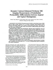
Fig. 1.—Diagramof two-compart menttissue model describing kinet cs of exchangeof contrastagent
Plasma Flow
betweenplasmaandinterstitialwa ter spaces in tumor. KPS(ml mind
cc@oftissue) is endothelialtransfer coefficient denoting clearance of contrast agent from plasmato inter stitial water and k (min1) is rate constant denoting fractional rate of reflux of contrast medium from in terstitial water back to plasma. Box around plasma compartment de
110—121, we hypothesize that MR imaging per
meability estimates based on MMCM-enhanced data should allow reliable differentiation of be nign from malignant tumors. As a corollary, we hypothesize that such differentiation should not be possible using permeabilities estimated from SMCM, since selective hyperpermeability to small-molecular solutes has not been demon stratedfor the micrc)vessels of malignanttumors, with the notable exception ofthc CNS [10]. The purposeofthis studywasto examinecor relations between histopathologic tumor grade and quantitative MR-derived estimates of tumor microva.scular permeability and plasma volume in a spectrum of mammary tumors induced in
Before MR imaging, the animals were anesthe tized by administration of pentobarbital, 50 mg/kg body weight i.p. A 23-gauge butterfly cannula (Abbott Laboratories, North Chicago, IL) was in
rodents.
serted in a tail vein for contrast medium injection.
A prototype
MMCM,
albumin-(Gd
DTPA)30(molecular weight, 92 kDa) was corn paredto a conventionalsmall-molecularcontrast medium, gadopentetate dimeglumine (0.5 Wa).
notesa forcingfunction.Kineticsof both compartmentstogether, corre sponding to Cr(t), concentration of gadolinium in tumor tissue at any
timet,aredescribedbyequation6.
study if no visible tumor was observed within 160 days or no tumor grew in the mammary fat pad.
After completion of imaging procedures using both MMCM and SMCM, animals were killed and tumors were excised for histologic analysis. ContrastMedia
Materials and Methods TumorInductionandAnimals N-ethyl-N-nitrosourea
(ENU) is an alkylating
agentand a potentcarcinogenthat inducestumorsof varyinggrade and locationin rats, dependingon the site of injection, the dose, and the age and sex of the
animal 113—151. Mammarycells are the primarytar get cells when ENU,45—180 mg/kg.is administered intraperitoneallyinto female rats I14. 151.Pharma cologically, ENU acts rapidly and without the neces sity of enzymatic activation on cellular targets, including DNA and certain species of RNA. The
histologic spectrum of mammary tumors induced using this model has been shownto be similarto the spectrumof histologiesencounteredin humanbreast tumors [14. 151. Following a single intraperitoneal (i.p.) administration of 180 mg/kg ENU, malignant
breast tumorsdevelopin up to 100%of rats after an averagelatent period of 92 days. At a lowerdose of 45 mg/kg.approximately42% of the rats developIi broadenomas [14, 151.For our study, ENU (ENU Isopack; Sigma Chemicals, St. Luis, MO). pur chased as a crystalline substance. was dissolved
shortly before administrationin 0.9% sterile saline to a concentrationof 10mg/mI. Fifty30-day-oldfemaleSpragueDawleyratswere
Albumin-(Gd-DTPA)30is a prototype MMCM with a molecular weight of 92 kDa and an initial distribution volume of 0.05 I/kg, which closely ap
was acquiredwithin30 sec. It was substitutedinto a
for the first few hours following bolus injection
(for postcontrastdata) [27]. Followingthe keyhole
with a half-life of approximately
data acquisition, 20 additional postcontrast three-di
3 hr [16]. Albu
min-(Gd-DTPA)30is prepared in our laboratory according to the method of Ogan et al. [171. Gadopentetate dimeglumine (Berlex Laborato
ries, Wayne,NJ) is a conventionalSMCM, 547 Da, with a distribution volume of 0.266 I/kg, approxi mating the extracellular fluid volume [18, 191. The
dose in this study was 0.1 mmol/kg. Plasma disap pearance is multiexponential and rapid with equili bration into the interstitial water space and with excretion exclusively through the urine [20]. Gado pentetate has been used extensively in clinical MR mammography studies [1—3.2 1—25]. MR Imaging
MR imaging was performed using a 2.0-1 sys tem (OmegaCSI-Il; Broker,Fremont,CA).Animals wereplacedsupinewithina “¿bird-cage― RFcoil(inner diameter,4.5 cm; length,7.6 cm).AxialTl-weighted
tings: TE, I.4 msec: one acquisition: field of view. SOxSOx 16mm: 128x 128matrix;andaneffective
lowedto continuegrowinguntiltheyreacheda diam eter of 1—1.5 cm. Animalswere eliminatedfrom the 942
fold by using a keyhole technique [26, 27]. The key hole data matrix was 128 x 32 x 16 and the image reference full-matrix data set, acquired either pm contrast (for precontrast data) or 20 mm postcontrast
ENU and group2 (ii = 15)received180mg/kgENU
research. Tumors, when visually apparent. were al
was shown to return to baseline levels in each animal
prior to administrationof the secondcontrast agent. The rapid biodistributionof gadopentetatedictateda higher temporal resolution.During the first 20 mm postcontrastthe acquisitionrate was increasedfour
proximates the blood volume [16]. The dose in
three-dimensional spoiled gradient-recalled (SPGR)
linesof the NationalInstitutesof Healthand withthe approval of the institutionalcommittee on animal
min-(Gd-DTPA)30enhancement, three initial pre contrastand 30 postcontrastimageswereacquiredat 2-mm intervalsover a I-hr period. Signal intensity
this study was 0.03 mmol of gadolinium per kilo gram. Plasma disappearance is monoexponential
randomly divided into two groups. Grnup 1 (a = 35) received a single i.p. dose of 45 nig/kg body weight i.p. The animals were treated according to the guide
msec. Either albumin-(Gd-DTPA)3()(0.03 mmol of gadolinium per kilogram) or gadopentetate (0.1 mmol of gadolinium per kilogram), randomly Se lected, was injected via the tail vein, with the other contrast agent administered 24—48hr later. For albu
sequences were acquired using the following set
slice thickness of 2 mm. Precontrast SPGR Se quences includedseveralseries withTR varyingbe tween 50 and 2000 msec for calculating baseline longitudinal relaxation rates (RI). Dynamic post contrast images were acquired with TR fixed at 50
mensionalSPGR imageswere acquiredat 2-mm in tervals as described above for macromolecular enhancedimages Tissue Analysis Postmortem, all tumors were removed, fixed in
10% formalin, embedded in paraffin, and sec tioned in the same place as the MR images. H and
E staining was performed for standard histologic analyses. All tumors, both benign and malignant,
were graded using the Scarf-Bloom-Richardson (SBR) method [28, 29]. Using this method each tumor is assigned score from 1 to 3 in each of three categories: gland formation. anaplasia, and mitoses. Microscopic observations are made on 10 high-power fields in the most mitotically active ar eas using a field diameter of 0.44 mm. The lowest
SBR score possible, even for a benign tumor, is 3. The highest SBR score, indicative of a poorly dif ferentiated malignancy, is 9. KineticAnalysisofDynamic MR Imaging Data
Average signal intensities for the tumor rim, blood within the vena cava, and an external corn oil phantom were measured in the central section
of the imaging volume using two to four operator defined regions of interest (minimum of 30 pixels per region). Kinetic analyses of tumor enhance AJR:171,October1998
SI@@@@(t)
Correlation
of Dynamic Contrast-Enhanced
Kinetic analysis of AR1(t) data from tissue and
ment responses were limited to the tumor rim be cause the tumor periphery is typically the most vascularized and least necrotic region and is not subject to the elevated interstitial pressure often
vena cava blood was performed similarly to that de scribed by Yeung et al. [34] for determination KPS in brain tumors using CT and iopamidol. A two-corn
observed in tumor centers [10, 30, 31]. Further more, previous MR imaging mammary tumor in
partmenttumor tissue model shownin Figure 1 as a special case of Patlak et al. [35] and composed of plasma and interstitial water equilibrating pools (whereexchangeis assumedto be based on passive
vestigations have shown the tumor rim to be the region most representative of angiogenic activity
[4] and most responsive to chemotherapy [32], ra diotherapy [33], and angiogenesis inhibition [7]. The signalintensitymeasurementswere acquired @
using a SPARC workstation (Sun Microsystems, Mountain View, CA) and an MRVision software pack age (MRVision, Stanford, CA). After correcting for potential spectrometer variation over time by dividing
terest at any time [i@iR1(t)],which are assumed to be directly proportional to the gadolinium concentration of the tissue, were calculated according to
@R1(t)= T1@0@@(t) T1pre
The differential equation describing the time varying concentration of gadolinium in the inter
dC1(t)
=
ps
K
C@(t)—kC1(t)
where Cp(t) is the concentration of gadolinium in
KPS is the endothelial transfer coefficient (ml
min@ cc1 of tissue), and k is rate constant of re flux from interstitial water back to plasma (min1). The solution to equation 4 in terms of the convolu tion integral where C1(t) = 0 at t = 0 is given by
C1(t) = KPSf@Cp(O)e@@tO)dO
(5)
Since the concentration of gadolinium in tumor
(2)
tissue at any time t, Cr(t) (mmol cc1 oftissue), is
Assuming that the fully relaxed signal intensity
does not vary significantlyon pm- and postcontrast SPGR images,valuesfor Ti@0@@(t) can be calculated from signal intensity measurements pre- and post contrast and Tipre when substituted into
the combination of gadolinium in both interstitial water and plasma spaces, its dynamic description is given by
Cr(t)
@
(6)
KPSJ@ Cp(O)e_@t
°@d8+ fpvCp(t)
TR
1 _eT@@1@) TR
51pre
@
l—e
where fp@@ is the fractional plasma volume of the tumor tissue (mmol cc1 of tissue). Theendothelialtransfercoefficient,KPS,is re lated to the permeability surface area product of
(3)
TI
10
Crone model of capillary permeability [36, 37] by the relationship PS
K@5 = FE = F 1 —¿ eF
(7)
which reduces to K@5 = PS
(8)
when E .9). All 10 benign tu AJR:171, October1998
Discussion Our analysis shows a strong positive correla tion between tumor microvascular permeability, estimated
from
dynamic
contrast-enhanced
macromolecular
permeability, a KPS value
above zero, emerges as a superior character istic by which to differentiate benign from
though both can be assessed microscopically. Althotigh
tumor grade and angiogenesis
angiogenesis. was grade for prediction
may
independent of tumor of metastatic rate and
patient survival. In the Weidner series, tumor microvascular density correlated weakly 945
Daldrup et al.
10
@RIbloodIulnor0.1
0
I
I
I
20
40
. .60 mm p.i.
4A
4B
5A
5B Figs.4 and5.—Representative Ti-weightedspoiledgradient-recalledimagesof subcutaneousmammarytumors—benign fibroadenoma(4Aand5A)andmalignantade nocarcinoma)4Band 5B)—beforeand 2, 10,and 60mmafter administrationof albumin-(Gd-DTPA)30 (0.03mmolgadoliniumper kilogram;4A and4B) or gadopentetate(0.1 mmolgadoliniumper kilogram;5A and5B).Also shownto right of MR imagesare correspondinggraphsof tumor and bloodAR1 responsesover initial 60mmafter contrast administration.AR1values are considereddirectly proportionalto tissue concentrationsof gadolinium-containingcontrast medium.Notethat using albumin-)Gd-DTPA)30,
dynamicresponsesin tumorandbloodarenear-parallel for benigntumor)4A),whilefor malignanttumor(4B),dynamictumorAR1responseincreaseswith respectto blood response,indicating substantial microvascular leak. Little difference in responsebetween benign and malignanttumors can be seen using gadopentetate)5A and
5B).i.p.= intraperitoneal. with tumor grade. Thus, it is appropriate to distinguish between the current study and previous investigations 4. 40. 41 ] demon strating positive correlations between dy
namic MR imaging data and angiogenic parameters. For instance, van Dijke and co workers (4) showed a significant correlation (r2 .82) between MMCM-estimated 946
microvascular permeahilities
density and in R3230
mammary cancers. Unlike the van Dijke in vestigation. topathologic
our current study focuses on his tumor grade and the MR
imaging differentiation of benign and malig nant tumors. The literature describing the microscopic morphology of tumor vessels provides insights and a possible mechanism for the observed re
lationship between endothelial permeability
and tumor grade 142—451.The endothelial lin ings and basement membranes in tumor blood
vessels vary considerably in organization, yet the vascular
organization
tends to correspond
with tumor cell differentiation. For example, nonfenestrated capillaries with a distinct intact basement membrane were found in highly differentiated hemangiopericytomas [44], while fenestrated capillaries. characterized AJR:171, October1998
@
@o
Correlation
of Dynamic Contrast-Enhanced
Fig. 6.—Typicalleast-squares fits (solid lines) ofmodeltoAR1 measuresofdynamic MR imagingdata obtainedfrom bloodin infe nor vena cava (triangles) andtumor (circles) following IV administration of albumin-(Gd DTPA)30(A) and gadopentetate (B). Model parametersfor fPVand KPSin this study for albumin-(Gd-DTPA)@are 0.028 ml cct of
MR Imaging with Histologic Tumor Grade Efforts were made to minimize the poten tial for bias in correlations of histology with MR imaging characteristics and in compari
ARI
sons of MMCM
and gadopentetate.
MR im
aging data acquisition, postprocessing, and analyses were performed independently of histopathologic evaluations. The pathologist was blinded to permeability measures ob tamed from kinetic analysis; the image ama lyzer was blinded to histopathologic grade. The keyhole modification of the three-di mensional SPGR technique, selected to ac quire gadopentetatedata by virtue of its rapid,
tissueand0.057mlmm1 100cc1 oftissue, respectively. Comparable values for gado pentetate are 0.032ml cct oftissue and4.21 mlmm1 100cc1 oftissue, respectively.
30-sec acquisition rate, could have introduced
errors which would not appear in the data sets acquired at 2-mm intervals [26, 27]. To exam
methispotential,twogadopentetate datasets
B by transendothelial gaps as large as 400—600 mor cell differentiation or grade may deter nm, are described in less differentiated Walker mine tumor vessel development and 256 carcinomas[44]and xenograftsof human organization, which, in turn, may determine renal cell carcinomas [46]. Even more per vessel permeability. Additional studies corre meable capillaries with discontinuous endo lating electron-microscopic observations of to thelium, resembling the hepatic sinusoids with mor microvessels with permeability measures wide interendothelial gaps and absent base ment membranes, have been observed in
poorly differentiated mammary [45] and renal cell carcinomas [44]. Representing the least organized tumor blood channels are certain melanomas and sarcomas in which no true mi crovessels exist; the blood “¿percolates― be tween and around tumor cells in the absence of endothelial cells [33, 43]. Thus, observations in the current study of increasing microvascu lar permeability to MMCM with increasingly malignant tumor grade can be explained function of microvascular disorganization.
@
@
@
@ @
Fig.7.—Correlation betweenMRima9ing estimatedpermeability,KF'S(mlmm' 100 cc1 of tissue),and histopathologictumor grade,assayedusing Scarf-Bloom-Rich ardson(SBR)scoringmethodinbenign(n= 10)andmalignant(n= 23)tumors. A, Correlation between@(PS afterIV injec
as a Tu
B,Samecorrelation asinAinsame tumors usinggadopentetate as contrast agent Note significantpositivecorrelationwith macromolecularcontrast agent and ab senceof correlationwith lower molecular
variations
which
were
unacceptably
high,
these coefficient of variations were reduced may help to establish these relationships. to acceptablelevels by replacing the value for The ENU method of tumor induction was fPv in the model by that from the albumin well suited for the current study. A broad study and refitting the gadopentetate data spectrum of tumors, benign through highly subject to this constraint. malignant, much like the spectrum of human General appeal for the use of gadopentetate breast tumors, is reported with ENU [13, 14] for enhancement of tumors includes its world and was confirmed in the current study. The wide commercial availability and the consider spontaneous tumors developed de novo and able body of accumulated experience with this therefore even the earliest stages of oncogen agent in human cancer characterizations.As esis were represented.Unlike xenografts, stated previously, gadopentetate-enhanced MR there was no chance that the tumor popula imaging is highly sensitivefor tumor detection tion had been selected by either in vitro or in but has been shown to lack specificity for can vivo passage. cer, for example, in breast tumors [47]. How
albumin-(Gd-DTPA)@
gadopentetate
0.20 .@
g@ Ii
r275Ap>.95025
r2=.75
;@ 0.15
p