EUKARYOTIC CELL, Apr. 2008, p. 602–609 1535-9778/08/$08.00⫹0 doi:10.1128/EC.00307-07 Copyright © 2008, American Society for Microbiology. All Rights Reserved.
Vol. 7, No. 4
Binding of the Wheat Germ Lectin to Cryptococcus neoformans Suggests an Association of Chitinlike Structures with Yeast Budding and Capsular Glucuronoxylomannan䌤† Marcio L. Rodrigues,1* Mauricio Alvarez,2 Fernanda L. Fonseca,1 and Arturo Casadevall2,3* Laborato ´rio de Estudos Integrados em Bioquı´mica Microbiana, Instituto de Microbiologia Professor Paulo de Go ´es, Universidade Federal do Rio de Janeiro, Rio de Janeiro 21941-590, Brazil,1 and Department of Microbiology and Immunology2 and Division of Infectious Diseases of the Department of Medicine,3 Albert Einstein College of Medicine, 1300 Morris Park Ave., Bronx, New York 10461 Received 19 August 2007/Accepted 11 November 2007
The capsule of Cryptococcus neoformans is a complex structure whose assembly requires intermolecular interactions to connect its components into an organized structure. In this study, we demonstrated that the wheat germ agglutinin (WGA), which binds to sialic acids and -1,4-N-acetylglucosamine (GlcNAc) oligomers, can also bind to cryptococcal capsular structures. Confocal microscopy demonstrated that these structures form round or hooklike projections linking the capsule to the cell wall, as well as capsule-associated structures during yeast budding. Chemical analysis of capsular extracts by gas chromatography coupled to mass spectrometry and high-pH anion-exchange chromatography suggested that the molecules recognized by WGA were firmly associated with the cell wall. Enzymatic treatment, competition assays, and staining with chemically modified WGA revealed that GlcNAc oligomers, but not sialic acids, were the molecules recognized by the lectin. Accordingly, treatment of C. neoformans cells with chitinase released glucuronoxylomannan (GXM) from the cell surface and reduced the capsule size. Chitinase-treated acapsular cells bound soluble GXM in a modified pattern. These results indicate an association of chitin-derived structures with GXM and budding in C. neoformans, which may represent a new mechanism by which the capsular polysaccharide interacts with the cell wall and is rearranged during replication. suggested by the reactivity of yeast cells with the wheat germ agglutinin (WGA) (12). Capsule expression is probably the most-studied virulence factor of C. neoformans (15, 18, 24, 25). It is generally accepted that the cryptococcal capsule is a very complex structure comprising different polysaccharides (4, 23, 24, 27, 30) and mannoproteins (15, 20). The major capsular polysaccharide of C. neoformans is GXM, which represents around 88% of the capsular mass (15, 23, 24). GXM is synthesized intracellularly (11, 13, 43) and secreted to the extracellular space in vesicles (35). Secreted GXM is used for distal capsular growth (23, 44) in a process that apparently involves divalent cation-mediated polysaccharide aggregation (29). The molecular mechanisms by which GXM is attached to the cell wall are still obscure. India ink staining is the simplest method for evaluating capsule expression in C. neoformans. In India ink preparations, the C. neoformans capsule presents a uniform aspect when examined by light microscopy. However, different regions of the C. neoformans capsule differ in density, sugar composition, and charge (45). Scanning electron microscopy of budding yeasts showed the existence of tunnel-like structures in the capsule at sites of nascent bud emergence (44). Further evidence for the existence of distinct structures within the capsule comes from the observation of capsular ringlike structures in India ink preparations (45). Although the molecular composition of the elements present at these capsular regions is still unknown, these findings strongly support the idea that the cryptococcal capsule is a highly complex structure that requires sophisticated mechanisms of assembly at the cell surface. In the present study, we report the existence of external cell
Cryptococcus neoformans is the etiological agent of cryptococcosis, a clinical syndrome associated with high indices of morbidity and mortality in immunosuppressed patients (41). The pathogenesis of C. neoformans involves the expression of several virulence factors, including pigment production (42), enzymatic activities (9, 10), regulation of signaling pathways (19), and synthesis of the capsular polysaccharides (15, 25). The cell wall of C. neoformans is a complex molecular network comprising polysaccharides, proteins, lipids, pigments, and bioactive enzymes (reviewed in reference 28). Cell wall polysaccharides play key roles in the physiology and pathogenicity of C. neoformans. For instance, glucuronoxylomannan (GXM) anchoring to the cryptococcal wall requires ␣-1,3-glucan (33, 34). In addition, it has been demonstrated that chitosan, the de-O-acetylated form of chitin, is required for cell wall integrity and maintenance of the correct assembly of the pigment melanin and the cryptococcal capsule (2). The presence of cell wall chitinlike oligomers in C. neoformans was
* Corresponding author. Mailing address for Marcio L. Rodrigues: Instituto de Microbiologia Professor Paulo de Go ´es, Universidade Federal do Rio de Janeiro, 21941590, RJ, Brazil. Phone: 55 21 2562 6740. Fax: 55 21 2560 8344. E-mail:
[email protected]. Mailing address for Arturo Casadevall: Department of Microbiology and Immunology, Albert Einstein College of Medicine, 1300 Morris Park Avenue, Bronx, NY 10461. Phone: (718) 430-2215. Fax: (718) 430-8968. E-mail:
[email protected]. † Supplemental material for this article may be found at http://ec .asm.org/. 䌤 Published ahead of print on 26 November 2007. 602
VOL. 7, 2008
CAPSULE-CELL WALL CONNECTIONS IN C. NEOFORMANS
wall structures protruding into and interacting with the capsule of C. neoformans. Several lines of evidence indicate that these structures contain chitinlike material that is expressed at the C. neoformans surface in close association with GXM and projected to the capsule during yeast budding. MATERIALS AND METHODS Fungal cells. The Cryptococcus cells used in this study included C. neoformans strains (strain H99, a serotype A clinical isolate, and strain Cap 67, an acapsular mutant of serotype D strain 3501) and one C. gattii isolate (strain NIH198, serotype B). Yeast cells were inoculated into 100-ml Erlenmeyer flasks containing 50 ml of a minimal medium composed of dextrose (15 mM), MgSO4 (10 mM), KH2PO4 (29.4 mM), glycine (13 mM), and thiamine-HCl (3 M) (pH 5.5). Fungal cells were cultivated for 2 days at 30°C with shaking. For the experiment whose results are shown in Fig. 2F and G, C. neoformans (strain H99) was cultivated in yeast extract-peptone-dextrose medium (YPD) for the same period. Yeast cells were obtained by centrifugation, washed in phosphate-buffered saline (PBS), and counted in a Neubauer chamber. The results presented in this report correspond to the results of experiments performed using strains H99 and Cap 67, but several analyses were also performed using the serotype D strain 24067, producing similar results. Data generated using the C. gattii isolate are presented in Fig. 2C. Fluorescent probes. WGA is a 36,000-Da lectin with known affinity for -1,4N-acetylglucosamine (GlcNAc) oligomers, present in the fungal polysaccharide chitin, and for sialic acids (1, 32). If modified with a succinyl group, the lectin loses affinity for sialic acids (26). Early reports indicated that WGA also recognized hyaluronic acid, although it has not been clearly demonstrated (39). Calcofluor white is a relatively small (916-Da) fluorescent dye that has been extensively used to stain chitin in fungal cells, due to its ability to recognize the GlcNAc--1,4-GlcNAc linkage. Monoclonal antibody (MAb) 18B7 is a mouse immunoglobulin G1 (IgG1) with high affinity for GXM of different cryptococcal serotypes (6). Fluorescence microscopy. Yeast cells (106) were suspended in 4% paraformaldehyde cacodylate buffer (0.1 M, pH 7.2) and incubated for 30 min at room temperature. Fixed yeast cells were washed twice in PBS and incubated in 1% bovine serum albumin in PBS for 1 h. The cells were then suspended in 100 l of a 5-g/ml solution of the Alexa Fluor 594 conjugate of WGA (Molecular Probes) and incubated for 30 min at 37°C. After being washed in PBS, the cells were incubated with 25 M calcofluor white (Invitrogen) under the same conditions. The cells were washed again and incubated for 1 h in the presence of MAb 18B7 (1 g/ml). After being washed in PBS, the cells were finally incubated with a fluorescein isothiocyanate (FITC)-labeled goat anti-mouse IgG (Fc specific) antibody (Sigma). For a negative control, we used an isotype-matched irrelevant IgG at the same concentrations used for MAb 18B7. To eliminate the possibility that the fluorescence pattern was derived from a specific sequential use of reagents, the order of the reagents was changed, and the results were the same (data not shown). Three-dimensional (3D) images were obtained after placing C. neoformans cell suspensions in mounting medium (50% glycerol and 50 mM N-propyl gallate in PBS) over glass slides. Z-series (116 sections for each cell) were obtained by capturing images every 0.25 m with a Leica AOBS laser scanning confocal microscope. The 3D images were finally processed using ImageJ (NIH; http://rsb.info.nih.gov/ij/) and Voxx (Indiana University; www .nephrology.iupui.edu/imaging/voxx/) software. Cell suspensions were mounted over glass slides as described above and analyzed under an Olympus AX70 microscope. Images were acquired by using a QImaging Retiga 1300 digital camera and processed using QCapture suite V2.46 software (QImaging, Burnaby, British Columbia, Canada). Transmission electron microscopy. After being washed in PBS, C. neoformans cells were fixed with 4% formaldehyde–1% glutaraldehyde for 3 h. The cells were then treated with 1% osmium tetroxide and these 1% uranyl acetate, followed by dehydration through a graded series of ethanol solutions and, finally, embedded resin (Electron Microscopy Science, Fort Washington, PA). Ultrathin sections of 70 to 80 nm were prepared in nickel grids. The grids were sequentially incubated in 10% H2O2 and a saturated solution of sodium periodate for 10 min. After being washed in PBS, the grids were blocked in 5% goat serum and incubated overnight at 4°C with a 5-g/ml solution of gold-labeled (30 nm) WGA (EY Laboratories). After sequential washes with PBS and distilled water and 10% uranyl staining, the sections were observed with a JEOL 100 CXII instrument at 80 kV. Staining of infected macrophages with WGA. Mouse RAW 264.7 macrophages were cultured in Dulbecco’s modified Eagle’s medium (Gibco-BRL, Gaithers-
603
burg, MD) supplemented with 2 mM L-glutamine and 10% complement-inactivated fetal bovine serum (Gibco-BRL, Gaithersburg, MD). C. neoformans cells were opsonized with MAb 18B7 (6), washed in PBS, and allowed to interact with phagocytes at a fungus/host cell ratio of 10:1 during 4 h at 37°C. Nonassociated fungi were then removed by washing, and the remaining cells fixed with cold methanol. After being washed in PBS, infected cells were incubated with fluorescent WGA as described above and observed with an Axioplan 2 (Zeiss, Germany) fluorescence microscope. Images were acquired by using a color view SX digital camera and processed with the software system analySIS (Soft Image System). WGA targets in C. neoformans. As detailed above, the potential targets of WGA binding in C. neoformans include sialic acids, chitin, chitin oligomers, and hyaluronic acid, all of them previously described as surface components of cryptococci (2, 3, 12, 16, 37). To evaluate whether the binding of WGA to C. neoformans involved sialic acids, yeast cells were incubated in the presence of tetramethyl rhodamine isocyanate-labeled, succinylated WGA (EY Laboratories) under the same conditions described above for the Alexa 594 conjugate. Alternatively, cryptococci were treated with sialidase under conditions described previously (37) and then incubated with the Alexa 594 WGA conjugate. To evaluate the affinity of WGA for chitinlike molecules in cryptococci, fungal cells were incubated with fluorescent WGA in the presence of a chitin suspension (1 mg/ml, Sigma, extracted from crab shells) or a mixture of chitooligosaccharides (100 g/ml, final carbohydrate content). The oligosaccharide mixture was prepared under the conditions described by Peumans and coworkers (31). Briefly, 9 mg of crab shell chitin was dissolved in 7 N HCl (3 ml) and partially hydrolyzed for 15 h at 40°C. HCl was then removed by evaporation; the residue was dissolved in PBS and diluted to a final concentration of 100 g/ml. Chitin hydrolysis into chitooligomers was confirmed by thin-layer chromatography (not shown). Incubations of C. neoformans with fluorescent WGA were also performed in the presence of 100 g/ml hyaluronic acid (purified from Streptococcus sp.; Calbiochem) or stachyose, a nonrelated tetraoligosaccharide (specificity control). Sugar analysis of capsular extracts. To analyze capsular material for the presence of potential WGA ligands in C. neoformans capsular extracts, polysaccharides were extracted from washed cells by using dimethyl sulfoxide (DMSO) or gamma radiation from radioisotope 137Cs as described previously (21). For DMSO extraction, C. neoformans cells (3 ⫻ 108) were suspended in DMSO (15 ml) and incubated for 15 min with shaking at room temperature. Supernatants containing released capsular polysaccharides were collected by centrifugation, and the pellet was again suspended in 15 ml DMSO for a second extraction under the same conditions. The supernatants were combined and extensively dialyzed against water for subsequent lyophilization and dry-weight determination. For the extraction of capsular polysaccharides by using gamma radiation, yeast cells (5 ⫻ 107) were suspended in water (15 ml) and irradiated for 90 min using a Shepherd mark I irradiator (J. L. Shepherd and Associates, San Fernando, CA) at 1,388 rads/min. The sugar composition was determined by gas chromatography-mass spectrometry (GC-MS) analysis of per-O-trimethylsilyl-derivatized monosaccharides from the polysaccharide film. Methyl glycosides were first prepared from the dry sample (0.3 mg) by methanolysis in methanol–1 M HCl at 80°C (18 to 22 h), followed by re-N-acetylation with pyridine and acetic anhydride in methanol for the detection of amino sugars. The sample was then per-O-trimethylsilylated by treatment with Tri-Sil (Pierce) at 80°C (0.5 h). GC-MS analysis of the per-O-trimethylsilyl derivatives was performed on an HP 5890 gas chromatograph interfaced to a 5970 MSD MS, using a Supelco DB-1 fused silica capillary column (30-m by 0.25-mm internal diameter). The carbohydrate standards used were arabinose, rhamnose, fucose, xylose, glucuronic acid (GlcA), galacturonic acid, mannose, galactose, glucose, mannitol, dulcitol, and sorbitol. The presence of sialic acids in irradiated or DMSO extracts was also analyzed by high-pH anion-exchange chromatography (HPAEC) after the hydrolysis of samples with 2 M acetic acid at 80°C for 3 h. A mix of sialic acid standards (N-acetyl neuraminic acid and N-glycol neuraminic acid) with a known number of moles was hydrolyzed at the same time as the samples. The sialic acids were analyzed using a Dionex DX500 system equipped with a GP40 gradient pump, an ED40 electrochemical detector, and a thermo-separations AS3500 autosampler containing a stainless steel needle. The individual sialic acids were separated by using a Dionex CarboPac PA20 (3 by 150 mm) analytical column with an amino trap. All methods were based on protocols described previously (14). GXM release after treatment of C. neoformans with trypsin and chitinase. After extensive washing with PBS, yeast cells (106) were suspended in 100 l of 0.01 M phosphate buffer (pH 6.0) containing chitinase (0 to 100 g/ml, purified from Streptomyces griseus; Sigma), followed by incubation at 37°C for 12 h. Alternatively, the cells were suspended in the same amount of phosphate buffer (pH 8.0) containing 500 g/ml trypsin (cell culture grade; Sigma), followed by
604
RODRIGUES ET AL.
similar conditions of incubation. The cell suspensions were incubated overnight at 37°C and centrifuged at 4,000 rpm for cell removal. The controls included cells treated in buffer containing no enzyme. The presence of GXM in the supernatants was determined by capture-enzyme-linked immunosorbent assay as previously described (5). Briefly, 96-well polystyrene plates were coated with a goat anti-mouse IgM. After the removal of unbound antibodies, a solution of MAb 12A1, an IgM MAb with specificity for GXM, was added to the plate, and this step was followed by blocking with 1% bovine serum albumin. Supernatants in different dilutions or purified GXM were added to the wells, and the plates were incubated for 1 h at 37°C. The plates were then washed five times with a solution of Tris-buffered saline supplemented with 0.1% Tween 20, followed by incubation with MAb 18B7 for 1 h. The plate was again washed and incubated with an alkaline phosphatase-conjugated goat anti-mouse IgG1 for 1 h. Reactions were developed after the addition of p-nitrophenyl phosphate disodium hexahydrate, followed by reading at 405 nm with a Multiscan MS (Labsystem, Helsinki, Finland). The antibodies used in this assay were all at a concentration of 1 g/ml. The effects of the enzyme treatments on surface-associated GXM were indirectly evaluated by measurements of capsule size in India ink-stained cells. Capsule sizes were defined as the distances between the cell wall and the outer border of the capsule. Capsule measurements were determined by using ImageJ software. GXM binding by acapsular cells. Acapsular C. neoformans cells (strain Cap 67, 106 cells) were suspended in 100 l of 0.01 M phosphate buffer (pH 6.0) supplemented or not with chitinase at 100 g/ml and incubated for 24 h at 37°C. The cells were washed and suspended in the same volume of purified GXM at 10 g/ml in PBS. The suspension was incubated for 12 h at 25°C and extensively washed with PBS, followed by fixation with 4% paraformaldehyde. The cells were further blocked by incubation for 1 h in PBS-bovine serum albumin and reacted with MAb 18B7 (1 g/ml) for 1 h at room temperature, followed by an FITClabeled goat anti-mouse IgG (Fc specific) to detect bound antibody (Sigma). Yeast cells were finally observed with an Axioplan 2 (Zeiss, Germany) fluorescence microscope.
RESULTS WGA recognizes cryptococcal structures connecting the cell wall to the capsule and around nascent buds. Staining of C. neoformans cells with fluorescent WGA, calcofluor white, and MAb 18B7 revealed that structures recognized by the lectin were limited to certain regions of the cell surface (Fig. 1A). These regions are apparently in contact with the cell wall, but WGA-positive areas were also found separated from the cell wall, in a spatial location that would correspond to the capsule. Analysis of yeast cells by transmission electron microscopy after incubation with the gold-labeled lectin (Fig. 1B) indicated that WGA interacted with cell wall sites at opposite poles of the cell, confirming the data from fluorescence microscopy. Interestingly, lectin binding extended into the capsular polysaccharide of C. neoformans. Similar results were obtained in experiments using a serotype D strain of C. neoformans (data not shown). The profile of C. neoformans reactivity with WGA shown in Fig. 1B resembled the capsular spots described by Zaragoza and coworkers after India ink staining (45). Hence, we investigated whether the areas of WGA staining corresponded to the India ink capsular spots. However, staining of C. neoformans with fluorescent WGA followed by India ink revealed that the regions recognized by the lectin and those described in the study by Zaragoza et al. did not colocalize (Fig. 1C). Actually, the regions of the cell surface recognized by the lectin formed 90° angles with the India ink-stained channels, supporting the idea that the capsule of C. neoformans manifests polarity in its architecture. To evaluate whether the profile of WGA staining of C. neoformans was also observed during infection of host cells, infected macrophages were incubated with the fluorescent lectin and analyzed by fluorescence mi-
EUKARYOT. CELL
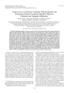
FIG. 1. Binding of WGA to surface structures of C. neoformans. Cells were treated with calcofluor (blue staining), MAb 18B7 (green staining), and WGA (red staining) and analyzed by fluorescence microscopy (A). WGA interacts with cryptococcal structures distributed in a polarized fashion, as demonstrated by fluorescence microscopy (A) and confirmed by TEM (B). The white asterisk in panel B denotes the capsule, and the black asterisk represents the cell wall. India ink staining of WGA-treated cells (C) reveals that the capsular channels of C. neoformans (arrowheads) do not correspond to the lectin-reactive structures (white arrows). (D) C. neoformans infecting macrophages is recognized by WGA (red staining) in similar patterns. Merged images from microscopic fields observed under differential interferential contrast are shown in panels C and D. Scale bars correspond to 10 m in panel A, 500 nm in panel B, 2 m in panel C and 10 m in panel D.
croscopy. The polarized pattern of lectin staining was also detected in macrophage-associated C. neoformans (Fig. 1D), indicating that the fluorescence profile observed in Fig. 1A was not a culture artifact. The structures recognized by WGA are associated with the cell wall but are visibly projected into the capsular network (see Movies S1 and S2 in the supplemental material). The structures recognized by the lectin appeared connected to the region stained by the anticapsular MAb 18B7 and formed round or hooklike projections. This purported association was supported by the 3D analysis of C. neoformans and C. gattii after sequential incubations with WGA, calcofluor, and MAb 18B7 (Fig. 2; see Movies S1 and S2 in the supplemental material). The cultivation of C. neoformans in YPD resulted in decreased capsule expression (Fig. 2F). Interestingly, the observation of WGA-reactive cell sites in this population was less frequent. For instance, WGA staining was observed only in 31% of the cryptococcal population grown in YPD, while 78% of the cells grown in minimal medium were stained after reaction with the lectin. These results suggest that the expression of the molecules recognized by the lectin is associated with capsule expression in C. neoformans. In approximately 40% of the dividing cells, the structures reacting with WGA appeared to form an interface between the capsule and bud necks (Fig. 2C; see Movie S2 in the supplemental material). The association of the capsule and molecules recognized by WGA in dividing cells was confirmed by the observation of septumlike structures extending from the bud neck to the capsular area (Fig. 2E). The septumlike structure, in fact, seemed to separate capsular structures from mother and daughter cells. To confirm this supposition, z-sections of a C. neoformans budding cell were obtained and analyzed sepa-
VOL. 7, 2008
CAPSULE-CELL WALL CONNECTIONS IN C. NEOFORMANS
FIG. 2. Confocal analysis of cryptococci after treatment with calcofluor (blue staining), MAb 18B7 (green staining), and WGA (red staining). Fungal cells were cultivated in minimal medium (A to E) or YPD (F and G). The results reveal the existence of structural elements reactive with the lectin between the cell wall and the capsule in C. neoformans (A, B, D, and E) and C. gattii (C). The existence of lectin-reactive structures inside the capsular area, concentrated around the bud neck, was also observed (E). Cultivation of cryptococci in YPD resulted in decreased capsule expression and a reduced number of lectin-reactive cells (F). In these cells, the structures recognized by the lectin can localize to outer layers of the capsule (arrows in panels F and G). Scale bars correspond to 2 m, except for panel F (4 m).
605
rately (Fig. 3). This analysis demonstrated that the molecules recognized by the lectin form a ringlike structure around the bud neck. WGA was previously shown to interfere with fungal growth (8), but despite binding to cell wall and capsular regions at the budding sites of C. neoformans, the presence of WGA in a suspension of replicating yeast did not influence cryptococcal growth (data not shown). Chemical analysis of released capsular polysaccharides. To evaluate whether the molecules recognized by WGA were covalently bound to the cell wall or associated with the capsule, C. neoformans cells were treated with DMSO or irradiated to release capsular polysaccharides. The various fractions were then analyzed by GC-MS and HPAEC for the presence of GlcNAc and sialic acids (Table 1). None of the monosaccharide components that are supposedly recognized by WGA were detected in the capsular extracts, despite efficient extraction of GXM from the capsule. On the other hand, WGA-stained, DMSO- and radiation-decapsulated cells were still recognized by the lectin, suggesting that the structures recognized by WGA were tightly associated to the cell wall and/or inner layers of the capsule (data not shown). Lectin binding to C. neoformans was unaffected by organic solvents (not shown), suggesting that lipids are not related to the structures reacting with WGA. WGA binds to GlcNAc-containing structures in C. neoformans. Sialic acids and -1,4-GlcNAc oligomers are recognized by WGA (1, 26, 32). We first investigated whether the WGAbinding molecules at the cryptococcal surface corresponded to
FIG. 3. Confocal microscopic analysis of C. neoformans. Sequential z-sections of C. neoformans after incubation with fluorescent WGA (red), calcofluor (blue), and MAb 18B7 (green) were taken, and the significant ones are shown here. WGA recognized a ringlike structure around the proximal bud periphery. This structure was most apparent after equatorial rotation of the nonsectioned image and x- and y-sectioning of budding cells (boxed image). Section numbers are shown for each image. Scale bar (presented in panel 70), 1 m.
606
RODRIGUES ET AL.
EUKARYOT. CELL
TABLE 1. Glycosyl composition analysis of polysaccharide extracts from C. neoformans cellsa Irradiated sample
DMSO-treated sample
Glycosyl residue Mass (g)
Arabinose Rhamnose Fucose Xylose GlcA Galacturonic acid Mannose Galactose Glucose N-Acetyl galactosamine N-Acetyl mannosamine GlcNAc Sialic acidb a b
ND ND ND 155.9 23.8 ND 259.6 6.9 3.1 ND ND ND ND
Mole (%)
39.1 4.6 54.2 1.4 0.7
Mass (g)
ND ND ND 203.8 63.0 ND 386.2 5.0 202.6 ND ND ND ND
Mole (%)
27.3 6.5 43.0 0.6 22.6
ND, not detected. Sialic acid analysis also included HPAEC.
sialic acids, since these sugars were previously reported as cell wall components of C. neoformans (37). Sialidase treatment did not affect WGA binding to C. neoformans (data not shown). Importantly, the pattern of binding of the succinylated lectin, which has no affinity for sialic acids, was similar to that observed after the incubation of C. neoformans with unmodified WGA (Fig. 4A). The identification of hyaluronic acid as the WGA target in C. neoformans was also discarded, since the affinity of WGA for C. neoformans cells was not influenced by the presence of the polysaccharide during the incubation of fungi with the lectin (not shown). These results were confirmed by the fact that only in a very small fraction of the cryptococcal population did we observe overlying fluorescence between the regions recognized by WGA and hyaluronic acid-binding protein (data not shown). The levels of WGA binding to C. neoformans were similar when incubations of fungal cells with the lectin were performed under standard conditions or in the presence of a chitin suspension (Fig. 4B). This observation is in full agreement with the data presented in Fig. 2 demonstrating that calcofluor and WGA recognize clearly different cellular sites and with a previous demonstration that the lectin binds chitooligosaccharides with more affinity than chitin (1, 32). In this context, a partial hydrolysis of chitin was performed to generate a mixture of oligosaccharides (31). This mixture was used in competition assays. In the presence of the oligosaccharide mixture, lectin binding to C. neoformans was clearly inhibited. WGA binding to cryptococci was unaffected by the presence of the nonrelated carbohydrate stachyose, a tetraoligosaccharide consisting of -D-fructofuranosyl-O-␣-D-galactopyranosyl-(136)-O-␣-Dgalactopyranosyl-(136)-␣-D-glucopyranoside. This observation supports the idea that the inhibition of lectin binding by the chitooligosaccharide mixture was specific. In combination, these results are interpreted as indicating that WGA interacts preferentially with GlcNAc-containing oligomers at the surface of C. neoformans, as described for bud scars in other fungal species (7, 8). Peptidase treatment did not affect WGA binding to cryptococci (not shown). Chitinase, but not trypsin, releases GXM from C. neoformans. The finding that the WGA-binding structures in C. neo-
formans were in close association with capsular components suggested a role for GlcNAc oligomers in capsule anchoring. These molecules, in principle, could be components of glycoproteins or represent small branches of chitin. To discriminate between the two hypotheses, we treated C. neoformans cells with trypsin and chitinase and measured capsule size and concentrations of released GXM. Trypsin treatment did not affect capsule size (not shown). Supernatants of cells treated with trypsin, but not with buffer alone, contained polypeptides in the range of 5 to 10 kDa, as determined by sodium dodecyl sulfate-polyacrylamide gel electrophoresis analysis (data not shown). The GXM levels in supernatants from trypsin-treated cells were similar to those detected in supernatants from cells incubated in buffer alone (Fig. 5). These results indicate that the structures recognized by WGA do not represent proteins and confirm the results of a previous report suggesting that structural proteins are not part of the capsular architecture (33). In contrast to the results with trypsin, the treatment of fungal cells with chitinase released GXM (Fig. 5A). A moredetailed analysis of chitinase-treated cells revealed a dosedependent release of GXM from C. neoformans, which was accompanied by a decrease in capsule expression (Fig. 5B). The comparison of control cells with yeast cells treated with the highest chitinase concentration suggests that the enzyme can cause a decrease of approximately 70% in the capsule size. Next, we examined the binding of WGA and GXM to acapsular C. neoformans treated with or without chitinase for 24 h. Chitinase treatment reduced the number of C. neoformans cells presenting the surface projections recognized by WGA by approximately 60% (Fig. 6). As expected, Cap 67 control cells incorporated GXM on their cell wall surfaces, resulting in binding of MAb 18B7 and WGA in overlying surface areas, as denoted by the green (MAb 18B7 alone) and orange (WGA and MAb 18B7) staining of fungal cells. Chitinase-treated cells were still able to bind GXM, although the pattern of polysaccharide binding differed from that observed in control yeast. After incubation with GXM, chitinase-treated cells presented a loose polysaccharide coat. The pattern of GXM binding
FIG. 4. WGA targets in C. neoformans. (A) WGA and its succinylated derivative bind to yeast cells in similar patterns. Scale bar, 1 m. (B) Incubation of WGA with cryptococci in the presence of competing carbohydrates reveals that a chitin hydrolyzate, but not chitin or the nonrelated oligosaccharide stachyose, inhibits lectin binding, suggesting that WGA recognizes chitooligosaccharides at the fungal surface. Scale bars, 10 m.
VOL. 7, 2008
CAPSULE-CELL WALL CONNECTIONS IN C. NEOFORMANS
607
FIG. 5. Release of GXM from the cryptococcal surface after treatment with chitinase. (A) Yeast cells were incubated in the buffers indicated in Materials and Methods (control, black bars) or with the enzymes named on the x axis (100 g/ml chitinase; 500 g/ml trypsin). (B) Treatment of cryptococci with chitinase caused a dose-dependent decrease of capsule size and a correlated increase in the detection of soluble GXM.
following chitinase digestion resembled that observed for glucanase-treated cells in a previous study (33). DISCUSSION The cell surface of C. neoformans is unique. The diverse composition of the cryptococcal cell wall, which includes lipids (36), polysaccharides (2, 3, 34), pigments (42), structural proteins, and bioactive enzymes (28), makes evident its complexity. This dense layer is surrounded by a polysaccharide coat that forms the cryptococcal capsule, which confers to the cryptococcal cell surface high levels of complexity in terms of molecular architecture (4, 15, 21, 23, 24). The study of capsule structure and polysaccharide release is important because the capsular phenotype appears to make the largest relative contribution to the virulence of C. neoformans (22). Despite the importance of this remarkable structure, very little is known
FIG. 6. Binding of GXM to control or chitinase-treated acapsular cells of C. neoformans. Yeast cells (Cap 67) were incubated in the presence of phosphate buffer (control) or chitinase and then GXM, followed by treatment with WGA and MAb 18B7. Labeling in red represents WGA binding, while green staining represents binding of the antibody to GXM. Orange staining denotes superposition between lectin and antibody binding to the cell wall. Scale bar, 1 m.
about the assembly process and the functions of capsular components other than GXM; the recent advances in understanding the structure and biophysical properties of GXM reinforce the idea that the capsular network of C. neoformans includes heterogeneous molecules (23, 29). The results of the present study, combined with the recent data of Zaragoza and coworkers (45), confirm that the C. neoformans capsule contains heterogeneous microenvironments despite a relatively homogenous appearance when visualized by India ink suspension and light microscopy. Our results establish that WGA staining molecules associated with the cell wall also project into the capsule. Furthermore, our results, in combination with the earlier report of India ink channels at equatorial locations in the capsule (45), imply that C. neoformans cells have a distinct geometry. If one considers the India ink channels to be at the equator of the cell, the WGA staining areas would then be at the cellular poles, such that the India ink-filled channels are at approximate right angles to the locations of nascent buds. The mechanisms by which this cellular geometry and polarity are maintained are not understood, but our ability to stain for these regions with WGA and India ink provides new tools for the study of capsular architecture. The lectin WGA has been used extensively in the last three decades as a probe to study surface components of different organisms, including C. neoformans (12). The lectin has affinity for sialic acids and -1,4-GlcNAc oligomers (1, 26, 32). On the basis of prior studies, we assumed that WGA would recognize sialic acid, which is produced by C. neoformans (37), or chitin, a -1,4-GlcNAc polymer that interacts very efficiently with the fluorescent dye calcofluor white. Our current data, however, show that sialic acids are not the target of the WGA lectin in this fungus. Competition assays indicated that the lectin recognizes GlcNAc oligomers at the interface between the capsule and the cell wall, which is compatible with the previously described specificities of WGA and calcofluor (1, 26, 32) and the different cellular sites recognized by these fluorescent
608
RODRIGUES ET AL.
probes (Fig. 2). More importantly, these results suggest that, besides the key roles played by chitosan and chitin in the architecture of the cryptococcal cell wall (2, 3), chitinlike oligomers can be important structures connecting cell wall to capsular components. Treatment of C. neoformans cells with chitinase released a substantial amount of GXM and altered the pattern of WGA binding to the fungal cell, with an apparent release of the projected structures together with capsular polysaccharides. Interestingly, the cell wall of chitinase-treated cryptococci was uniformly recognized by the lectin, suggesting a preferential affinity for WGA in binding to cell wall structures exposed after the partial removal of chitin. Our current results and those of previous studies (1) indicate that, rather than binding to chitin, WGA recognizes -GlcNAc oligomers. We therefore hypothesize that the uniform profile of lectin binding observed after chitinase treatment is a result of the generation of cell wall chitooligosaccharides after partial enzymatic hydrolysis of chitin, as described in other models (17). Based on the fact that GXM was released from the cryptococcal surface after the treatment of yeast cells with chitinase, but not peptidase, we believe that WGA is indeed interacting with outer chitin branches or chitinlike structures in C. neoformans. By “chitinlike” structures, we refer to C. neoformans components that manifest properties similar to those of chitin, such as GlcNAc composition and susceptibility to chitinases. Chitinlike material may include chitosan, the deacetylated form of chitin. WGA has been previously described as recognizing bud scars in yeast cells (7, 12), whose content is supposedly enriched in chitin. In the present study, a relationship between yeast budding and WGA binding was also suggested. However, lectin staining was also detectable outside bud necks in dividing cells. This observation may suggest that chitinlike oligomers are only associated with bud necks at the final stages of cell division and then become components of bud scars. In this regard, cell wall chitooligomers could be formed from the chitinase-mediated hydrolysis of chitin during the cell wall rearrangement which is required for cell division. The cryptococcal capsule is proposed to undergo local rearrangement during budding, possibly producing a tunnel for the bud to emerge (24, 44). In this context, the capsules of the mother and daughter cells have been proposed to be distinct, so that separation can occur, a finding demonstrated by scanning electron microscopy (44). The mechanisms involved in the physical separation of the capsules of dividing cells remain unknown. Given that the chitinlike projections identified here are found at the budding sites and that their stringy appearance resembles the types of structures that may be expected to form the type of tunnels visualized by electron microscopy (44), we propose that chitinlike oligomers form a septumlike structure that originates in the bud neck, extends to the capsular region, and helps to separate the capsules of budding cells. This observation suggests that the interaction between chitooligomers and capsular components is among the mechanisms involved in capsule separation during the replication of C. neoformans. This notion is consistent with the report that chitosan is involved not only with cell wall integrity and bud separation but also with the maintenance of normal capsule width (2). Since WGA was previously demonstrated to interact with chitosan (38), we cannot rule out the hypothesis that this
EUKARYOT. CELL
polysaccharide, alone or in association with chitin, is also recognized by the lectin in our model. ␣-1,3-Glucan is required for capsule anchoring at the surface of cryptococci (33, 34). Cryptococcal cells with disrupted alpha glucan synthase genes shed capsular material but lacked surface capsule (34). In addition, glucanase-treated acapsular cells bound to GXM in a defective manner (33). Indeed, cell wall glucans can anchor other polysaccharides at the fungal cell wall (28), which may explain the fact that chitinase treatment did not fully remove the cryptococcal capsule. In addition, the hypothesis that other components connect glucans and capsular components cannot be discarded, since loss of cell wall glucan would disturb cell wall assembly and, consequently, capsule anchoring. Our results indicate that chitinlike structures could also be relevant to direct GXM binding in C. neoformans. Chitinase-treated acapsular cells still bind soluble GXM, but they do so in a manner that forms a loose polysaccharide coat at the surface of C. neoformans. Chitin synthesis and distribution and the relationship of chitins with other surface structures in C. neoformans remain poorly understood processes. Eight putative chitin synthase genes have been identified in cryptococci (3), and strains with any one chitin synthase deleted were viable at 30°C. Melanization in C. neoformans is regulated through the expression of several genes, including the chitin synthase-encoding gene CHS3 (40). Although this observation suggests a link between virulence and chitin synthesis, an association between chitin and capsular polysaccharide has not been previously made. The association between chitin-derived material and capsular components could be due to ionic interactions, since chitosan, the most-prevalent form of chitin in cryptococci (2, 3), is a polycation at neutral-to-acid pHs that could interact with the polyanionic polysaccharide GXM. Alternatively, it has been recently reported that C. neoformans produces glycosyltransferases that link GlcNAc to GlcA residues (16), which are putatively used to form hyaluronic acid. In theory, the same enzymes could be used to bond GlcNAc residues in chitin to GlcA residues in GXM, providing C. neoformans with an additional mechanism for anchoring GXM to the fungal wall. ACKNOWLEDGMENTS M.L.R. is supported by grants from Coordenac¸˜ao de Aperfeic¸oamento de Pessoal de Nı´vel Superior (CAPES, Brazil), Fundac¸˜ao Carlos Chagas Filho de Amparo a Pesquisa no Estado do Rio de Janeiro (FAPERJ), and Conselho Nacional de Desenvolvimento Cientı´fico e Tecnolo ´gico (CNPq, Brazil). A.C. is supported by NIH grants AI033142, AI033774, AI052733, and HL059842. Carbohydrate analyses were performed at the Complex Carbohydrate Research Center, University of Georgia (Atlanta), which is supported in part by the Department of Energy-funded (DE-FG-9-93ER-20097) Center for Plant and Microbial Complex Carbohydrates. We are indebted to Susana Frases for help with the radiation analyses, Andre Nicola for discussions regarding lectin specificity, and the analytical image facility (Albert Einstein College of Medicine, Bronx, NY) staff for help with electron and confocal microscopy. We also thank Eliana Barreto-Bergter for the donation of competing carbohydrates and Leonardo Nimrichter for helpful comments and constant collaboration. REFERENCES 1. Allen, A. K., A. Neuberger, and N. Sharon. 1973. The purification, composition and specificity of wheat-germ agglutinin. Biochem. J. 131:155–162. 2. Baker, L. G., C. A. Specht, M. J. Donlin, and J. K. Lodge. 2007. Chitosan, the
VOL. 7, 2008
3.
4.
5.
6.
7.
8.
9.
10.
11.
12.
13.
14.
15. 16.
17.
18.
19.
20.
21.
22.
23.
CAPSULE-CELL WALL CONNECTIONS IN C. NEOFORMANS
deacetylated form of chitin, is necessary for cell wall integrity in Cryptococcus neoformans. Eukaryot. Cell 6:855–867. Banks, I. R., C. A. Specht, M. J. Donlin, K. J. Gerik, S. M. Levitz, and J. K. Lodge. 2005. A chitin synthase and its regulator protein are critical for chitosan production and growth of the fungal pathogen Cryptococcus neoformans. Eukaryot. Cell 4:1902–1912. Bose, I., A. J. Reese, J. J. Ory, G. Janbon, and T. L. Doering. 2003. A yeast under cover: the capsule of Cryptococcus neoformans. Eukaryot. Cell 2:655– 663. Casadevall, A., J. Mukherjee, and M. D. Scharff. 1992. Monoclonal antibody based ELISAs for cryptococcal polysaccharide. J. Immunol. Methods 154: 27–35. Casadevall, A., W. Cleare, M. Feldmesser, A. Glatman-Freedman, D. L. Goldman, T. R. Kozel, N. Lendvai, J. Mukherjee, L. A. Pirofski, J. Rivera, A. L. Rosas, M. D. Scharff, P. Valadon, K. Westin, and Z. Zhong. 1998. Characterization of a murine monoclonal antibody to Cryptococcus neoformans polysaccharide that is a candidate for human therapeutic studies. Antimicrob. Agents Chemother. 42:1437–1446. Chen, C., and R. Contreras. 2004. The bud scar-based screening system for hunting human genes extending life span. Ann. N. Y. Acad. Sci. 1019:355– 359. Ciopraga, J., O. Gozia, R. Tudor, L. Brezuica, and R. J. Doyle. 1999. Fusarium sp. growth inhibition by wheat germ agglutinin. Biochim. Biophys. Acta 1428:424–432. Cox, G. M., H. C. McDade, S. C. A. Chen, S. C. Tucker, M. Gottfredsson, L. C. Wright, T. C. Sorrell, S. D. Leidich, A. Casadevall, M. Ghannoum, and J. R. Perfect. 2001. Extracellular phospholipase is a virulence factor in experimental cryptococcosis. Mol. Microbiol. 39:166–175. Cox, G. M., J. Mukherjee, G. T. Cole, A. Casadevall, and J. R. Perfect. 2000. Urease as a virulence factor in experimental cryptococcosis. Infect. Immun. 68:443–448. Feldmesser, M., Y. Kress, and A. Casadevall. 2001. Dynamic changes in the morphology of Cryptococcus neoformans during murine pulmonary infection. Microbiology 147:2355–2365. Foster, A. J., R. A. Bird, S. L. Kelly, K. Nishimura, D. Poyner, S. Taylor, and S. N. Smith. 2004. FITC-lectin avidity of Cryptococcus neoformans cell wall and capsular components. Mycologia 96:1–8. Garcia-Rivera, J., Y. C. Chang, K. J. Kwon-Chung, and A. Casadevall. 2004. Cryptococcus neoformans CAP59 (or Cap59p) is involved in the extracellular trafficking of capsular glucuronoxylomannan. Eukaryot. Cell 3:385–392. Hardy, M. R., and R. R. Townsend. 1994. High-pH anion-exchange chromatography of glycoprotein-derived carbohydrates. Methods Enzymol. 230:208– 225. Janbon, G. 2004. Cryptococcus neoformans capsule biosynthesis and regulation. FEMS Yeast Res. 4:765–771. Jong, A., C. H. Wu, H. M. Chen, F. Luo, K. J. Kwon-Chung, Y. C. Chang, C. W. Lamunyon, A. Plaas, and S. H. Huang. 2007. Identification and characterization of CPS1 as a hyaluronic acid synthase contributing to the pathogenesis of Cryptococcus neoformans infection. Eukaryot. Cell 6:1486– 1496. Kneipp, L. F., A. F. Andrade, W. de Souza, J. Angluster, C. S. Alviano, and L. R. Travassos. 1998. Trichomonas vaginalis and Tritrichomonas foetus: expression of chitin at the cell surface. Exp. Parasitol. 89:195–204. Kozel, T. R., G. S. Pfrommer, A. S. Guerlain, B. A. Highison, and G. J. Highison. 1988. Role of the capsule in phagocytosis of Cryptococcus neoformans. Rev. Infect. Dis. 10:S436–S439. Lengeler, K. B., R. C. Davidson, C. D’souza, T. Harashima, W. C. Shen, P. Wang, X. Pan, M. Waugh, and J. Heitman. 2000. Signal transduction cascades regulating fungal development and virulence. Microbiol. Mol. Biol. Rev. 64:746–785. Levitz, S. M., and C. A. Specht. 2006. The molecular basis for the immunogenicity of Cryptococcus neoformans mannoproteins. FEMS Yeast Res. 6:513–524. Maxson, M. E., E. Dadachova, A. Casadevall, and O. Zaragoza. 2007. Radial mass density, charge, and epitope distribution in the Cryptococcus neoformans capsule. Eukaryot. Cell 6:95–109. McClelland, E. E., P. Bernhardt, and A. Casadevall. 2006. Estimating the relative contributions of virulence factors for pathogenic microbes. Infect. Immun. 74:1500–1504. McFadden, D. C., M. De Jesus, and A. Casadevall. 2006. The physical properties of the capsular polysaccharides from Cryptococcus neoformans suggest features for capsule construction. J. Biol. Chem. 281:1868–1875.
609
24. McFadden, D., O. Zaragoza, and A. Casadevall. 2006. The capsular dynamics of Cryptococcus neoformans. Trends Microbiol. 14:497–505. 25. Monari, C., F. Bistoni, and A. Vecchiarelli. 2006. Glucuronoxylomannan exhibits potent immunosuppressive properties. FEMS Yeast Res. 6:537–542. 26. Monsigny, M., C. Sene, A. Obrenovitch, A. C. Roche, F. Delmotte, and E. Boschetti. 1979. Properties of succinylated wheat-germ agglutinin. Eur. J. Biochem. 98:39–45. 27. Moyrand, F., T. Fontaine, and G. Janbon. 2007. Systematic capsule gene disruption reveals the central role of galactose metabolism on Cryptococcus neoformans virulence. Mol. Microbiol. 64:771–781. 28. Nimrichter, L., M. L. Rodrigues, E. G. Rodrigues, and L. R. Travassos. 2005. The multitude of targets for the immune system and drug therapy in the fungal cell wall. Microbes Infect. 7:789–798. 29. Nimrichter, L., S. Frases, L. P. Cinelli, N. B. Viana, A. Nakouzi, L. R. Travassos, A. Casadevall, and M. L. Rodrigues. 2007. Self-aggregation of Cryptococcus neoformans capsular glucuronoxylomannan is dependent on divalent cations. Eukaryot. Cell 6:1400–1410. 30. Pericolini, E., E. Cenci, C. Monari, M. De Jesus, F. Bistoni, A. Casadevall, and A. Vecchiarelli. 2006. Cryptococcus neoformans capsular polysaccharide component galactoxylomannan induces apoptosis of human T-cells through activation of caspase-8. Cell. Microbiol. 8:267–275. 31. Peumans, W. J., M. De Ley, H. M. Stinissen, and W. F. Broekaert. 1985. Isolation and partial characterization of a new lectin from seeds of the greater celandine (Chelidonium majus). Plant Physiol. 78:379–383. 32. Privat, J. P., F. Delmotte, G. Mialonier, P. Bouchard, and M. Monsigny. 1974. Fluorescence studies of saccharide binding to wheat-germ agglutinin (lectin). Eur. J. Biochem. 47:5–14. 33. Reese, A. J., and T. L. Doering. 2003. Cell wall alpha-1,3-glucan is required to anchor the Cryptococcus neoformans capsule. Mol. Microbiol. 50:1401– 1409. 34. Reese, A. J., A. Yoneda, J. A. Breger, A. Beauvais, H. Liu, C. L. Griffith, I. Bose, M. J. Kim, C. Skau, S. Yang, J. A. Sefko, M. Osumi, J. P. Latge, E. Mylonakis, and T. L. Doering. 2007. Loss of cell wall alpha(1–3) glucan affects Cryptococcus neoformans from ultrastructure to virulence. Mol. Microbiol. 63:1385–1398. 35. Rodrigues, M. L., L. Nimrichter, D. O. Oliveira, S. Frases, K. Miranda, O. Zaragoza, M. Alvarez, A. Nakouzi, M. Feldmesser, and A. Casadevall. 2007. Vesicular polysaccharide export in Cryptococcus neoformans is a eukaryotic solution to the problem of fungal trans-cell wall transport. Eukaryot. Cell 6:48–59. 36. Rodrigues, M. L., L. R. Travassos, K. R. Miranda, A. J. Franzen, S. Rozental, W. de Souza, C. S. Alviano, and E. Barreto-Bergter. 2000. Human antibodies against a purified glucosylceramide from Cryptococcus neoformans inhibit cell budding and fungal growth. Infect. Immun. 68:7049–7060. 37. Rodrigues, M. L., S. Rozental, J. N. N. Couceiro, J. Angluster, C. S. Alviano, and L. R. Travassos. 1997. Identification of N-acetylneuraminic acid and its 9-O-acetylated derivative on the cell surface of Cryptococcus neoformans: influence on fungal phagocytosis. Infect. Immun. 65:4937–4942. 38. Senstad, C., and B. Mattiasson. 1989. Purification of wheat germ agglutinin using affinity flocculation with chitosan and a subsequent centrifugation or flotation step. Biotechnol. Bioeng. 34:387–393. 39. Toda, N., A. Doi, A. Jimbo, I. Matsumoto, and N. Seno. 1981. Interaction of sulfated glycosaminoglycans with lectins. J. Biol. Chem. 256:5345–5349. 40. Walton, F. J., A. Idnurm, and J. Heitman. 2005. Novel gene functions required for melanization of the human pathogen Cryptococcus neoformans. Mol. Microbiol. 57:1381–1396. 41. Waters, L., and M. Nelson. 2005. Cryptococcal disease and HIV infection. Expert Opin. Pharmacother. 6:2633–2644. 42. Williamson, P. R. 1997. Laccase and melanin in the pathogenesis of Cryptococcus neoformans. Front. Biosci. 2:e99–e107. 43. Yoneda, A., and T. L. Doering. 2006. A eukaryotic capsular polysaccharide is synthesized intracellularly and secreted via exocytosis. Mol. Biol. Cell 17: 5131–5140. 44. Zaragoza, O., A. Telzak, R. A. Bryan, E. Dadachova, and A. Casadevall. 2006. The polysaccharide capsule of the pathogenic fungus Cryptococcus neoformans enlarges by distal growth and is rearranged during budding. Mol. Microbiol. 59:67–83. 45. Zaragoza, O., E. E. McClelland, A. Telzak, and A. Casadevall. 2006. Equatorial ring-like channels in the Cryptococcus neoformans polysaccharide capsule. FEMS Yeast Res. 6:662–666.