[Downloaded free from http://www.ijo.in on Tuesday, June 26, 2018, IP: 178.205.13.20] Case Reports July 2018
Endogenous Cryptococcus neoformans endophthalmitis with subretinal abscess in a HIV‑infected man Joveeta Joseph, Savitri Sharma, Raja Narayanan1 To report a rare case of Cryptococcus neoformans endogenous endophthalmitis with subretinal abscess in a 36‑year‑old HIV‑positive man, referred with progressive blurred vision in his right eye for the last 6 months. Vitreous biopsy followed by intravitreal ganciclovir did not result in significant improvement. Microbiology revealed the presence of C. neoformans, and intravitreal amphotericin B was then administered. The patient was treated aggressively with systemic and intravitreal antifungals but had a poor visual and anatomical outcome. A high degree of clinical suspicion combined with microbiological evaluation helped to arrive at an appropriate diagnosis. Key words: Cryptococcus, endophthalmitis, immunocompromised, subretinal abscess
Endogenous endophthalmitis is a rare entity that accounts for 5%–7% of endophthalmitis cases.[1] Fungal etiology is reported in 50%–62% of cases,[2] with Candida being the most common isolate, followed by Aspergillus sp.[2] Cryptococcus neoformans is ubiquitous‑encapsulated yeast found in soil. Although various types of ocular cryptococcosis have been reported in the literature, in most cases, the diagnosis was made presumptively on clinical evidence or histopathological diagnosis.[2,3] Definite microbiological identification was lacking in most of them, including direct microscopy of the vitreous sample.[2] We report a case of C. neoformans endogenous endophthalmitis with subretinal abscess in a young immunocompromised male who was treated promptly based on positive microscopic examination of the vitreous.
Case Report A 36‑year‑old patient was referred for painful gradual decrease of vision for 6 months in the right eye. There were no associated systemic complaints or any complaints in the left eye (OS). He was HIV seropositive for the last 15 years Access this article online Quick Response Code:
Website: www.ijo.in DOI: 10.4103/ijo.IJO_60_18 PMID: *****
Jhaveri Microbiology Centre, Brien Holden Eye Research Centre, L. V. Prasad Eye Institute, 1Smt. Kanuri Santhamma Centre for Vitreo‑Retinal Diseases, L. V. Prasad Eye Institute, Hyderabad, Telangana, India Correspondence to: Dr. Joveeta Joseph, Jhaveri Microbiology Centre, Brien Holden Eye Research Centre, L. V. Prasad Eye Institute, Hyderabad ‑ 500 034, Telangana, India. E‑mail:
[email protected] Manuscript received: 03.03.18; Revision accepted: 16.04.18
1015
with CD4 counts 105 cells/μl (normal 383–1347 cells/μl) and had taken highly active antiretroviral treatment for about 4 years and discontinued thereafter. On examination, the patient was denying light perception in the right eye with intraocular pressure 14 mm Hg in the right eye and 15 mm Hg in the OS. The conjunctiva showed congestion, and the pupil was round and middilated with reacting relative afferent pupillary defect, Grade 2. Fundus examination revealed the presence of patches of retinal whitening, which were hazily seen. Retina appeared attached and optic disc was pale. A presumptive diagnosis of acute retinal necrosis or endogenous endophthalmitis of possible viral etiology with subretinal abscess was made, and a complete core vitrectomy was performed along with intravitreal ganciclovir (2 mg/0.1 mL), and oral valganciclovir (900 mg two times a day for 3 days) was administered. The vitreous biopsy sample was sent for microbiological investigation. Other investigations including hemoglobin, random blood sugar, blood urea, and serum creatinine were within normal limits. Postoperative B‑scan ultrasound showed persistent moderate intensity echoes all throughout the vitreous cavity [Figs. 1]. The microbiological processing of the vitreous biopsy included direct microscopy with 0.1% calcofluor white, Gram and Gomori methenamine silver stains [Fig. 2a and b], and culture on 5% sheep blood agar, chocolate agar, thioglycollate broth, brain heart infusion broth, Sabouraud dextrose agar (SDA), and potato dextrose agar (PDA). All smears showed round, capsulated, budding yeast cells. Large, cream, mucoid, yeast‑like colonies grew on blood agar, chocolate agar, SDA, and PDA after 72 h [Fig. 3a and b]. The fungus was later identified as C. neoformans using YST strip of VITEK 2 compact system. Molecular tests included polymerase chain reaction for eubacterial 16S rDNA[4] and panfungal 28S rDNA as described earlier.[5] Fungal DNA was detected in the vitreous sample of this patient [Fig. 4] and the sample was negative for bacterial DNA. At 1‑week follow‑up, the patient had no light perception, and fundus showed dense vitreous exudates with optic atrophy. The patient was started on systemic amphotericin B as well as intravitreal injection of amphotericin B 5 μg/0.1 mL with close follow‑up, and a repeat intravitreal antifungal (amphotericin B 5 μg/0.1 mL) was administered after a gap of 2 weeks. Media cleared sufficiently and vitreous echoes were reduced but his best‑corrected visual acuity remained no light perception in the right eye at the final visit at 1½ months. As the patient was comfortable, treatment was discontinued, and the extremely poor visual prognosis with risk of phthisis bulbi was explained.
Discussion This case of endogenous cryptococcal endophthalmitis is noteworthy for the following reasons: It highlights the importance This is an open access journal, and articles are distributed under the terms of the Creative Commons Attribution-NonCommercial-ShareAlike 4.0 License, which allows others to remix, tweak, and build upon the work non-commercially, as long as appropriate credit is given and the new creations are licensed under the identical terms. For reprints contact:
[email protected] Cite this article as: Joseph J, Sharma S, Narayanan R. Endogenous Cryptococcus neoformans endophthalmitis with subretinal abscess in a HIV-infected man. Indian J Ophthalmol 2018;66:1015-7.
[Downloaded free from http://www.ijo.in on Tuesday, June 26, 2018, IP: 178.205.13.20]
1016
Indian Journal of Ophthalmology
a
Volume 66 Issue 7
b
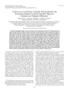
Figure 2: (a) Direct smear prepared from the vitreous biopsy examined after staining with Gomori Methenamine Silver stain demonstrates characteristic budding yeast cells and unstained capsule (×400), (b) Gram‑stained smear of vitreous biopsy showing encapsulated budding yeast cells engulfed within a macrophage (×1000)
Figure 1: B‑scan ultrasound showing low echo spike opacities in the vitreous cavity with localized elevations in all quadrants and submembranous opacities suggestive of exudates in the first visit
a
b
Figure 3: Photograph of (a) chocolate agar plate showing confluent growth of yeast‑like colonies on the drop inoculum of vitreous sample after 48 h incubation at 37°C, (b) potato dextrose agar slant showing characteristic mucoid colonies after 10 days incubation which was identified as Cryptococcus neoformans by ViTEK 2
of investigating vitreous biopsy including direct microscopy in the diagnosis of progressive endogenous endophthalmitis. Using simple Gram and GMS staining techniques, capsulated and budding forms were easily identified and were diagnostic of Cryptococcus. Given the difficulty of obtaining tissue from the intraocular space, clues to the cause of endogenous endophthalmitis will generally need to come from peripheral blood cultures and serum immunologic testing, in addition to urine and sputum culture. Most cases of confirmed cryptococcal endophthalmitis in the literature are from autopsy or enucleation,[6,7] fine needle aspiration biopsy,[8,9] and rarely after vitrectomy.[6] Intraocular Cryptococcus may take the form of chorioretinitis, multifocal choroiditis, neuroretinitis, vitritis, uveitis, or endophthalmitis.[4,6,8,9] Subretinal abscess formation in endogenous endophthalmitis is rare. Aspergillus was identified as the most frequent fungal cause of subretinal abscess.[10] Cryptococcal endophthalmitis is an infrequent entity, and treatment usually consists of amphotericin B or flucytosine, with therapeutic failure or relapses being reported in approximately 33% of cases.[8,9,11] There are some case reports of cryptococcal meningitis and endophthalmitis being successfully treated with voriconazole, especially to patients who had a treatment failure with amphotericin B alone or accompanied by fluconazole.[12,13] Sheu et al.[6] have reported
Figure 4: Agarose gel showing amplicon of 259 bp obtained with the polymerase chain reaction of the vitreous sample (target: 28S rRNA gene). NC: Negative control; Lane 1: Negative another patient sample; Lane 2: Current patient sample (positive); PC: Positive control; MW‑100 base pairs‑molecular weight ladder
that systemic amphotericin B‑fluconazole and two doses of intravitreous amphotericin B injection eliminated the cryptococcal infection successfully; however, in our case, in spite of a quick microbiological diagnosis and aggressive medical and surgical management, there was a poor visual and anatomical outcome possibly due to the immune status of the patient as well as late presentation to the clinic.
Conclusion Although endogenous C. neoformans endophthalmitis is commonly considered as a possible etiology in
[Downloaded free from http://www.ijo.in on Tuesday, June 26, 2018, IP: 178.205.13.20] Case Reports July 2018
immunocompromised hosts, it should also be included in the differential diagnosis of endophthalmitis with subretinal abscesses. Late presentation by the patient may lead to extensive inflammation and heavy load of organisms may account for poor outcome despite specific treatment. Acknowledgment We would like to thank Hyderabad Eye Research Foundation, Hyderabad. Declaration of patient consent The authors certify that they have obtained all appropriate patient consent forms. In the form the patient(s) has/have given his/her/their consent for his/her/their images and other clinical information to be reported in the journal. The patients understand that their names and initials will not be published and due efforts will be made to conceal their identity, but anonymity cannot be guaranteed. Financial support and sponsorship Nil. Conflicts of interest There are no conflicts of interest.
1017
3. Jarvis JN, Harrison TS. HIV‑associated cryptococcal meningitis. AIDS 2007;21:2119‑29. 4. Reddy GS, Aggarwal RK, Matsumoto GI, Shivaji S. Arthrobacter flavus sp. nov., a psychrophilic bacterium isolated from a pond in McMurdo Dry Valley, Antarctica. Int J Syst Evol Microbiol 2000;50 Pt 4:1553‑61. 5. Sandhu GS, Kline BC, Stockman L, Roberts GD. Molecular probes for diagnosis of fungal infections. J Clin Microbiol 1995;33:2913‑9. 6. Sheu SJ, Chen YC, Kuo NW, Wang JH, Chen CJ. Endogenous cryptococcal endophthalmitis. Ophthalmology 1998;105:377‑81. 7. Hiss PW, Shields JA, Augsburger JJ. Solitary retinovitreal abscess as the initial manifestation of cryptococcosis. Ophthalmology 1988;95:162‑5. 8. Henderly DE, Liggett PE, Rao NA. Cryptococcal chorioretinitis and endophthalmitis. Retina 1987;7:75‑9. 9. Hiles DA, Font RL. Bilateral intraocular cryptococcosis with unilateral spontaneous regression. Report of a case and review of the literature. Am J Ophthalmol 1968;65:98‑108. 10. Halperin LS, Roseman RL. Successful treatment of a subretinal abscess in an intravenous drug abuser case report. Arch Ophthalmol 1988;106:1651‑2.
References
11. Rostomian K, Dugel PU, Kolahdouz‑Isfahani A, Thach AB, Smith RE, Rao NA, et al. Presumed multifocal cryptococcol choroidopathy prior to specific systemic manifestation. Int Ophthalmol 1997;21:75‑8.
1. Okada AA, Johnson RP, Liles WC, D’Amico DJ, Baker AS. Endogenous bacterial endophthalmitis. Report of a ten‑year retrospective study. Ophthalmology 1994;101:832‑8.
12. Yao Y, Zhang JT, Yan B, Gao T, Xing XW, Tian CL, et al. Voriconazole: A novel treatment option for cryptococcal meningitis. Infect Dis (Lond) 2015;47:694‑700.
2. Essman TF, Flynn HW Jr., Smiddy WE, Brod RD, Murray TG, Davis JL, et al. Treatment outcomes in a 10‑year study of endogenous fungal endophthalmitis. Ophthalmic Surg Lasers 1997;28:185‑94.
13. Vela JI, Díaz‑Cascajosa J, Sanchez F, Roselló N, Buil JA. Management of endogenous cryptococcal endophthalmitis with voriconazole. Can J Ophthalmol 2009;44:e61‑2.
Culture‑positive unilateral panophthalmitis in a serology‑positive case of dengue hemorrhagic fever
Dengue fever, a mosquito‑borne disease commonly found in the tropics, is one of the most prevalent forms of Flavivirus infection in humans. Symptomatically, it is characterized by fever, arthralgia, headache, and rash. Ophthalmic manifestations can involve both the anterior and posterior segment. Panophthalmitis is rare in dengue hemorrhagic fever, and there is no report of culture‑positive panophthalmitis in this setting. Here, we report a case of a serology‑positive 33‑year‑old male patient of dengue hemorrhagic fever who developed sudden onset pain, redness, and proptosis in the right eye. The patient subsequently developed panophthalmitis in his right eye, and Bacillus cereus was isolated from eviscerated sample. This case provides unique insights into pathogenesis of panophthalmitis in dengue and highlights the management options.
Richa Kamal, Dhaivat Shah, Satish Sharma, Madharuvasal Krishnan Janani1, Arindam Kar2, Kumar Saurabh, Rupak Roy, Hajib Narahari Rao Madhavan1 Access this article online Quick Response Code:
Website: www.ijo.in DOI: 10.4103/ijo.IJO_113_18 PMID: *****
Department of Vitreoretinal Services, Aditya Birla Sankara Nethralaya, 2 Department of Critical Care, Media Superspeciality Hospital, Kolkata, West Bengal, 1Department of Molecular Microbioloy, Sankara Nethralaya Referral Laboratory, Chennai, Tamil Nadu, India Correspondence to: Dr. Rupak Roy, Aditya Birla Sankara Nethralaya, 147, Mukundapur, EM Bypass, Kolkata ‑ 700 099, West Bengal, India. E‑mail:
[email protected] Manuscript received: 22.01.18; Revision accepted: 07.03.18
Key words: Bacillus cereus, dengue, panophthalmitis
Dengue is a virus‑borne infection endemic in South‑East Asia, Central America, and South America, which is characterized This is an open access journal, and articles are distributed under the terms of the Creative Commons Attribution-NonCommercial-ShareAlike 4.0 License, which allows others to remix, tweak, and build upon the work non-commercially, as long as appropriate credit is given and the new creations are licensed under the identical terms. For reprints contact:
[email protected] Cite this article as: Kamal R, Shah D, Sharma S, Janani MK, Kar A, Saurabh K, et al. Culture-positive unilateral panophthalmitis in a serology-positive case of dengue hemorrhagic fever. Indian J Ophthalmol 2018;66:1017-9.