Plexiglas box by use of a Philips hard-X-ray therapy unit (RT305). ...... Lab. Immunol. 1:67-72. 25. Powles, R. L., A. Z. Barrett, H. Clink, H. E. M. Kay,. J. Sloane ...
Vol. 32, No. 3
INFECTION AND IMMUNITY, June 1981, p. 1164-1175 0019-9567/81/061164-12$02.00/0
Selective Induction of Immunological Tolerance in Antiviral T Killer Cells of Inbred Mice After Treatment with Cyclosporin A I?IETER ARMERDING Immunology Department, Sandoz Forschungsinstitut, Vienna, Austria Primary anti-influenza A cytotoxic thymus-derived (T) and bone marrow (B) lymphocyte-dependent responses in inbred mice were used as an in vivo model system to study the effects of the immunosuppressive fungus metabolite cyclosporin A (CyA). Five consecutive daily oral applications of CyA, with the first being given 1 or 2 h before virus inoculation of the animals, caused a complete blockage of induction of anti-influenza T killer cells and a partial reduction of cytotoxic B lymphocyte activities. Adoptive cell transfer experiments revealed that incapability to respond was due neither to humoral factors nor to the generation of suppressor cells. The tolerance state appeared to be specific for influenza A; cytotoxic T lymphocytes against allogeneic cell surface determinants could be stimulated in immunosuppressed mice. CyA treatment abolished virusspecific and cross-reactive anti-influenza killer T cell responses. Suppression was of short duration: less than 1 week for B cell-dependent functions, and between 1 and 2 weeks for T killer cell responses. Animals appeared to be normal with regard to both of these cellular activities for 4 weeks after tolerance induction. Thus, the data indicate that CyA exerted preferential effects on killer T cells. Moreover, evidence was presented that CyA treatment during an ongoing influenza infection did not increase sensitivity to that virus. Mice with no measurable cytolytic anti-influenza T killer cell activities but significant B cell responses, although partially diminished by the drug, were completely protected against the lethal effects of influenza infection.
Immunological tolerance to soluble or cellular antigens can be achieved in several ways and has been shown to involve quite complex and different cellular and subcellular mechanisms (reviewed in references 15, 18, and 27). Cumulative evidence led to the understanding of the refractory state as being in some cases only the extreme result of regulatory events involved in normal immune responses. Interference with any particular process involved in the maintenance of the finely tuned balance between opposing effector mechanisms and signals will just lead to either immunoenhancement or immunosuppression. The understanding of such basic concepts of immunoregulation has motivated the search for pharmacologically active compounds which might enable one to manipulate the immune system. Of particular interest are substances which could induce and maintain transplantation tolerance, especially since the surgical techniques for performing organ transplantations have been perfected. However, all of the currently existing means for preventing allograft rejection have been hampered by serious side effects of the intervening measures and by their nonspecific action.
Hence, Borel and colleagues (2-4, 31) discovered that the fungus metabolite cyclosporin A (CyA) induces immunological nonresponsiveness in experimental systems. This finding started a new wave of investigations on immunosuppression, particularly with regard to transplantation tolerance (10, 21, 23, 29). The most striking features of CyA are its nontoxicity for myelopoiesis, the antigen specificity of its effects, and the ease with which the suppressed immune status can be achieved (2-4, 17, 24, 28, 29). Nonetheless, mechanism(s) of action and the cellular target(s) have not yet been satisfactorily identified. Hypotheses have suggested clonal abortion (17, 30), induction of suppressor cells (7, 19), and functional impairment of certain lymphocyte subsets (2, 3) depending on the experimental system employed. Since CyA is already in use in human clinical trials (24, 28), it is of utmost importance to resolve this state of confusion and to delineate the cellular and molecular events affected and induced by this drug in the relevant in vivo situations. The aims of this study were to analyze in an animal system the effects of CyA on various lymphocyte populations and to identify its func-
1164
VOL. 32, 1981
TOLERANCE INDUCTION IN T KILLER CELLS
tional target(s) and mechanism(s) of action. I chose antiviral (influenza A) immune responses in inbred mice for several reasons: (i) antiviral immune reactions resemble basically those against transplantation antigens; (ii) the influenza system in particular allows us to assess effects on the function of various lymphocyte populations, such as antibody-mediated antiviral responses, cytolytic T cell activity, and helper T cell functions (1); and (iii) the existence of serologically defined determinants on the influenza virus and on virus-infected cells permits one to elucidate the antigen-specific action of CyA. The latter feature of the system is of particular interest since, in a primary situation, influenza A-induced killer T cells exhibit broad cross-reactivity against all respective virus-subtypes known (1, 5, 13, 14), whereas B cell responses are usually substrain restricted (1). The studies presented here demonstrate that T killer cell responses are most sensitive to the suppressive activity of CyA. However, B lymphocyte activity was also in part inhibited. Yet, 1 week after the tolerance treatment, no defect of antiviral B cell functions could be detected anymore, whereas cytotoxic T cells still remained in a state of unresponsiveness.
MATERIALS AND METHODS Animals. Mice of the inbred strain BALB/c/A Bom were derived from Gi (Bomholtgard Ltd., Ry, Denmark). C3H/HeHan miice were purchased from Zentralinstitut fur Versuchstierzucht, Hannover, West Germany. Only mice 10 to 14 weeks old were used. Cell lines. Exponentially growing L929 and P815 mastocytoma cells, expressing H-2k and H-2d mouse cell surface antigens, respectively, were used in the cytotoxicity assays. They were maintained in tissue culture. Viruses. AOPR8 (A/PR8/34) (HON1) and A/HK (A/Hong Kong/68) (H3N2) influenza A strains were grown in 10-day embryonated eggs and stored as allantoic fluid at -70°C. Virus titration was done in hemagglutination assays. Immunizations and immunosuppressive treatments. Mice were primed and boosted intravenously with infectious virus. The virus dose injected was equivalent to a hemagglutination titer of about 60. Primary responses were assayed on day 6 after sensitization, and secondary responses were assayed on day 5. Alloreactive lymphocytes were induced in BALB/c (H-2d) mice with spleen celis (5 x 10⁷ cells per mouse) from semiallogeneic (C3H x BALB/c)F1 (H-2d-)) mice which were bred in our own animal facilities. Immunosuppressive treatment consisted of five successive daily oral applications of CyA in olive oil. CyA was dissolved at a concentration of 25 mg/mil of ordinary olive oil by stirring at 60°C for about 20 min. Mice received 0.2 ml of this preparation. CyA (OL 27-400 N, lot no. 79806) was a generous gift from J. F. Borel (Biological and Medical Research Division, Sandoz Ltd., Basel, Switzerland). Solubilization and mode of
1165
application of the drug have been suggested by J. F. Borel. The galenical form and doses of CyA used herein were analogous to those applied in human clinical trials (25). Live virus was administered 2 h after the first CyA dose. Control mice received olive oil alone. Since no significant effect on the antiviral immune responses analyzed was achieved by the oil treatment, data were omitted from most experimental results presented. The immune status of the mice was examined in the spleen cell population from three to five animals per group. Cells were pooled and used as effector cells in the cytotoxicity assays. Adoptive cell transfers. Spleen cells obtained from normal, influenza-sensitized, untreated, or CyAtreated BALB/c mice (for details, see below) were prepared. Single-cell suspensions were washed once with minimal essential medium (Eagle) and injected intravenously into irradiated, syngeneic recipient mice (5 x 10⁶ to 10 x 106 cells per mouse). Irradiation was done by whole-body exposure of the animals in a Plexiglas box by use of a Philips hard-X-ray therapy unit (RT305). The absorbed dose rate was 50 R/min. Mice received a dose of 400 R 4 to 5 h before cell transfers. The recipient mice were challenged intravenously about 1 h after injection of the donor cells. Cytotoxicity assays. T and B lymphocyte-mediated cytotoxic responses were assayed by the 5"Cr release method (8) with the modifications detailed previously (1). Briefly, virus-specific lytic activity caused by T cells was estimated by isotope release from influenza-infected H-2-compatible tumor target cells (P815 for BALB/c and L929 for C3H mice) in the absence of complement. B cell-dependent cytotoxicity was assayed by using H-2-incompatible target cells and a complement source (selected normal rabbit serum) which was added to the cultures 2 h before harvest of the culture supernatants. The incubation times for the target-effector cell mixture yielding the highest sensitivity of the assay system were about 4 h at 37°C and overnight at 30°C in an atmosphere of 5% CO2 in air. The complement-dependent lysis was induced afterwards at 37°C. Results are expressed as percent specific 51Cr release calculated from the geometric means of the counts per minute of four replicas, using the following formula: percent specific 51Cr release = [(E - LC)/(HC - LC)] x 100. E represents the total 51Cr released by the target cells in the presence of sensitized effector cells. LC is the low control determined by the activity of cells from normal mice or, in the case of adoptive transfer experiments, of cells from mice receiving normal cells and no antigen. HC is the amount of isotope released from the target cells after an incubation with 10% Triton X-100 (Serva Feinbiochemica, Heidelberg, Germany) for 15 min at 37°C.
The standard error of the mean was usually less than 1.05. Cytolysis measured in the presence of nonsensitized cells was around 20% in the absence of complement and rose to 40% after addition of complement. Experiments were performed usually at least three times, yielding similar results. Estimation of sensitivity of mice against influenza A. The mouse-adapted A/HK virus was used to assess sensitivity of BALB/c mice during or after treatment with CyA. The lethal doses were established in pilot experiments (data not shown). Mice were
1166 ARMERDING
INFECT. IMMUN.
infected intranasally by aerosol with virus preparations of various concentrations (see below) and checked for mortality over a period of 21 days. Each experimental group consisted of 10 to 15 mice.
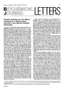
RESULTS CyA suppresses primary cytotoxic T and B lymphocyte responses against influenza A. The tolerance treatment used throughout all experiments presented in this paper consisted of five daily orally administered doses of CyA and antigen application 2 h after the first dose (see above). In the first series of experiments, we attempted immunosuppression for two serologically non-cross-reactive influenza A strains, H3N2 and HON1. Control mice did not receive CyA but were injected with the same virus substrains. Presence of virus-specific primary killer T lymphocytes and antibody-producing B cells was examined on day 6 after sensitization. The results from two representative experiments are depicted in Fig. 1. Cytotoxic responses were analyzed against P815 and L929 tumor cells which were infected with either HON1 or H3N2 virus. Activity against infected P815 cells in the absence of complement represents T cell responses; that against infected L929 cells in the presence of a complement source represents B
lymphocyte reactivity (1). The following findings were obtained. (i) Good T cell responses were induced in non-CyA-treated mice in both experiments, exhibiting complete cross-reactivity. (ii) Respective B cell activities in both groups of mice which were primed with either HON1 or H3N2 were high in experiment 1 and low in experiment 2. Responses were also crossreactive, although a clear preference for the sensitizing virus was expressed. (iii) CyA treatment drastically diminished or completely abrogated primary T killer cell induction. Suppression concerned cross-reactive and, if present, substrain-specific responses as well. (iv) B cell activity was less effectively reduced by the tolerance course. Inhibition ranged between 20 and 50% in most experiments with regard to the homotypic responses. A tendency to a more pronounced suppression was noticeable in crossreactive cytotoxic activities. Extent of suppression did not appear to be different in potentially high- or low-responding animal groups. Thus, it was shown that CyA treatment impaired induction of primary cytotoxic T cell responses more effectively than the corresponding B lymphocyte activities. Both homotypic and heterotypic immunities were depressed. It should further be noted here that CyA given
TARGET RATIO
100:1 50:1
EFFECTOR CELLS TREATMENT PRIMING ANTIGEN
TARGET CELLS
HONi
NONE
H3N2
HONW CyA
P815-HONI
H3N2
HONI NONE
H3N2
HONI CyA
P815-H3N2
H3N2
HONI NONE
H3N2
CyA
H3N2
HON1
L929-HON1 PLUS C
HONW NONE
CyA
H3N2
HON1
L929-H3N2
PLUS C'
H3N2 01020 30 40 50607080 90100 %SPECIFIC S"Cr-RELEASE
0102030 40 50 60 70 80 90100
FIG. 1. Effect of CyA on induction of primary anti-influenza cytotoxic T and B cell responses. BALB/c mice were either treated with CyA plus HONI or H3N2 virus, respectively, or inoculated with the virus substrains alone. Spleen cells were assayed on day 6 after sensitization or CyA treatment for cytotoxicity against H-2-compatible (P815, without complement) or incompatible (L929, with complement) target cells infected with either HONI or H3N2 virus.
VOL. 32, 1981
TOLERANCE INDUCTION IN T KILLER CELIS
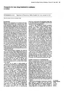
alone did not have any effect on the capability of mice to mount specific primary T and B cell responses if the antigen was administered only 2 days (or later) after the last CyA dose (data not shown). Feeding of olive oil alone did not affect immunity regardless of whether the animals were primed during or after the treatment (data not shown). Only cytotoxic anti-influenza T cells fail to respond to secondary stimulation in CyA-treated mice. Secondary in vivo responses to influenza are a little difficult to study because of a phenomenon mentioned by Doherty et al. (13) and Braciale and Yap (6) and studied in detail by us (D. Armerding and E. Liehl, Cell. Immunol., in press). Effective primary sensitization always blocks induction of secondary lymphocytes to the homologous virus because of the presence of neutralizing antibodies. Successful stimulation is only possible with heterologous influenza substrains, in which case cross-reactive T and B cell populations are induced. There are basically two ways to assess existence of specific memory cells in primed animals. First, one can study secondary in vitro responses or, second, perform in vivo adoptive cell transfer experiments. I studied in a first set of experiments anamnestic cytotoxic T and B cell responses induced with heterologous influenza substrains in animals treated before with CyA and influenza virus. In a further series of studies, I transferred lymphocytes from CyA tolerance-induced mice into irradiated recipients and focused on the capability of these cells to mount secondary killer T cell responses. In both investigations, secondary sensitization was performed 1 week after prming and onset of CyA treatment. All mice examined for secondary immunity were derived from the respective experimental groups tested for primary responses and described above. Figure 2 shows the results from two experiments designed to delineate the influence of the CyA treatment on the establishment of a heterotypic memory. The tolerance-inducing antigen was H3N2, and boosting viruses were HONi and H3N2. Control groups did not receive CyA but were previously primed with H3N2. Secondary anti-influenza responses were compared with primary antiviral activities in non-CyA-treated mice. Cytotoxicity was assayed against P815 and L929 target cells infected with either HON1 or H3N2 in the absence or presence ofcomplement, respectively. The following results were obtained. (i) As expected (see above), H3N2 virus did not induce significant T and B lymphocyte responses in H3N2-primed mice. However, in experiment 2, low and completely cross-reactive B cell-mediated cytolysis could be observed in
1167
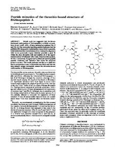
CyA-treated mice. Note the substrain restriction of the respective primary B cell activities and the cross-reactivity of the T killer population. (ii) Cross-boost with heterologous HON1 virus did generate cytotoxic anti-influenza T and B cell responses in untreated H3N2-primed mice. The level of stimulation obtained resembled that induced in the non-presensitized control groups. However, the secondarily provoked B cell responses were, in contrast to the primary cytotoxic activities, highly cross-reactive. (iii) Most importantly, treatment of animals with CyA with H3N2 resulted in nonreduring sponsiveness with regard to secondary induction of T killer cells with heterologous HON1 virus. Nevertheless, B cell activity was stimulated effectively in the same mice and exhibited a high degree of cross-reactivity. Figure 3 shows the responses of mice after adoptively receiving cells from donors which were tolerance treated with CyA plus H3N2 or HON1. Control mice obtained lymphocytes from donors which were either primed, but not CyA treated, or unprimed and nontreated. Challenge viruses were H3N2 and HON1. Cytotoxic responses were analyzed on day 5 after boost. It is obvious from the results that under the conditions employed, no primary T cell responses were obtained after adoptive transfer of splenic lymphocytes from normal mice. Significant killer cell activity was induced in recipients that received HON1- or H3N2-sensitized cells and were stimulated with both the homologous or the heterologous virus. On the contrary, responses of animals adoptively acquiring immunocytes from CyA-treated mice were suppressed. In this experiment, tolerance induced with HON1 was superior to that induced with H3N2. Furthermore, B cell responses measured in parallel did not reveal any indication of suppression (data not shown). Taken collectively, the data presented in this chapter indicate that CyA impairs generation or recall of T killer memory cells, or both. Suppressive status of the T killer population for in situ responses against heterologous virus substrains is not bound to humoral factors. Specificity of tolerance induction. Thus far, it has been demonstrated here that oral administration of CyA during the inductive phase of primary anti-influenza responses preferentially reduces expression ofT killer cell functions in the immune defense against influenza. It is important to show specificity of the suppressive treatment. Table 1 presents evidence that CyA impairs induction of cytotoxic T cell activity only to that antigen inoculated during exposure to the drug. BALB/c mice which were either unprimed, sen-
1168
ARMERDING
INFECT. IMMUN. EFFECTOR:TARGET RATIO 100:1
25:1 EFFECTOR CELLS TREATMENT PRIMING ANTIGEN HONI NONE
SECONDARY ANTIGEN NONE
H3N2
NONE
NONE
H3N2
H3N2
CyA
H3N2
HON1 H3N2
NONE
HONI H3N2
NONE NONE
NONE
H3N2
HONI H3N2
CyA
H3N2
HONW H3N2
HON1
TARGET CELLS
EXPERIMENT II
EXPERIMENT
I
P H815-ONI
I_L
IE
r
H P815-H3N2
I-
L11
IIIIIi NONE
H3N2
NONE NONE
NONE
H3N2
HONW H3N2
CyA
H3N2
HONW H3N2
NONE
HONI H3N2
NONE NONE
NONE
H3N2
HON1 H3N2
CyA
H3N2
iI L929-HON1 PLUS
C'
I
II
Ii
L 929-H3N2
PLUS C'
HON1 H3N2 0 10 20 30 40 50 60 70 80 90 100 0 10 20 30 40 50 60 70 80 90 100
%SPECIFIC 5'Cr-RELEASE
FIG. 2. Effect of CyA on induction of anti-influenza cytotoxic T and B lymphocyte memory. Effector cells were derived from three groups of BALBIc mice: (i) mice primed with either HONJ or H3N2 virus 5 days before the assay; (ii) mice primed with H3N2 12 days before the test receiving secondary virus (HON1 of H3N2) 7 days after pre-sensitization; and (iii) mice immunized as in (ii) above but treated with CyA for 5 consecutive days starting 2 h before priming. Spleen cells were assayed for cytotoxicity against H-2-compatible (P815, without complement) or incompatible (L929, with complement) targets infected with either HONJ or H3N2 virus.
sitized to H3N2, or treated with CyA during sensitization to this virus were immunized 1 week after onset of the treatment with semiallogeneic spleen cells from (BALB/c x C3H)F1 hybrids. The mice examined here were derived from larger groups. Primary responses against influenza of mice from these groups were illustrated in Fig. 1 (experiment 1) and secondary responses are shown in Fig. 2 (experiment 1). I demonstrated above that CyA treatment did, in fact, result in anti-influenza immunosuppression. The animals were tested 5 days after injection of H-2-incompatible cells for their capabil-
ity to generate T cells for cytotoxic L929 tumor target cells which express cell surface alloantigens identical to those on the stimulator cells. P815 targets which were H-2 compatible with the BALB/c mice served as negative controls in the assay. The data from a representative experiment are depicted in Table 1 (experiment 1). It is obvious from the results that alloantigen-reactive killer cells have been equally raised in all three experimental groups. Although one could argue that in my system CyA might not affect alloantigen-specific activation of T-killer cells, in any case, I tried to suppress such responses by
TOLERANCE INDUCTION IN T KILLER CELLS
VOL. 32, 1981
1169
EFFECTOR: TARGET RATIO 100:1 50:1
25:1
DONORS: TREATMENT PRIMING
ANTIGEN
RECIPIENTS: TARGET ANTIGEN
CELLS
H3N2 NONE
NONE
HON1 H3N2
H3N2
HONI
NONE H3N2
HON1
HONI
III iiI
P815-H3N2
H3N2 H3N2
HONW
CoA H3N2 HONI
HONW H3N2
NONE
NONE
HON1 H3N2
H3N2
W. ME"
HONi
i
NONE H3N2
HONI
HONI
PSIS-HONI
H3N2 H3N2
HOWN
CPA H3N2
HONI
HONI 50 40 30 10 '20 %SPECIFIC S'Cr-RELEASE_
0
6O
FIG. 3. Cytotoxic anti-influenza T cell response of adoptively transferred lymphocytes from CyA-treated mice. Donor (BALBIc) mice were either unprimed, primed with H3N2 or HON1, or treated with CyA plus H3N2 or HON1 virus. Spleen cells were transferred into irradiated (400 R) syngeneic recipients (5 x 10⁷ cells per host) on day 7 after sensitization or start of CyA treatment. Hosts were challenged 1 h after transfer with either HON1 or H3N2 virus. Spleen cells were assayed on day 5 for T cell cytotoxicity against P815 targets infected with HONI or H3N2 virus.
administering the drug during priming with H2-incompatible cells. Table 1, experiment 2, demonstrates that CyA is in fact capable of suppressing alloantigen reactivity. Furthermore, mice made nonresponsive for C3H transplantation antigens were able to mount normal antiinfluenza responses (data not shown). The results from this chapter demonstrate that CyA acts in an antigen-specific fashion since only T cell activities to immunogens present during CyA treatment were suppressed. Time-dependent escape from T cell tolerance. Having demonstrated failure to induce anamnestic antiviral killer T cell responses 1 week after tolerance treatment against influenza A it was of interest to determine duration of the T cell tolerance. BALB/c mice were sensitized with H3N2 and left either untreated or fed with CyA. Groups of mice were boosted with H3N2 or HON1 1, 2, or
4 weeks after treatment. Capacity to generate anti-influenza killer T cells was tested. The results from one typical experiment are illustrated in Fig. 4. Three points about the data presented are important. (i) Cross-stimulation was unsuccessful 1 week after the start of the tolerance treatment but induced killer T cells already another week later. (ii) These effector T cells expressed clear preference for the secondary antigen HON1. The anti-HON1 response of the CyA-treated mice was not significantly different from that of non-CyA-manipulated HON1-primed mice. (iii) At 4 weeks, cross-boost of CyA-treated mice yielded normal cytotoxic responses. T cell activity of these mice was as cross-reactive as that of the control group and similarly effective. Furthermore, it is surprising that homotypic stimulation also resulted in cytotoxic effector cells, although one would expect that mice with normnal B cell functions create
11 70 ARMERDING
INFECT. IMMUN.
TABLE 1. Influence of CyA on induction of alloantigen-reactive killer ceilsa % specific 61Cr release at following effector/target ratio: Expt
Treatmentb
p.
Scond antiary'
b
p815d
L929
gen
1
None None CyA
H-2k H3N2 H3N2
100:1
50:1
25:1
100:1
50:1
25:1
None
15.4
H-2k H-2k
13.9 12.9
NDe ND ND
8.7 7.5 7.8
2.0