International Journal of Bioelectromagnetism Vol. 9 No. 1 2007
Design and Implementation of a New System for Whole-Atrial Epicardial Mapping Cuiwei Yang, Weijia Lu, Xiaomei Wu, Zuxiang Fang Department of Electronic Engineering, Fudan University, Shanghai 200433, China E-mail:
[email protected]
analysis both amplitude and phase information. Simultaneous mapping overcomes some of the limitations encountered with sequential mapping by recording electrical information from several or even hundreds of locations at the same time [2]. To record whole atrial electrical activity at the same time, a new experimental research tool for studying AF was developed. The objective was to implement 128channel epicardial mapping system to show the electrical signals on the epicardial surface of atrium and store these data for subsequent analysis.
Abstract Atrial fibrillation (AF) is the most common arrhythmia and is becoming more prevalent each year. Its morbidity and mortality is increasing with the growth of age. Most in vivo experimental research on AF is performed by simultaneous epicardial mapping technique. This paper describes a whole-atrial epicardial mapping system that can record electrical information from 128 locations at the same time, with a spacing of 3.5-5.0mm. The purpose of this research is to show the electrical signals on the epicardial surface of atrium and store these data for subsequent analysis. 128 unipolar electrodes arranged in eight flexible patches were adopted to detect signal on the surface of whole atrium. Each electrode patch was designed to adaptable to the corresponding epicardial surface of canine atrium. Animal tests were operated on four mongrel dogs, in which two were models of chronic AF. Through amplifier contained 128 isolated channels and USB based data acquisition card, random acquisition duration of interested data during sinus or AF rhythm can be recorded. Result shows that this system can perform successfully in a surgical setting, making this system a special and portable tool for electrical mapping of whole-atrium and therefore useful for studying AF.
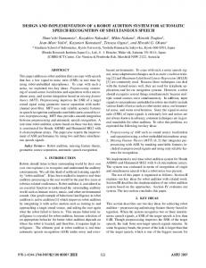
2. Methods 2.1. Electrodes of whole-atrium 128 unipolar electrodes arranged in eight flexible patches were adopted to detect signals by suturing into the atria. Each electrode patch was soft and was designed to adaptable to the corresponding epicardial surface of canine atrium. Figure 1 shows the distribution of electrodes. The unipolar electrodes, 1.2mm in diameter and 3.5mm~5.0mm apart, were carefully arranged in these flexible patches to match the size of canine atrium in a surgical setting. On each patch several sites used as suture are pre-perforated.
2.2. Data acquisition
1. Introduction The system provided 128 amplifier channels filtered with a fixed bandwidth of 40-600Hz. Signals were amplified by two-stage amplifiers with a selected gain of 625, 1250 or 2500 by manual switching. Amplified and filtered signals were passed to A/D converter through USB, and converted to 16-bit digital values with a sampling rate of 2.5KHz. The system is capable of saving continuously to disk and allow virtually unlimited sampling durations. Hence random acquisition duration of interested data during sinus or AF rhythm can be recorded.
Atrial fibrillation (AF) is a common and complex arrhythmia and is becoming more prevalent each year. Its crude rate in China is 0.77% and is growing with aging [1]. Further investigation into the electrophysiological properties in AF is required to develop an appropriate treatment, though radiofrequency ablation has opened the new era of therapies for AF. In order to be used as an important role in characterizing complete conduction through atrium, whole-atrial epicardial mapping system may
19
International Journal of Bioelectromagnetism Vol. 9 No. 1 2007
A
B
D3
D4
D2 D1
C’ C
Figure 3. Original waveform of sampling atrial potentials during sinus rhythm.
Figure 1. A photograph of whole-atrial electrode. Eight patches outfitted on atrial surface wre illustrated. The wholeatrial-electrode was composed of five parts: (1) A: anterior LA (ALA, 32 sites) and (2) B: anterior RA (ARA, 32sites) and (3) C: posterior LA (PLA, 16 sites) and (4) C’: posterior RA (PRA, 16 sites) and (5) D1-D4: pulmonary veins (PVs, 64 sites).
Figure 4. Original waveform of sampling atrial potentials during AF (AF was induced by embedded pacemaker).
4. Discussion and future work Result shows that this system can perform successfully in a surgical setting, making this system a special and portable tool for electrical mapping of whole-atrium. The data communication based on USB benefits this system easy to use for end user and having single model for cabling and connectors. Further research may be done to produce activation maps [4] during surgery and therefore make this system reliable and useful for studying AF.
Figure 2. A photograph showing the amplifier (lower) and A/D converter (top middle). This side contained 64 isolated amplifier channels and another 64 channels were on the posterior side of PCB.
Acknowledgment
2.3. User Interface (UI)
This research was supported by NSFC (National Natural Science Foundation of China, No. 30400102). We also thank Dr. Xingpeng Liu for arranging animal experiments and performing AF models.
The designation of UI was very important to provide a user-friendly control for easy management of the system. The support computer was DELL 9300 notebook PC with processor P-M 770 (2.13GHz). In order to make waveform scrolling smoothly, some special techniques were used. Vertical blanking interval of television signal is defined as a time period. In this period nothing will be displayed in the screen, and it is perfect for repainting work. Also DirectDraw interface was used here.
References [1] Zhou Z Q, Hu D Y, Chen J et al, “An epidemiological survey of atrial fibrillation”, Chin J Intern Med, 2004 (43), pp. 491–494. [2] Robert A.M., Nicolle K, Benjamin S et al, “Advances in electrical and mechanical cardiac mapping”, Physiol. Meas., 2005 (26), pp. R1–R14. [3] Diao H Y, Ma C S, Li S M et al, “Establishment of a canine atrial fibrillation model with chronic rapid atrial pacing”, Chinese Journal of Cardiac Pacing and Electrophysiology, 2001 (15), pp.98–100. [4] Wu X M, Fang Z X, and Yang C W, “Using active depolarization wave image to register and analyze epicardial electrical activities of atrial fibrillation”, Chin J of Cardiac Pacing and Electrophysiology, 2004 (18), pp. 148-150.
3. Results Animal tests were operated on four mongrel dogs, in which two were normal and another two were models of chronic AF [3]. The system could display waveform successively and interested data of episodes of random duration could be stored and replay for subsequent analysis (see figure 3-4).
20