Aug 10, 1984 - Most signal sequences are found to vary considerably in length and primary sequence, but possess some common structural features. Analysis ...
317
Biochem. J. (1984) 224, 317-325 Printed in Great Britain
Design and synthesis of a consensus signal sequence that inhibits protein translocation into rough microsomal vesicles Brian M. AUSTEN, John HERMON-TAYLOR, Mustak A. KADERBHAI and David H. RIDD Department of Surgery, St. George's Hospital Medical School, Cranmer Terrace, London SWJ7 ORE, U.K. (Received 20 June 1984/Accepted 10 August 1984) Most signal sequences are found to vary considerably in length and primary sequence, but possess some common structural features. Analysis of known signal sequences has led to the design of a 19-residue sequence that, although not a naturally occurring signal, possesses the structural features that commonly occur in pre-proteins. This peptide has been synthesized by solid-phase methods, and has been shown to inhibit, in a concentration-dependent manner, the processing in vitro of nascent pre-prolactin, pre-forms of pancreatic digestive enzymes, and pre-placental lactogen. The peptide acts at the cytoplasmic surface of microsomal vesicles added to the protein translation system, preventing translocation of the nascent chains to the lumenal space of vesicles where signal peptidase normally cleaves to remove the signal from nascent preproteins.
The amino acid sequences of more than 50 Nterminal extensions (signal sequences) found on the nascent chains or initially-translated precursors of secreted or membrane-bound proteins from prokaryotic and eukaryotic sources are known (Blobel et al., 1979; Chan et al., 1979; Austen & Ridd, 1981). Signal sequences are believed to assist transfer of proteins across membranes, and in eukaryotic cells are thought to act by enabling the nascent chain-polysomal complex to bind to an 11 S ribonucleoprotein complex (Walter & Blobel, 1981), which in turn binds to a specific protein, termed docking protein, associated with the membrane of the endoplasmic reticulum (Meyer et al., 1982). These interactions are followed by vectorial transfer of nascent chains into the lumen of this organelle. Although no obvious primary sequence homologies are apparent among known signal sequences, a number of common structural features have been identified (Austen, 1979; Austen & Ridd, 1981; Blobel et al., 1979; Gamier et al., 1980; Chan et al., 1979). Signal sequences commonly possess an uneven distribution of charged and uncharged residues, so that there exists a hydrophobic core of at least nine consecutive uncharged residues, bordered by basic amino acid residues at the Nterminus, and basic and acidic residues at the Cterminus. There is specificity in the region at which proteolytic cleavage takes place to give rise Abbreviations used: Boc, t-butoxycarbonyl; SDS, sodium dodecyl sulphate; PTH, parathyroid hormone.
Vol. 224
to a mature protein, and predictions of secondary structure have demonstrated high potential for adopting a-helical structure in the central hydrophobic core, and f-turns close to the cleavage sites. In order to test the importance of these common structural features in effecting translocation of nascent secretory proteins into the endoplasmic reticulum, we have synthesized a peptide which is not a naturally occurring one, but represents a consensus of known signal sequences. This peptide has been tested for its activity in inhibiting translocation of nascent pre-proteins in cell-free protein translation systems in vitro into dog pancreatic microsomal vesicles present during translation.
Materials and methods Materials Dichloromethane (Fisons, Loughborough, U.K.), dried and distilled grade, was stored over molecular sieve (4A). Dimethylformamide (Fisons) and di-isopropylethylamine (Fluorochem, Glossop, Derbyshire, U.K.) were distilled from ninhydrin in vacuo. Trifluoroacetic acid (KochLight, Slough, Berks., U.K.) was redistilled. Protected amino acid derivatives, from Vega Biochemicals, Tucson, AZ, U.S.A., were tested for purity on t.l.c. and recrystallized if necessary. [35S]Methionine (>800Ci/mmol) and [4,53H]leucine were from Amersham International, Amersham, Bucks., U.K. Synthetic y-endorphin and human parathyroid hormone (residues 28-48) were obtained from Cambridge Research Biochemicals,
318
B. M. Austen, J. Hermon-Taylor, M. A. Kaderbhai and D. H. Ridd
Cambridge, U.K. Chloromethylated polystyrene (1% cross-linked SX1, 200-400 mesh, 1.29mequiv./g) from Bio-Rad, Watford, Herts., U.K., was substituted with Boc-Glu(O-benzyl) as its caesium salt (Gisin, 1973) to a level of 0.65mmol/g, and additional reactive groups were blocked by treatment with benzoyl chloride (0.2 ml/g) and pyridine (0.18ml/g). Aminopeptidase M was from Rohm, Darmstadt, Germany, and chymotrypsin and tosylphenylalanylchloromethane ('TPCK')-treated trypsin from Millipore, Freehold, NJ, U.S.A. Oligo(dT)-cellulose and biochemicals used in cellfree translation were from Sigma, Poole, Dorset, U.K., and X-ray film from Kodak, Hemel Hempstead, Herts., U.K. RNA from human placenta was obtained from NEN Chemicals, Dreieich, Germany. All other materials were of the highest grade available. Peptide synthesis The 19-residue consensus peptide and a control pentapeptide, Leu-Ala-Tyr-Val-Ala, were synthesized on solid phase (Erickson & Merrifield, 1976). All amino acids were protected at the aamino positions with Boc groups, and the following side chain protectants were used: Lys, N-2chlorobenzyloxycarbonyl; Asp, O-benzyl; Cys, S-4-methylbenzyl; Ser, O-benzyl; Tyr, 0-2,6dichlorobenzyl. After two alternating washes with dichloromethane and propan-2-ol, three washes with dichloromethane, deprotection with 50% (v/v) trifloroacetic acid in dichloromethane for two cycles, one of 5min, the next of 25 min, was performed. Then, after four washes with dichloromethane, the resin was neutralized with three washes of 5% (v/v) di-isopropylethylamine in dichloromethane. After four washes with dichloromethane, two with propan-2-ol and five with dichloromethane, protected amino acids (4-fold molar excess) were coupled with dicyclohexylcarbodi-imide (3-fold molar excess) for 2h. Coupling was monitored with a ninhydrin test (Kaiser et al., 1970). The coupling step was repeated at step 8 (Val), step 9 (Tyr), step 11 (Leu), step 12 (Ala), step 13 (Leu) and step 14 ([4,5-3H]Leu). At step 12, the repeat coupling was performed with the addition of a 3-fold molar excess of 1-hydroxybenzotriazole in dimethylformamide. Boc-[4,53H]Leu was incorporated at step 13 to provide a convenient radioactive marker. After coupling, the resin was washed with alternate cycles of dichloromethane (nine times), dimethylformamide (twice), and methanol (twice), and any unreacted peptide terminated by reaction with a 5-fold molar excess of N-acetylimidazole in dichloromethane. Peptides were removed from resin supports and deprotected with HF, freshly distilled from CoF3, for 1 h at 0°C in the presence of anisole and ethane-
dithiol, and extracted into 30% (v/v) acetic acid. The acetic acid was removed by rotary evaporation, and a portion of the residue arising from synthesis of the 19-residue peptide (0.96mmol) taken up in 0.1 M-NH4HCO3 (pH 8.9)/6M-guanidinium chloride (26ml), and incubated for 48h with sodium sulphite (10mmol) and sodium tetrathionate (5mmol). Acetic acid was then added to give 30% (v/v), and the sulphonylated peptide was purified by gel filtration on Sephadex G-50, ionexchange chromatography on SP (sulphopropyl)Sephadex, and finally reverse-phase h.p.l.c. on SynChroPak RP-P (Synchron, Linden, IN, U.S.A.) in 25% (v/v) acetic acid adjusted to pH 2.5 with 1 M-NH40H, eluting with a linear gradient of acetonitrile. The pentapeptide was purified by gel filtration on Sephadex G-25 in 30% (v/v) acetic acid. The sulphonylated peptide (60nmol) in formic acid (0.05 ml; 70%, v/v) was cleaved by incubation with CNBr (30pmol) overnight at room temperature. The incubate was diluted with water and lyophilized. Sulphonylated peptide (60nmol) was also digested with trypsin (7 pg) in 0.1 M-NH4HCO3 (pH 8.0) (0.2ml) for 12h at 22°C, and lyophilized after addition of water. Peptide fragments were separated by t.l.c., eluted from the strip with 30% (v/v) acetic acid, and identified by amino acid analysis after acid hydrolysis. The CNBr fragments and tryptic fragments were added to cellfree translation systems after desulphonylation as described below. Analyses of synthetic peptides Small samples of peptides were hydrolysed in vacuo in 6M-HCl containing 0.01% mercaptoacetic acid and 0.01% phenol at 1 10°C for 18 h. Peptides (70nmol) were digested with aminopeptidase M (50g) and chymotrypsin (5pg) for 2h 30°C in 0.1 M-NH4HCO3 (0.1 ml). Amino acid analyses were performed on an LKB 4400 amino acid analyser, using a standard citrate programme for elution. Peptides were subjected to high-voltage paper electrophoresis for 30min at 3kV at pH 1.9. T.l.c. was performed on cellulose in butan-lol/water/pyridine/acetic acid (15 :12 :10 :3, by vol.) Cell-free translations Poly (A)-containing RNA was isolated from dog pancreas and bovine pituitaries by extraction into guanidine thiocyanate (Chirgwin et al., 1979) and chromatography on oligo(dT)-cellulose (Bantle et al., 1976), and translated in a nuclease-treated rabbit reticulocyte lysate (Pelham & Jackson, 1976). Dog rough microsomal membranes were prepared, and portions stripped by treatment with micrococcal ribonuclease, while other portions 1984
319
Synthesis and properties of a signal peptide
chymotrypsin (100pg/ml) and trypsin (100pg/1) in the presence or absence of sodium deoxycholate (1%, w/v). Control samples were also incubated further without any additions. After 1 h, Trasylol (5500 k.-i.u./ml) was added. Proteins were analysed after addition of 3 vol. of 0.1 M-Tris/HCl (pH 6.8), 10% (w/v) SDS and 1 Mmercaptoethanol and boiling for 5 min, by subjecting them to electrophoresis on polyacrylamide, followed by fluorography of dried gels (Laskey & Mills, 1975), and quantification on a Kratos model 3000 spectrodensitometer fitted with a computing integrator. Gels were either 15% (w/v) in polyacrylamide, or were gradients of 10-17.5%, as described in Figure legends.
were stripped by EDTA, as described by Scheele et al. (1980). The integrity of vesicles was examined under the electron microscope. Before adding to cell-free translations, synthetic peptides were incubated in 0.2M-dithiothreitol/20mM-Hepes [4-(2hydroxyethyl)-l-piperazine-ethane sulphonic acid]/ KOH (pH 7.4) at concentrations of 50mM at 37°C for 2 h to remove the sulphonyl protecting group, then lyophilized. Desulphonylated peptides were redissolved in 20mM-Hepes/KOH (pH7.4)/2mM dithiothreitol, their concentration measured by radioactivity, and then added to 3 vol. of stripped microsomes to give them a concentration of 25A260 units/ml together with concentrations of peptide up to 250puM. After 3min at 22°C, 4Md aliquots of the membranes with peptide were transferred to rabbit reticulocyte translation mixtures (16p1) that 1 min previously had been initiated in protein synthesis by addition of poly(A)-containing RNA. The final concentration of microsomes within the cell-free system was 5A260 units/ml, poly(A)-containing RNA concentrations were 45 yg/ml (bovine pituitary) or 70ug/ml (dog pancreas), peptide concentrations ranged between 3 and 50 m, and other constituents were [35S]methionine (750pCi/ml), ATP (0.94mM), GTP (0.18mM), KCI (80mM), MgCl2 (1.5mM). Hepes/KOH (pH7.4; 7.5mM) dithiothreitol (2mM), creatine phosphate (9.4mM), creatine phosphokinase (0.23 ug/ml), unlabelled amino acids without methionine (20-120Mm) and haemin (12 gM), and translation was continued for 60min at 30°C. Human placental RNA was translated in reticulocyte lysate supplied by NEN Chemicals as described by the supplier. To examine the state of protection of 35Slabelled products at the end of translation, samples were incubated further at 0°C with a mixture of
+
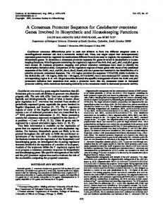
Results The structure of the consensus signal peptide is shown in Fig. 1. Amino acid analysis of the purified synthetic peptide (Table 1) and electrophoresis of the peptide on paper at pH 1.9 (RASP 1.12) indicated that the required peptide had been synthesized, and was homogeneous. Complete cleavage by CNBr into two peptides, residues 1-9 (RASP 1.12, RF 0.79 on t.l.c.) and residues 10-19 (RASP 1.66, RF 0.29 on t.l.c.), showed that the methionine residue was not oxidized. Tryptic digestion yielded three peptides, Lys-Lys (RASP 2.94) Ala-Asp-Lys-Glu (RASP 1.72) and a 1 3-residue peptide (residues 3-15 inclusive), as expected from its sequence. The synthetic consensus peptide was tested for its effect on the processing of the products of translation of mRNA isolated from three different sources in translation systems in vitro derived from nuclease-treated rabbit reticulocyte lysates supplemented with stripped rough microsomes isolated
+
+
+
Lys-Lys-Ser-Ala-Leu-Leu-Ala-Leu-Met-Tyr-Val-Cys-Pro-Gly-Lys-Ala-Asp-Lys-Glu h
h
i
H
H
H
H
H
H
b
h
i
B
B
h
H
I
h
H
fib
b
b
i
h
h
i
h
h
H
H
h
B
b
b
i
B
b
B
8.1
x
10-4
a
Pt
=
H.1 = 2.0
Fig. 1. Structure of the consensus signal sequence The peptide sequence was designed by examination of common structural features occurring in 52 known signal sequences (Austen & Ridd, 1981). The influence of residues on predicted secondary structure: aH, strong a-former; ah, a-former; aI, weak a-former; ai, indifferent; ab, a-breaker; aB, strong a-breaker; PH, strong fl-former; Ph, /1former; fli, indifferent; fib, fl-breaker; fiB, strong P-breaker. Potential for P-turn (PF) was calculated according to Chou & Fasman (1978). The average hydrophobicity (Hi) was derived as described by Segrest & Feldman (1974).
Vol. 224
320
B. M. Austen, J. Hermon-Taylor, M. A. Kaderbhai and D. H. Ridd Table 1. Amino acid analyses of consensus signal peptide N.D., not detected Composition (mol/mol of peptide) Residue
Theoretical
Acid hydrolysis (20h) 1.03 0.90 1.11 0.98 1.07 2.99 1.00 0.97 2.88 0.93 3.97 N.D.
Asp Ser Glu Pro Gly Ala
Val. Met Leu
Tyr Lys Cys
e-prolacTifl
Aminopeptidase/chymotrypsin (2h) 0
0.98 0 0 0 1.84 0.57 0.72 2.16 0.59 2.02 0
I
3 3
4
.__ j
UiAj3IJin_ _
-
Prolactin~ ::~ ~ ~ ~~~~~~ ~.. ...:.... .... .::
Track
no.
1
2
3
4
5
6
7
8
9
10
11
12
Fig. 2. Blockage of processing of bovine pre-prolactin by consensus signal peptide Translation of bovine pituitary mRNA was performed in the presence of pancreatic microsomal membranes and peptide, and 35S-labelled proteins were analysed by electrophoresis on polyacrylamide (15%) in SDS. Track 1, -EDTA membranes, -peptide; track 2, +EDTA membranes, -peptide; track 3, +EDTA membranes, +peptide (5gM final concentration); track 4, +EDTA membranes, +peptide (25 gM); track 5, +EDTA membranes, +peptide (50 Mm); track 6, +nuclease membranes, -peptide; track 7, +nuclease membranes, +peptide (3Mm); track 8, +nuclease membranes, +peptide (7.5 Mm); track 9, +nuclease membranes, +peptide (12.5 gM); track 10, +nuclease membranes, +peptide (20Mm); track 11, +nuclease membranes, +peptide (35 gM); track 12, +nuclease membranes, +peptide (50 Mm).
from dog pancreas. Under the electron microrough microsomes were seen to consist entirely of vesicles from about 200 to 600Mm in diameter, studded with ribosomes. The latter were lost after treatment with EDTA, but the membranes retained their vesicular appearance. In the presence of EDTA-stripped rough microsomes, the major product of translation of bovine pituitary mRNA, preprolactin (Fig. 2, track 1), was converted into a form which migrates faster on SDS/polyacrylamide gels, and has been shown to be identical to the mature secreted form of prolactin (Shields & Blobel, 1977) (Fig. 2, track 2). The scope,
extent of processing determined by densitometry
achieved with nuclease-treated microsomes (35%) was less than that achieved with EDTA-stripped microsomes (70%) (compare tracks 6 and 2). Processing of nascent pre-prolactin was inhibited in a dose-dependent fashion by 3-50 gM concentrations of the desulphonylated peptide with both EDTA-stripped rough microsomes (tracks 3-5) and nuclease-treated rough microsomes (tracks 7-12). The peptide was also tested for its inhibitory action against processing of products of translation of mRNA obtained from two other sources. The 1984
321
Synthesis and properties of a signal peptide
and bands in the region 27000-33000 are the preforms of serine zymogens, of which the member of lowest Mr has been found to be cationic pretrypsinogen (Ridd et al., 1982). A smaller protein of Mr 16000 is pre-prophospholipase. When EDTA-stripped microsomes were included in the translation, significant changes occurred in the banding pattern, and smaller proteins were produced, clearly seen in the production of cationic trypsinogen from pre-trypsinogen and prophospholipase from its pre-form (track 6). The addition of consensus signal peptide again caused inhibition of this change, being more potent at 48 Mm (track 7) than 24Mm (track 8). To investigate further the specificity of the action of the consensus sequence in blocking processing of pre-forms into mature forms of secreted proteins, effects of control peptides were studied. Peptides formed by cleavage of the consensus sequence by CNBr and trypsin, an additional hydrophobic pentapeptide, a hydrophobic peptide fragment from human PTH (residues 28-48), and the 17-residue peptide y-endorphin were tested for their efficiency at inhibiting the cotranslational processing of bovine pre-prolactin. The extent of inhibition of processing, measured by densitometry of X-ray films, in the presence of various peptides are shown in Fig. 4. Whereas 50 M of the whole sequence completely prevented processing, the tryptic fragments together at 50 M
35S-labelled products produced by translation of human placental mRNA in a rabbit reticulocyte lysate is shown in Fig. 3 (track 1). A major radiolabelled protein with an apparent M, of 25 000 was produced. A second high-Mr 35S-labelled product seen on the gel (tracks 1-4) was also produced by reticulocyte lysates not programmed with exogenous mRNA, and results from ribosome-independent addition of [35S]methionine to a pre-existing protein (Jackson & Hunt, 1983). When EDTAstripped microsomes were present during translation, a smaller labelled protein was produced (track 2), which has previously been shown to correspond to authentic human placental lactogen (Szczesna & Boime, 1976). Consensus peptide inhibits this conversion progressively at concentrations of 24pM (track 4) and 48pM (track 3). 35Slabelled products yielded by translation of dog pancreatic mRNA in a rabbit reticulocyte lysate are shown in Fig. 3 (track 5). Assignments have been made by comparison with the results of Scheele et al. (1980), and by changes yielded in the banding pattern by including enterokinase, (EC 3.4.21.9), a proteinase which selectively cleaves trypsinogen, in the translation mixture (Ridd et al., 1982). Products of Mr 55000 and 57000 are preamylase and pre-lipase, bands in the region of 45 000-48 000 include the pre-forms of procarboxypeptidases plus the [35S]methionine-labelled protein mentioned above and also seen in tracks 1-4,
_ I_ _nS~'u.w *:
4I._...: ..
_-Pre ~ arnylase pre-lipase
-Pre-carboxypeptidases
_t_
Pre trypsinoqen
Pre-placental lactogen-
-.Pre
_
;
1
proelastase
chyrnotrypsinogen -.TPre-trypsinogen 2 Pre
Trypsinogent
2
Placental lactoqeni-
Pre-prophospholipase -Prophospholipase
-
Track rno.
2
3
4
5
6
7
8
Fig. 3. Blockage of processing of human placental lactogen and dog pancreatic pre-digestive enzymes by consensus signal peptide The effects of desulphonylated signal peptide on processing achieved in the presence of EDTA-stripped microsomes were examined in translation systems programmed with full-term human placental RNA (25.ug/ml) (tracks 1-4) or dog pancreatic RNA (70pg/ml) (track 5-8) as described in the Materials and methods section. 35S-labelled proteins were separated on gradient polyacrylamide gels (10-17.5, w/v) in SDS. Tracks 1 and 5 show 35S-labelled proteins translated by mRNA alone, tracks 2 and 6 the 35S-labelled proteins yielded in the presence of microsomes, and other tracks the effects of signal peptide at 24pM (tracks 3 and 7) or 48Mm (tracks 4 and 8) concentrations.
Vol. 224
B. M. Austen, J. Hermon-Taylor, M. A. Kaderbhai and D. H. Ridd
322
80
/
(' 60-
CNBr fraogieiils
0
°
40 0
Tryptic fragmenllts
.20
C~20
~~
y-Endorphin
PTH-(28 48) pepti(de (A) _ _ -~~~~~~~~~~0 LAYVA (C))
10
20
30
40
50
[Peptidel (,M) Fig. 4. Effects on processing of bovine pre-prolactin by fragments of consensus signal peptide, and control peptides y-Endorphin, PTH-(28-48)-peptide, combined CNBr peptides, and tryptic peptides released by digestion of the consensus signal sequence, and a hydrophobic synthetic pentapeptide Leu-Ala-TyrVal-Ala (LAYVA), were incubated with nucleasetreated dog rough microsomes, and added to a translation system programmed with poly (A)containing RNA from bovine pituitary as described in the Materials and methods section. 35S-labelled proteins were separated by electrophoresis on polyacrylamide (15%, w/v) in SDS, fluorographed, and quantified by densitometric scanning of the X-ray film.
reduced processing by two-thirds, and the CNBr fragments by one-fifth. The hydrophobic pentapeptide Leu-Ala-Tyr-Val-Ala and PTH-(28-48)peptide were not inhibitory at 5OpM concentrations, while y-endorphin was slightly inhibitory. The observed inhibition of proteolytic processing, which gives rise to mature proteins of lower Mr than the pre-forms, does not allow us to ascertain whether the peptide inhibits binding ofthe nascent pre-proteins to microsomal membranes, inhibits passage of nascent pre-proteins across microsomal membranes, or inhibits signal peptidase, the enzyme which proteolytically removes the signal sequence and gives rise to a decrease in Mr. It has been shown (Blobel & Dobberstein, 1975) that proteolysis of nascent protein chains by signal peptidase only occurs if first the protein is translocated across the microsomal membrane, presumably because the active site of the peptidase is confined to the lumenal surface of microsomes. Thus, post-translational limited proteolysis was performed in order to ascertain the location of the precursor that accumulates in the presence of signal peptide. The results are shown in Fig. 5. In the presence of the consensus sequence, inhibition of processing occurs (track 1). In the presence of deoxycholate, chymotrypsin and trypsin added at
the end of translation cause disappearance of both pre-prolactin and its mature form (track 2). In the absence ofdetergent, almost all of the pre-form disappears, while the mature form is resistant (track 3). The results of the same conditions of limited proteolysis applied to 35S-labelled proteins translated in the absence of inhibition by peptide (tracks 4, 5 and 6) shows that again the mature form is resistant to proteolysis in the absence of detergent, but that the pre-form is susceptible. Tracks 7, 8 and 9 show the results obtained when post-translational proteolysis is performed on unprocessed [35S]pre-prolactin translated in the absence of microsomes. As expected, the pre-form is completely degraded. Thus, because the preform is still accessible to proteinases when translated in the presence of peptide, the predominant mode of inhibition occurs on the cytoplasmic surface of the microsomal residues, although additional inhibition of signal peptidase cannot be excluded. The results also show that, although the peptide contains both hydrophobic and hydrophilic regions, it does not cause inhibition by detergent-like disruption of the membranes which would expose the processed form of prolactin to exogenous proteinases. Discussion The 19-residue consensus sequence was devised by consideration of common structural features contained in known signal sequences, as described previously (Austen & Ridd, 1981). Calculated average hydrophobicity over the uncharged stretches of amino acid residues found in many naturally-occurring signal sequences was 2.31 + 0.49 calculated according to the index of Segrest & Feldman (1974). The hydrophobicity index of the uncharged stretch in the consensus signal (residues 3-14) was 2.0, in keeping with the range of values found in natural sequences (Fig. 1). In keeping with the double amphiphilic structures commonly found, the consensus peptide was made with two consecutive lysine residues at the Nterminus and a mixture of basic and acidic residues at the C-terminus (Fig. 1). A sequence Gly-LysAla-Asp was chosen at the C-terminal end of the consensus peptide as this would be in keeping with the specificity found at the region at which signal peptidase cleaves. This has been summarized as Aaa-Xaa-Baa-Yaa (Austen & Ridd, 1981), where Yaa is the N-terminus of the mature protein, Xaa is any residue, Aaa is an uncharged residue, and Baa is an uncharged residue with a small side chain. The consensus sequence would thus be anticipated to offer a potential cleavage site to signal peptidase between residues 16 and 17. An N-terminal methionine residue was not included, as this is 1984
323
Synthesis and properties of a signal peptide
Pre-prolactin
-l.
Prolactin -.........
1
2
3
4
Signal peptide
+
+
+
_
Microsomes
+
+
+
+
Deoxycholate
-
+
Proteinases
-
+
Track
no.
5
6
7
8
9
+
+
_
+
+
+
+
-
+
+
Fig. 5. Examination of the state of protection ofprocessed bovine prolactin to post-translational proteolysis Three translations of bovine pituitary poly(A)-containing RNA were performed, one in the presence of EDTAstripped microsomes plus desulphonylated signal peptide at 50pM final concentration (tracks 1-3), one in the presence of microsomes (tracks 4-6) and one with no additions apart from compensating amounts of Hepes/KOH buffer or 0.25M-sucrose (tracks 7-9), as described in the Materials and methods section. After translation, each incubation mixture was divided into three; one part was further incubated with proteinases and detergent (tracks 2, 5 and 8), one part with proteinases alone (tracks 3, 6 and 9), and one with no additions (tracks 1, 4 and 7). After addition of Trasylol, 35S-labelled proteins were separated on polyacrylamide (15%, w/v) in SDS.
known to be removed from nascent polypeptides after about 20 residues have been biosynthesized (Palmiter et al., 1978). The 19-residue peptide we have synthesized has been shown to compete effectively with naturally occurring signal sequences contained in bovine pre-prolactin, human pre-placental lactogen and the pre-forms of dog pancreatic digestive enzymes, leading to inhibition of processing of these preforms in vitro. The concentration at which it exerts inhibition, i.e. from 3 to 50pm, is similar to the concentrations at which a synthetic peptide encompassing the pre- and pro-parts of pre-proparathyroid hormone was found to inhibit processing of hormone pre-proteins in vitro (Majzoub et al., 1980), and those at which a peptide resembling the signal of dog pretrypsinogen has been found to inhibit processing of dog pancreatic pre-proteins Vol. 224
(Austen & Ridd, 1981). The fact that the nineteenresidue peptide represents a consensus of known signal peptides, yet acts in a similar way to peptides that are naturally-occurring sequences, shows that it is the common structural features incorporated that are important for translocation, and not precise, highly conserved primary sequences of the type originally discovered in the signal sequences of dog pancreatic zymogens (Devillers-Thiery et al., 1975). In keeping with this idea is the demonstration that the consensus sequence inhibits with similar potency the processing of pre-proteins of three different species from three different types of cells. Thus, it is unlikely that each cellular type is equipped with its own subset of microsomal receptors or other proteins involved in translocation, but rather that very similar protein trans-
324
B. M. Austen, J. Hermon-Taylor, M. A. Kaderbhai and D. H. Ridd
locators exist in different cells. Moreover, it has 'been shown that microsomal membranes from one cell type can process pre-proteins from a variety of different sources, including prokaryotic organisms (Muller et al., 1982). It has also been found that microsomal membranes from different sources process the same pre-protein so that it is likely that protein translocators have been closely conserved in the course of evolution. The precise mechanism whereby the synthetic signal peptide blocks translocation has yet to be determined. Studies of the effects of post-translational proteolysis indicate that interference occurs on the cytoplasmic surface of microsomal vesicles. Recent studies have shown that an 1251labelled form of the consensus signal peptide binds to a saturable receptor that is present in stripped rough microsomal membranes, with KD of about 1 X 1O-7M (Austen & Ridd, 1983). Our present studies show that much higher concentrations of this peptide are required to achieve blocking in the translocation system; the differences could, perhaps, be explained if the affinity of the receptor was altered by components present in the lysate translation system. Alternatively, the high-affinity binding sites discovered with 125I-labelled signal peptides may not be involved in translocation. We have not yet been able to demonstrate direct binding of the consensus signal peptide to the 11 S ribonucleoprotein complex isolated by Walter &
Blobel (1981), although we cannot exclude the possibility that observed inhibition of processing involves interaction with this complex. The structural features that are essential for interactions with microsomal translocators have not been fully elucidated. However, it is clear that a short hydrophobic sequence would not necessarily be active as the peptide Leu-Ala-Tyr-Val-Ala, which has a hydrophobicity index, as defined by Segrest & Feldman (1974), of 2.6, does not block processing in vitro. A relatively long section of uncharged, hydrophobic residues would appear to be important, as cleavage in the central core of the consensus sequence by CNBr results in marked loss of inhibitory protency. The typical double amphiphilic structure of sequences containing a basic region separated from a region of mixed charges by an uncharged, hydrophobic region would appear to be important in the function of signals (Austen & Ridd, 1981). The tryptic fragments of the consensus sequence do not possess this double amphiphilic structure. The reduced inhibition of processing by these fragments may result from the hydrophobic nature of the fragment peptide containing residues 3-15, and it is noteworthy that Inouye et al. (1982), have recently shown that change of basic residues at the Nterminus of the signal of a bacterial membrane prelipoprotein slows, but does not prevent, assembly of the processed form of this protein into mem-
Fig. 6. Molecular model of the consensus signal peptide The 19-residue peptide, built with CPK precision space-filling models, is shown. Residues 1-11 are a-helical, and a P-turn exists between residues 12 and 15. The hydrogen bond between the carbonyl oxygen of Cys to the amido nitrogen of Ala is marked with an arrow, holding the tetrapeptide sequence in a type 1 f-turn, which produces a reversal in the direction of the polypeptide chain.
1984
Synthesis and properties of a signal peptide branes. y-Endorphin, which also possesses a double amphiphilic structure, but is different in that it contains two short stretches of hydrophobic uncharged residues separated by charged residues positioned in the centre of the peptide, is only weakly inhibitory. The lack of significant inhibition by human PTH-(28-48)-peptide shows that pronounced hydrophobicity in a relatively long uncharged sequence (11 residues) is insufficient in the absence of the typical charge distribution and potential for folding incorporated in the consensus sequence. Human PTH-(28-54)-peptide has previously been shown not to block processing of preprolactin (Majzoub et al., 1980). Predictions of conformation, and calculations of conformational energy, have shown that signal sequences are remarkably similar, despite differences in primary structure (Pincus & Klausner, 1982; Austen, 1979; Gamier et al., 1980), and that their internal cores of uncharged hydrophobic residues are likely to adopt a-helical conformation, terminated by a fl-turn. These empirical considerations have been supported by chiroptical measurements on synthetic peptides (Rosenblatt et al., 1980), and it has been shown that an antibody directed against a particular signal peptide can recognize other signal peptides, possibly by acting as a conformational probe (Baty & Lazdunski, 1979). It is likely that, in the course of evolution, conformation of signals has been conserved rather than primary sequence. The consensus sequence has been designed to optimize the potential for folding into the common preferred conformation, shown in a space-filling model in Fig. 6. The calculated potential for a-helix and fl-turn in the sequence (Chou & Fasman, 1978) is depicted in Fig. 1. The model represents the type of structure that is recognized by the cytoplasmic surface of endoplasmic reticulum in secretory cells. The nature of the components in these membranes that interact with signals has not been elucidated, but this soluble signal peptide should provide a useful probe for further studies. We gratefully acknowledge the support of the Wellcome Trust, and Miss M. Moss for examination of microsomal vesicles with the electron microscope.
References Austen, B. M. (1979) FEBS Lett. 103, 308-313 Austen, B. M. & Ridd, D. H. (1981) Biochem. Soc. Symp. 46, 235-258 Austen, B. M. & Ridd, D. H. (1983) Biochem. Soc. Trans. 11, 160-161
Vol. 224
325 Bantle, J. A., Maxwell, I. H. & Hahn, W. E. (1976) Anal. Biochem. 72, 413-427 Baty, D. & Lazdunski, C. (1979) Eur. J. Biochem. 102, 503-507 Blobel, G. & Dobberstein, B. (1975) J. Cell Biol. 67, 852862 Blobel, G., Walter, P., Chang, C. N., Goldman, B. M., Erikson, A. H. & Lingappa, V. R. (1979) Symp. Exp. Biol. 33, 9-36 Chan, S. J., Patzelt, C., Duguid, J. R., Quinn, P., Labreque, A., Boyes, B., Keim, P., Heinrikson, R. L. & Steiner, D. F. (1979) Miami Winter Symp. 16, 361378 Chirgwin, J. M., Przybyla, A. E., MacDonald, R. J. & Rutter, W. J. (1979) Biochemistry 18, 5294-5299 Chou, P. Y. & Fasman, G. D. (1978) Annu. Rev. Biochem. 47, 251-276 Devillers-Thiery, A., Kindt, T., Scheele, G. & Blobel, G. (1975) Proc. Natl. Acad. Sci. U.S.A. 72, 5016-5020 Erickson, B. W. & Merrifield, R. B. (1976) Proteins 3rd Ed. 2, 349-351 Gamier, J., Gaye, P., Mercier, J.-C. & Robson, B. (1980) Biochimie 62, 231-239 Gisin, B. (1973) Helv. Chim. Acta 56, 1476-1482 Inouye, S., Soberon, X., Franceschini, T., Nakamura, K., Itakura, K. & Inouye, M. (1982) Proc. Natl. Acad. Sci. U.S.A. 79, 3438-3441 Jackson, R. J. & Hunt, T. (1983) Methods Enzymol. 96, 50-74 Kaiser, E., Colescott, R. L., Bossinger, C. D. & Cook, P. 1. (1970) Anal. Biochem. 34, 595-598 Laskey, R. A. & Mills, A. D. (1975) Eur. J. Biochem. 56, 335-341 Majzoub, J. A., Rosenblatt, M., Fennick, B., Maunus, R., Kronenberg, H. M., Potts, J. T. & Habener, J. F. (1980) J. Biol. Chem. 255, 11478-11483 Meyer, D. I., Krause, E. & Dobberstein, B. (1982) Nature (London) 297, 647-650 Muller, M., Ibrahimi, I., Chang, C. N., Walter, P. & Blobel, G. (1982) J. Biol. Chem. 257, 11860-11863 Palmiter, R. D., Gagnon, J. & Walsh, K. A. (1978) Proc. Natl. Acad. Sci. U.S.A. 79, 3413-3417 Pelham, H. R. B. & Jackson, R. J. (1976) Eur. J. Biochem. 67, 247-256 Pincus, M. R. & Klausner, R. D. (1982) Proc. Natl. Acad. Sci. U.S.A. 79, 3413-3417 Ridd, D. H., Hermon-Taylor, J. & Austen, B. M. (1982) Biochem. Soc. Trans. 10, 46-47 Rosenblatt, M., Beudette, N. V. & Fasman, G. D. (1980) Proc. Natl. Acad. Sci. U.S.A. 77, 3983-3987 Scheele, G., Jacoby, R. & Came, T. (1980) J. Cell Biol. 87, 611-628 Segrest, J. P. & Feldman, R. J. (1974) J. Mol. Biol. 87, 835-838 Shields, D. & Blobel, G. (1977) Proc. Natl. Acad. Sci. U.S.A. 74, 2059-2063 Szczesna, E. & Boime, I. (1976) Proc. Natl. Acad. Sci. U.S.A. 73, 1179-1183 Walter, P. & Blobel, G. (1981) J. Cell Biol. 91, 557561