International Journal of
Molecular Sciences Article
The Acute Inflammatory Response to Absorbed Collagen Sponge Is Not Enhanced by BMP-2 Hairong Huang 1 , Daniel Wismeijer 1 , Ernst B. Hunziker 2 and Gang Wu 1, * 1
2
*
Department of Oral Implantology and Prosthetic Dentistry, Academic Centre for Dentistry Amsterdam (ACTA), University of Amsterdam and Vrije Universiteit Amsterdam, Amsterdam Movement Sciences, Gustav Mahlerlaan 3004, 1081LA Amsterdam, The Netherlands;
[email protected] (H.H.);
[email protected] (D.W.) Departments of Osteoporosis and Orthopaedic Surgery, Inselspital (DKF), University of Bern, Murtenstrasse 35, 3008 Bern, Switzerland;
[email protected] Correspondence:
[email protected]; Tel.: +31-20-598-0866; Fax: +31-20-598-0333
Academic Editor: Allison Cowin Received: 1 January 2017; Accepted: 16 February 2017; Published: 25 February 2017
Abstract: Absorbed collagen sponge (ACS)/bone morphogenetic protein-2 (BMP-2) are widely used in clinical practise for bone regeneration. However, the application of this product was found to be associated with a significant pro-inflammatory response, particularly in the early phase after implantation. This study aimed to clarify if the pro-inflammatory activities, associated with BMP-2 added to ACS, were related to the physical state of the carrier itself, i.e., a wet or a highly dehydrated state of the ACS, to the local degree of vascularisation and/or to local biomechanical factors. ACS (0.8 cm diameter)/BMP-2 were implanted subcutaneously in the back of 12 eight-week-old Sprague Dawley rats. Two days after surgery, the implanted materials were retrieved and analysed histologically and histomorphometrically. The acute inflammatory response following implantation of ACS was dependent of neither the presence or absence of BMP-2 nor the degree of vascularization in the surrounding tissue nor the hydration state (wet versus dry) of the ACS material at the time of implantation. Differential micro biomechanical factors operating at the implantation site appeared to have an influence on the thickness of inflammation. We conclude that the degree of the early inflammatory response of the ACS/BMP-2 may be associated with the physical and chemical properties of the carrier material itself. Keywords: bone morphogenetic protein-2 (BMP-2); absorbed collagen sponge (ACS); inflammation; vascularization; biomechanical
1. Introduction Recombinant human bone morphogenetic protein-2 (BMP-2), a member of the transforming growth factor beta (TGF-β) superfamily, is in clinical use since more than a decade [1,2]. It is used in clinical practice for spinal fusion [3] and for treatment of non-unions to enhance bone formation processes and to accelerate the bony healing response; in dental practice it is used for oral and maxillofacial reconstruction [4,5]. Even though the clinical use of BMP-2 is very successful, its clinical application is associated with some serious unwanted effects such as heterotopic bone formation [6], bone resorption (by osteoclast activation) and formation of cyst-like bone voids [7], as well as postoperative inflammatory swelling [8,9] and neurological symptoms, etc. BMP-2 is clinically applied as a free factor (Infuse® (USA), Inductos® (Europe)) together with an ACS as a carrier. BMP-2 of this product is used in very high dosage, and it is believed that it is this high dosage level of BMP-2 that leads to extensive inflammatory responses. This use-associated Int. J. Mol. Sci. 2017, 18, 498; doi:10.3390/ijms18030498
www.mdpi.com/journal/ijms
Int. J. Mol. Sci. 2017, 18, 498
2 of 11
inflammation is one of the main reasons why several of the above-described unwanted effects do occur. It is also believed by many authors that BMP-2 itself contributes significantly to the enhancing of the inflammatory response during and after the implantation of the construct in this kind of a tissue engineering approach. Indeed, several publications report that BMP-2 itself enhances the swelling and the inflammatory response in the surrounding soft tissues in conjunction with the carrier material (ACS) [10]. Seroma formation is, for example, a frequently observed side effect of BMP-2-use, encountered most commonly in the first week postoperatively, as described in several studies [8,11]. Rihnet et al. [12] found that lumbar seromas occurred in 1.2% of rhBMP-2 treated patients compared to 0% in the control patient population. Robin et al. [8] described postoperative seroma formations associated with BMP-2 use in the surrounding soft tissues in the cervical region that led to bilateral paresthesia of the upper extremities. In clinical cases with BMP-2-induced seromas, elevated serum levels of inflammatory cytokines were found, such as those of IL-6, IL-8, and TNF-α [13], as well as those of IL-10 [10]. Indeed in the publication of Lee et al. [10], a dose-dependency of the inflammatory response to high dosage levels of BMP-2 was found. However, in a report of Wu et al. [14] it was described that BMP-2, in particular when delivered in a slow release system, is able to attenuate inflammatory responses. In other in vivo animal experiments [15], microcomputed tomography and histological analyses confirmed that PCL/PLGA/collagen/rhBMP-2 scaffolds (long-term delivery mode) showed the best bone healing quality at both weeks 4 and 8 after implantation without inflammatory response. Thus, conflicting data are encountered in the scientific literature respecting the role of BMP-2 and it use-associated inflammation. The purpose of this study was to investigate if the use of BMP-2, when applied at high concentrations as a free factor together with a carrier material (ACS), is indeed associated with a pro-inflammatory response in the acute phase of the body response, i.e., in the initial two days after implantation of this growth factor with the carrier material. It is, indeed, conceivable that it is not the BMP-2 itself that triggers the intensive inflammatory response, but that the inflammation may be elicited by a number of other factors operating in close topographical vicinity to the deposited collagen carrier. Such candidate factors may be the degree of tissue vascularity, or the local micromechanical conditions of different physiological stress fields, i.e., depend on differences in the local biological environment (differential niche biology). Another role may be played by the physical state in which the collagen carrier itself is deposited, i.e., inserted in a dry state or in a wet state into the living tissue spaces. Since burst release of BMP-2 (in surgical practise poured onto the ACS sponge) does readily occur, among others due to mechanical manipulation of the construct itself during surgical implantation [16], we set up in our experiments a specific control group in which the collagen carrier was kept in a dry state to assess the possible role of such mechanical stress-modulated release profiles of BMP-2 in the inflammatory response. In order to clarify the possible role of these various candidate factors, the Sprague Dawley (SD) rat was used as the animal model. ACS carrier material was implanted in the subcutaneous space in the back area (lumbar level). By this set up the deposited collagen carrier patch is exposed on one side towards the skin, where the skin muscles of the rat generate a continuous instability situation, i.e., a high biomechanical instability [17]. On the opposite side of the collagen patch, facing the large underlying lumber muscle package, a relatively stable micromechanical environment is present. In addition, the two different biomechanical niches around these implants are also characterized by specific differential densities of blood vessels. The differential blood vessel densities at these two opposite locations (skin side versus lumbar body side) were quantified in this study in order to elucidate their possible proinflammatory contribution.
Int. J. Mol. Sci. 2017, 18, 498
3 of 11
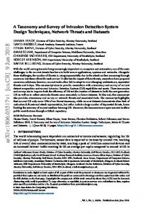
2. Results Figure 1A–D illustrated that already on the 2nd day after implantation, all collagen implants were surrounded by498 a capsule of inflamed tissue (delineated by a red line), and was highly vascularized. Int. J. Mol. Sci. 2017, 18, 3 of 11 The inflammatory response involved large numbers of macrophages around each of the implanted collagen sponges 1E). The border thethe inflammation border collagen implant wasby a collagen implant was(Figure delineated byouter a red line of and inner border of of thetheinflammatory zone delineated by a red line and the inner border of the inflammatory zone by a yellow line (Figure 1A–D). yellow line (Figure 1A–D).
Figure 1. Microscopic findings following of:(A) (A) dry Absorbed Collagen Figure 1. Microscopic findings followingsubcutaneous subcutaneous implantation implantation of: dry Absorbed Collagen Sponge (ACS), (B) (B) drydry bone morphogenetic (BMP-2)/ACS, ACS, (D) ACS/BMP-2; wet ACS/BMPSponge (ACS); bone morphogenetic protein-2 protein-2 (BMP-2)/ACS; (C)(C) wetwet ACS; (D) wet highmagnification magnification of of inflamed inflamed zone. zone waswas 2, (E)(E) high zone. Red Red arrow: arrow:macrophage. macrophage.The Theinflammatory inflammatory zone delineated by two different lines: outer border red, theinner innerborder borderin in yellow. yellow. Bar delineated by two different lines: thethe outer border in in red, the Bar == 500 500 µm µm (in (in A–D). The upper side is skin side and the lower side is lumber body side. Numerous macrophages A–D). The upper side is skin side and the lower side is lumber body side. Numerous macrophages were identified in the highly vascularized inflamed zone (cf. 1E, bar = 20 µm). were identified in the highly vascularized inflamed zone (cf. 1E, bar = 20 µm).
The degree of inflammation activitywas wasgauged gauged by by estimation of the implanted The degree of inflammation activity estimationofofthe thevolume volume of the implanted sample and the volume of the inflamed tissue. As Figures 2 and 3 showed, the volumes of the implanted sample and the volume of the inflamed tissue. As Figures 2 and 3 showed, the volumes of the collagen sample and the inflammation area were increased when the carrier (ACS) was loaded with implanted collagen sample and the inflammation area were increased when the carrier (ACS) was BMP-2. However, there were no significant differences observed between the collagen sponge volumes loaded with BMP-2. However, there were no significant differences observed between the collagen in the presence or absence of BMP-2, nor if implanted in a wet or a dry (dehydrated) state. sponge volumes in the presence or absence of BMP-2, nor if implanted in a wet or a dry (dehydrated) state. As Figure 4 illustrates, the mean thickness of the inflamed tissue at the skin side and the lumbar body side is different, and significant differences were indeed found around the dry ACS implants in the absence of BMP-2 (p = 0.001), and in the wet ACS groups in the presence (p = 0.0009) or absence
Tissue volume of collagen implant Tissue volume of collagen implant 3 3 (mm (mm ) )
higher on the body side than on side, was notgroup, significantly to a high degree of lumbar variation; cf. SEM-error barthe in skin Figure 5), but in the wet the areadifferent density (due of blood high degree variation; SEM-error bar in Figure 5), than in thethat wetongroup, theside area(pdensity blood vessels on theoflumbar bodycf.side was significantly higher the skin = 0.032).ofFigure on the lumbar body side wasvessel significantly higher than that onon thethe skin side (p =(Figure 0.032).6A, Figure 6vessels illustrates typical areas and blood densities as encountered skin side C) 6 illustrates typical blood and the lumbar bodyareas side and (Figure 6B,vessel D). densities as encountered on the skin side (Figure 6A, C) Int. J.the Mol.lumbar Sci. 2017, body 18, 498 side (Figure 6B, D). 4 of 11 and 25 25 20 20 15 15 10 10 5 5 0 0
n.s. n.s.
ACS ACScollagen Dry
n.s. n.s.
ACS/BMP-2 ACS/BMP-2 Wet collagen
Dry collagen
Tissue volume of inflammation Tissue volume of inflammation 3 3) (mm ) (mm
Wet collagen Figure 2. Mean volumes of collagen implants. No significant differences were found between dry Figure 2. Mean volumes of collagen implants. No significant differences were found between dry ACS Figure Mean volumes nor of collagen differences dry ACS and2.dry ACS/BMP-2 betweenimplants. wet ACS No andsignificant wet ACS/BMP-2. Data were were found presentbetween as Means ± and dry ACS/BMP-2 nor between wet ACS and wet ACS/BMP-2. Data were present as Means ± SEM. ACS and ACS/BMP-2 nor between wet ACS and wet ACS/BMP-2. Data were present as Means ± SEM. n.s.: dry no significant difference. n.s.: no significant difference. SEM. n.s.: no significant difference. 15 15 10 10
n.s. n.s.
n.s. n.s.
5 5 0 0
ACS ACScollagen Dry
ACS/BMP-2 ACS/BMP-2 Wet collagen
Dry collagen
Wet collagen
Figure 3. Periimplant inflammation volume. No significant differences were found between dry ACS
Figure 3. Periimplant inflammation volume. No significant differences were found between dry ACS and dry ACS/BMP-2 nor between wet ACS and wet ACS/BMP-2. Data were present as Means ± SEM. Figure Periimplantnor inflammation Nowet significant differences werepresent found between ACS and dry3.ACS/BMP-2 between wetvolume. ACS and ACS/BMP-2. Data were as Meansdry ± SEM. n.s.: no significant difference. and no drysignificant ACS/BMP-2 nor between wet ACS and wet ACS/BMP-2. Data were present as Means ± SEM. n.s.: difference. n.s.: no significant difference.
As Figure 4 illustrates, the mean thickness of the inflamed tissue at the skin side and the lumbar body side is different, and significant differences were indeed found around the dry ACS implants in the absence of BMP-2 (p = 0.001), and in the wet ACS groups in the presence (p = 0.0009) or absence (p = 0.009) of BMP-2. The differential blood vessel densities at these two opposite locations (skin side versus lumbar body side) were quantified in this study in order to elucidate their possible role to contribute to the proinflammatory response. As Figure 5 shows, the area density of blood vessels on both sides were different, the area density of blood vessels in the dry group without BMP-2 on the lumbar body side was significantly higher than that on the skin side (p = 0.014) but no significance was found in the wet group. In the group with BMP-2, the area density of blood vessels in the dry group was found to be higher on the lumbar body side than on the skin side, but was not significantly different (due to a high degree of variation; cf. SEM-error bar in Figure 5), in the wet group, the area density of blood vessels on the lumbar body side was significantly higher than that on the skin side (p = 0.032). Figure 6 illustrates typical areas and blood vessel densities as encountered on the skin side (Figure 6A,C) and the lumbar body side (Figure 6B,D).
Thickness of inflammation zone Thickness Thickness of of inflammation inflammation zone zone (μm) (μm) (μm)
Int. J.J. Mol. Mol. Sci. 2017, 2017, 18, 498 498 Int. Int. J. Mol.Sci. Sci. 2017,18, 18, 498
600 600
of 11 11 55 5of of 11
*** ***
n.s. n.s.
****
*** ***
400 400
200 200
00
dry dryACS ACS
dry dryACS/BMP-2 ACS/BMP-2 wet wetACS ACS
skin skinside side
wet wetACS/BMP-2 ACS/BMP-2
body bodyside side
Area density of blood vessels Area Area density density of of blood blood vessels vessels (percentage) (percentage) (percentage)
Comparison of of the mean mean thickness of of the inflammation inflammation zone on on the skin skin side and and Figure Figure4.4.Comparison Comparison ofthe the meanthickness thickness ofthe the inflammationzone zone onthe the skinside side andthe thebody body are significant differences ininthe thickness of of thethe inflammatory zones between the skin side side. There are significant differences the thickness inflammatory zones between the side. There are significant differences in the thickness of the inflammatory zones between theskin skin and the lumbar body side side in the dry ACS implant group without BMP-2, and in both the wet ACS side sideand andthe thelumbar lumbarbody body sideininthe thedry dryACS ACSimplant implantgroup groupwithout withoutBMP-2, BMP-2,and andininboth boththe thewet wet groups with with orwith without BMP-2. **: p