MEDINFO 2015: eHealth-enabled Health I.N. Sarkar et al. (Eds.) © 2015 IMIA and IOS Press. This article is published online with Open Access by IOS Press and distributed under the terms of the Creative Commons Attribution Non-Commercial License. doi:10.3233/978-1-61499-564-7-1079
1079
Design of a Graph-Based System for Similar Case Retrieval of Pulmonary Nodules José Raniery Ferreira Juniora, Marcelo Costa Oliveiraa, Paulo Mazzoncini de Azevedo-Marquesb a b
University Hospital (HUPAA/EBSERH), Institute of Computing, Federal University of Alagoas, Maceió, Alagoas, Brazil Internal Medicine Department, Ribeirão Preto Medical School, University of São Paulo, Ribeirão Preto, São Paulo, Brazil
Abstract Due to difficulty of diagnosing lung cancer, it is important to integrate computational tools in the imaging interpretation process. Content-Based Image Retrieval (CBIR) can provide decision support to specialists by allowing them to find nodules from a database that are similar to a reference one. However, CBIR systems still face problems visualizing multidimensional images. This paper presents the design of a graphbased system for the retrieval of similar temporal computed tomography (CT) scans of pulmonary nodules, in order to optimize the visualization of multidimensional images in a CBIR system. Temporal Image Registration has been used to compare reference and retrieved previously segmented nodules. A rooted tree graph is employed to visualize retrieved cases, and it is deployed in a web system for portability and usability purposes. Keywords: Pulmonary nodule; image retrieval; multidimensional data.
Introduction Pulmonary nodules are the most common manifestation of lung cancer, the most deadly of all cancers [1]. Lung cancer diagnosis is a challenging task, even for qualified specialists [2]. Furthermore, for the early diagnosis of lung cancer, it is important to measure accurately the change in size, edge and texture of pulmonary nodules from two time-separated CT scans [1]. In order to aid specialists in the diagnosis of lung cancer, it is interesting to integrate the computer-based assistance to the imaging interpretation process. CBIR can provide computer-aided diagnostic support by allowing radiologists to find images from a database that are similar to the images they are interpreting [2]. However, there has been limited investigation into visualization methods for CBIR systems. An effective method of showing the images to the user is a critical aspect for such systems [3]. The goal of this paper is to present the design of a multiplatform visualization CBIR system, for the retrieval of similar multidimensional images of pulmonary nodules in temporal CT scans, using a horizontaloriented rooted-tree-based graph approach.
Materials and Methods The CBIR algorithm was initially implemented by Oliviera and Ferreira [2]. The temporal image database developed for this work is composed by CT lung scans with nodules ≥ 4 mm, with at least 2 time-separated scans. Each nodule of this database is described in four dimensions (x, y, z, t) and will be automatically detected and segmented. After the user selects
the reference case, the system performs the similarity analysis, through Temporal Image Registration, and retrieves the most similar cases, according to Mutual Information metrics. The user can define the number of cases to retrieve. The system displays the most similar cases using a horizontal-oriented rooted-tree-based graph. A regular rooted tree is disposed in a vertical orientation, but for visualization purposes, the tree is presented horizontally. The proposed system is being developed with the multiplatform frameworks HTML5, Java and D3.js for platform portability and user usability purposes.
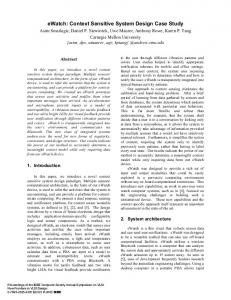
Results and Conclusions The root of the displayed tree is the reference case and its height is 3 (Figure 1). In the first level, each vertex of the tree represents a similar case. The higher the vertex is, the more similar the case is. Users can select one vertex, using a mouse click, to see temporal samples. By clicking a similar case vertex, it expands and the tree reaches to the second level. The second level vertices are the temporal samples. By clicking a temporal sample vertex, it expands and the tree reaches to the third level. The third level vertices are the CT scans with segmented nodules. We expect that the visualization of retrieved similar cases of pulmonary nodules can be improved by the rooted-tree-based graph, and that temporal CBIR visualization systems will employ our approach in their end-user tools.
Figure 1 – Rooted tree graph for nodule temporal retrieval.
References [1] Reeves AP, Chan AB, Yankelevitz DF, Henschke CI. On Measuring the Change in Size of Pulmonary Nodules. IEEE T Med Imaging. 2006: 25(4): 435-450. [2] Oliveira MC, Ferreira JR. A Bag-of-Tasks Approach to Speed Up the Lung Nodules Retrieval in the BigData Age. In: Proceedings of the 15th IEEE International Conference on E-Health Networking, Application & Services (Healthcom 2013); Lisbon, Portugal. 2013: 632–636. [3] Kumar A, Kim J, Cai W, Fulham M, Feng D. ContentBased Medical Image Retrieval: A Survey of Applications to Multidimensional and Multimodality Data. J Digital Imaging. 2013: 26(6): 1025-1039. Address for correspondence:
[email protected]