Microbiology (2009), 155, 2603–2611
DOI 10.1099/mic.0.028712-0
Detection and identification of specific bacteria in wound biofilms using peptide nucleic acid fluorescent in situ hybridization (PNA FISH) Sladjana Malic,1 Katja E. Hill,1 Anthony Hayes,2 Steven L. Percival,3 David W. Thomas1 and David W. Williams1 1
Correspondence
Tissue Engineering and Reparative Dentistry, School of Dentistry, Cardiff University, Heath Park, Cardiff CF14 4XY, UK
Sladjana Malic
[email protected]
2
School of Biosciences, Cardiff University, Park Place, Cardiff CF10 3US, UK
3
School of Medicine, University of West Virginia, Morgantown, WV 26506, USA
Received 26 February 2009 Revised
19 May 2009
Accepted 22 May 2009
Biofilms provide a reservoir of potentially infectious micro-organisms that are resistant to antimicrobial agents, and their importance in the failure of medical devices and chronic inflammatory conditions is increasingly being recognized. Particular research interest exists in the association of biofilms with wound infection and non-healing, i.e. chronic wounds. In this study, fluorescent in situ hybridization (FISH) was used in combination with confocal laser scanning microscopy (CLSM) to detect and characterize the spatial distribution of biofilm-forming bacteria which predominate within human chronic skin wounds (Pseudomonas aeruginosa, Staphylococcus aureus, Streptococcus sp. and Micrococcus sp.). In vitro biofilms were prepared using a constant-depth film fermenter and a reconstituted human epidermis model. In vivo biofilms were also studied using biopsy samples from non-infected chronic venous leg ulcers. The specificity of peptide nucleic acid (PNA) probes for the target organisms was confirmed using mixed preparations of planktonic bacteria and multiplex PNA probing. Identification and location of individual bacterial species within multi-species biofilms demonstrated that P. aeruginosa was predominant. CLSM revealed clustering of individual species within mixed-species biofilms. FISH analysis of archive chronic wound biopsy sections showed bacterial presence and allowed bacterial load to be determined. The application of this standardized procedure makes available an assay for identification of single- or multi-species bacterial populations in tissue biopsies. The technique provides a reliable tool to study bacterial biofilm formation and offers an approach to assess targeted biofilm disruption strategies in vivo.
INTRODUCTION Chronic wounds are an important and often unrecognized cause of disease and disability of the elderly population (Davies et al., 2007; Howell-Jones et al., 2006). These wounds harbour a diverse microflora, which contributes both directly and indirectly to their non-healing phenotype (Stephens et al., 2003; Wall et al., 2002). Much attention has recently been focused on the ability of the bacteria within chronic wounds to form and exist within a biofilm (James et al., 2008). Bacterial biofilms consist of a complex microenvironment of single- or mixed-species bacteria attached to each other or attached to surfaces, being encased within extracellular polymeric substances. Abbreviations: CLSM, confocal laser scanning microscopy; CDFF, constant-depth film fermenter; CVLU, chronic venous leg ulcer; FISH, fluorescent in situ hybridization; PNA, peptide nucleic acid; RHE, reconstituted human epidermis.
028712 G 2009 SGM
Printed in Great Britain
The moist chronic wound surface with its proteinaceous substrate, and supply of nutrients, represents an ideal environment for biofilm development. Many researchers have demonstrated that bacteria within the wound environment possess the ability to form biofilms in both acute and chronic wounds (Bjarnsholt et al., 2008; Davis et al., 2008; Mertz, 2003; Percival & Rogers, 2005; Serralta et al., 2001). Such biofilms may play an important role in resistance to host immune responses (Leid et al., 2005) and conventional treatment in these wounds (Rhoads et al., 2007). To understand more of the relationship of the biofilm to the disease, there is a need to better characterize the bacterial communities which exist within the wound. As conventional microbiological techniques (using currently available growth media) identify ,5 % of bacterial species, the development of alternative methods for the identifica2603
S. Malic and others
tion of bacterial populations and communities within the human disease states has become increasingly important (Amann et al., 2001; Moter & Goebel, 2000). To this end, molecular microbiological techniques are being increasingly employed in the study of the wound microflora (Andersen et al., 2007; Davies et al., 2001, 2004; Hill et al., 2003). The characterization of pathogenic bacteria within disease-associated biofilms has become an important area of current research, the effective monitoring of organisms within a biofilm being crucial to assessing possible management strategies (Gu et al., 2005). Studies of oral biofilms have demonstrated the relationship between the presence of particular bacterial species and the absence/presence of dental caries, suggesting that changes in the local oral environment (e.g. nutrition, oxygen, pH, long-term use of medication) alter gene expression and favour disease-associated organisms (ecological plaque hypothesis; Marsh 2003). Dental caries is associated with a lowering of the environmental pH and an associated increase in the proportion of acidogenic and aciduric bacteria (e.g. Streptococcus mutans, Streptococcus sobrinus and lactobacilli), which demineralize the enamel. In periodontal disease, the number of anaerobic bacteria, including Gram-negative proteolytic species, is increased (Socransky et al., 1998). These studies of plaque biofilm highlight the importance of analysing the diversity and distribution pattern of pathogenic organisms in biofilmassociated human infections. Fluorescent in situ hybridization (FISH) permits the visualization and identification of individual bacteria in human disease states in situ (Moter & Goebel, 2000). Traditionally labelled DNA probes hybridize to their complementary nucleic acid targets, obeying Watson– Crick base-pairing rules (Moter & Goebel, 2000; PerryO’Keefe et al., 2001b). Peptide nucleic acid (PNA) probes may also be utilized; these are DNA analogues (pseudopeptides), with an uncharged polyamide backbone instead of conventional sugar phosphates. Compared with traditional DNA probes, PNA probes have superior hybridization characteristics, including higher specificity and improved hybridization kinetics, which result from the uncharged chemical backbone of the PNA probe. As hybridization with PNA probes can be performed in a low-
salt buffer, a decrease in the stability of the rRNA secondary structure can be induced, facilitating the hybridization of PNA probes to less-accessible targets (Perry-O’Keefe et al., 2001b). The objectives of the current study were to utilize validated PNA probes in combination with confocal laser scanning microscopy (CLSM) to examine the spatial organization of chronic wound bacterial species in biofilms generated in a constant-depth film fermenter (CDFF), on human epithelium (ex vivo) and in skin biopsies from chronic leg wounds. Through the use of these approaches the aim was, for the first time, to characterize the species distribution of in vitro biofilms composed of chronic wound bacteria, and to demonstrate the applicability of this technique to tissue from chronic wounds. Such an approach could also in the future be employed to evaluate biofilm management strategies using in vitro models and in clinical situations.
METHODS Bacterial strains. Staphylococcus aureus D76, Streptococcus oralis B52, Micrococcus luteus B81 and Pseudomonas aeruginosa D40, isolated from non-infected chronic venous leg ulcers (CVLUs), were used in this study (Davies et al., 2004, 2007; Hill et al., 2003). Previous work from our group had already indicated that Staphylococcus and Pseudomonas species predominate by culture and molecular analysis in these wounds (Davies et al., 2004). The other two strains were selected on the basis of growth rate (to compete in mixed culture) as well as colony morphology (results not shown). Bacteria were routinely subcultured on blood agar no. 2 (BA; Lab M) supplemented with 5 % (v/v) sheep blood and then cultured in brain heart infusion broth (BHI; Oxoid) for experiments. PNA probes. The PNA probes and their hybridization conditions are summarized in Table 1. Two of the probe sequences used (Psaer-FITC and Sta-CY5) were designed to specifically target the 16S rRNA of two bacterial species (P. aeruginosa, Staph. aureus) and the third 16S rRNA probe (Bac-Uni1-CY3), was a universal bacterial probe. All probes were manufactured by Boston Probes for Applied Biosystems and were labelled with fluorescein isothiocyanate (FITC), cyanine 5 (CY5) or cyanine 3 (CY3). Probe sequences were pre-validated using a PNA probe designer software (Applied Biosystems) before synthesis. Detection of fixed, planktonic bacteria using PNA probes. The
specificity of the PNA probes for planktonic bacteria in both singlespecies and mixed bacterial populations was confirmed using all three
Table 1. PNA probes and their conditions of use Probe
Target
Nucleotide sequence (5§–3§)
Hybridization conditions: temp/formamide
Fluorescent Working label concn (nM)
Excitation wavelength (nm)
References
Coull & HyldigNielsen (2003) Perry-O’Keefe et al. (2001b) Perry-O’Keefe et al. (2001b)
Psaer
PNA P. aeruginosa
AACTTGCTGAACCAC
55 uC/no formamide
FITC
300
488
Sta
PNA Staph. aureus
GCTTCTCGTCCGTTC
55 uC/30 % formamide
CY5
500
633
BacUni1
PNA universal probe (Eubacteria)
CTGCCTCCCGTAGGA
55 uC/no formamide
CY3
150
561
2604
Microbiology 155
Detection of wound bacteria by PNA FISH PNA probes. In these experiments, fixed and unfixed bacteria were tested in a similar method to that previously described by PerryO’Keefe et al. (2001a). Briefly, overnight bacterial cultures were pelleted by centrifugation (16 000 g, 5 min) and resuspended in PBS (7 mM Na2HPO4, 7 mM NaH2PO4, 130 mM NaCl). Bacterial cell suspensions were centrifuged again (16 000 g, 5 min), and fixed by resuspension in PBS with 4 % (w/v) paraformaldehyde (Sigma) for 1 h. Fixed bacteria were rinsed in PBS, resuspended in 50 % (v/v) ethanol and incubated for at least 30 min at 220 uC prior to probe hybridization. For PNA-FISH, a 100 ml volume of prepared cells was concentrated by centrifugation and the pellet rinsed with PBS and resuspended in 100 ml hybridization buffer [25 mM Tris/HCl, pH 9.0; 100 mM NaCl; 0.5 % (w/v) SDS] containing 150–500 nM PNA probe (Table 1). The cells were incubated at 55 uC for 30 min, centrifuged at 16 000 g for 5 min and resuspended in 500 ml wash solution (10 mM Tris/HCl pH 9.0, 1 mM EDTA). After a further incubation at 55 uC for 10 min, the cells were pelleted by centrifugation. This was repeated twice for a total of three washes. The bacteria were then resuspended in 100 ml wash solution, and 2 ml of the suspension was spread on to a microscope slide and allowed to air-dry. Cells were mounted and visualized using a Leica TCS SP2 AOBS spectral confocal microscope. Construction of CDFF biofilms. Biofilms were prepared in a CDFF
(Pratten & Wilson, 1999; Vroom et al., 1999). Wound bacteria (approx. 108 c.f.u.) were cultured overnight at 37 uC in 10 ml BHI broth, and 5 ml of each culture was added to 1 l of BHI medium. This inoculum was recirculated through the CDFF for 24 h to seed the system. The 15 CDFF plugs were recessed to a depth of 400 mm and overlaid with a thin PTFE disk. The CDFF turntable housing the plugs was rotated at 20 r.p.m. at 37 uC. After the 24 h seeding time, the inoculum was disconnected and fresh medium fed into the CDFF. Fresh medium was delivered at 30 ml h21, using a peristaltic pump (Watson-Marlow). Biofilm samples were taken for analysis every 24 h over a period of 7 days. The PTFE disks containing the biofilms were placed in molten agarose (2 %, w/v; Sigma), fixed in 2 % (v/v) paraformaldehyde for 24 h and then embedded in paraffin-wax using standard histological techniques. Construction of in vitro biofilms on a reconstituted human epidermis. Cultivated 0.5 cm2 reconstituted human epidermis
(RHE) was obtained from SkinEthic Laboratories. These tissues had previously been cultured for 17 days in a chemically defined culture medium lacking antibiotics. The RHE consisted of normal human keratinocytes (human foreskin-derived) with a well-differentiated epidermis consisting of basal, spinous, granular layers and a stratum corneum. Bacteria were initially cultured on BA overnight at 37 uC. The resultant growth was used to inoculate 10 ml BHI, which was incubated at 37 uC for 24 h. Bacterial cells were harvested by centrifugation and washed three times with PBS. Pelleted bacteria were resuspended in RHE chemically defined medium MCDB 153, containing 5 mg insulin ml21 and 1.5 mM calcium chloride with no antibiotics (SkinEthic Laboratories). Two hundred microlitres of the resulting bacterial suspension was added to the RHE and this was incubated for 24–28 h at 37 uC in a humidified atmosphere, enriched with 5 % CO2. A non-infected control was included for comparison. To investigate the effect of epithelial disruption on biofilm formation, experimental skin wounds were created on the surface of the RHE model using a sterile scalpel. After incubation, the entire RHE was fixed in 10 % (v/v) formalin for 24 h and then embedded in paraffin wax using standard histological techniques. Processing of biopsies from chronic wound patients. Wound
informed written consent from non-infected CVLU patients attending the Wound Healing Research Unit Clinic at the University Hospital of Wales, Cardiff, between 1999 and 2002; these biopsies had been maintained at 280 uC (Davies et al., 2007). Biopsies were fixed in 10 % (v/v) formalin for 24 h and then embedded in paraffin using standard histological techniques. PNA FISH analysis on processed specimens. Prior to FISH, sections (20 mm) of processed specimens were placed on
Histobond+-coated microscope slides (Raymond A. Lamb, East Sussex, UK), de-waxed and processed through xylene and ethanol to water before probe hybridization. To hybridize the PNA probes to bacterial 16S rRNA, sections were directly treated with 100 ml lysozyme (10 mg ml21; Sigma) and incubated at 37 uC for 30 min. Sections were then briefly washed in pre-warmed wash solution (10 mM Tris/HCl pH 9.0, 1 mM EDTA) prior to application of the probe. Pre-warmed hybridization buffer [150 ml; 25 mM Tris/HCl, pH 9.0; 100 mM NaCl; 0.5 % (w/v) SDS] containing 150–500 nM fluorescently labelled PNA probe (Table 1), was then added to each biofilm or wound section (20 mm) and the sample was placed in a dark, humidified chamber and incubated at 55 uC for 90 min. The stringency was adjusted by adding 30 % (v/v) formamide to the hybridization buffer for the Staph. aureus-specific probe (Sta-CY5). After incubation, each slide was washed with pre-warmed wash solution using a magnetic stirrer for 30 min. RHE sections (5 mm) were also Gram-stained to demonstrate bacterial presence on the tissues. Staining of RHE keratinocytes. For nuclear context, RHE and biopsy sections were counterstained with Hoechst 33258 dye (2 mg ml21;
Sigma) for 20 min, before washing in wash-solution and mounted using Vectashield fade-retarding mountant (Vector Laboratories). Confocal laser scanning microscopy (CLSM). Sections hybridized
with the PNA probes were viewed and analysed by CLSM using a Leica TCS SP2 AOBS spectral confocal microscope. The sections were scanned through the full depth using appropriate settings for single-, double- or triple-channel fluorescence recordings of FITC, CY5, CY3 or Hoechst 33258 as detailed in Table 2. For multi-channel recordings, fluorochromes were scanned sequentially to eliminate spectral overlap between probes. Selected images were presented either as single confocal optical sections or maximum intensity type reconstructions. Quantitative analysis of bacterial populations within tissue sections. In an attempt to assess the bacterial composition of CDFF-
generated biofilms, a confocal Z series was imported into Image J 1.42l software (NIH, USA; http://rsbweb.nih.gov/ij/) and the images’ scale bar used to calibrate the ImageJ area measurement algorithm. Image stacks were separated into their constituent red (universal Cy-3 probe), green (P. aeruginosa FITC probe) and blue channels (Staph.
Table 2. Wavelength scan parameters used for the simultaneous excitation and detection of the four probes used for CLSM Fluorochrome
Laser excitation line (nm)
Emissions detected (nm)
Hoechst 33258 FITC CY3 CY5
405 488 543 633
410–485 498–540 550–610 635–700
biopsies were previously obtained with ethical approval and patient http://mic.sgmjournals.org
2605
S. Malic and others aureus Cy-5 probe) and these were intensity thresholded to discriminate the labelled bacterial populations from the background. The area of each of the resultant binary threshold masks was quantified for each confocal optical section within the Z-stack. The area occupied by each of the labelled bacterial populations was then expressed as a percentage of the total area of labelled bacteria (i.e. the total area occupied by red, green and blue label in composite images). To assess the number of bacteria within CVLU biopsy sections, automated bacterial cell counting was employed using the ITCN (image-based tool for counting nuclei) plug-in; version 1.6 (http:// www.bioimage.ucsb.edu/downloads/automatic-nuclei-counter-plugin-for-imagej). Particles were counted that had a width of 5 pixels, a minimum distance of 2.5 pixels and a threshold value of 10. These threshold values were selected based on typical bacterial sizes.
RESULTS PNA probe specificity To verify PNA probe specificity, planktonic preparations of bacteria were first analysed before applying the probe to biofilms and biopsy sections. All bacteria (in mixed-culture samples) were successfully detected using the PNA universal probe (Bac-Uni1-CY3) (Fig. 1a). Using the Psaer-FITC probe, only a proportion of the population stained green, identifying only P. aeruginosa (Fig. 1b). Cells which did not stain with the Psaer-FITC probe clearly exhibited a distinct ‘coccus-type’ morphology. When all the CLSM channels were overlaid, detection of all bacteria was evident with the three different fluorescent markers (Fig. 1c). P. aeruginosa cells are represented as yellow in this overlay as a result of hybridizing with both the BacUni1-CY3 and with the Psaer-FITC probes. Blue/purple cells represent Staph. aureus, as a result of hybridizing with both the Bac-Uni1-CY3 and the Sta-CY5 probes. Red cells in this overlay were either M. luteus or Strep. oralis, as these hybridized only with the Bac-Uni1-CY3 probe. Multiplex PNA staining demonstrated that these combined probe preparations were effective for species identification. Analysis of CDFF-generated biofilms using PNA FISH Both single- and multi-species biofilms in the CDFF were successfully detected with the PNA FISH probes. In mixedspecies biofilms (hybridized with all three PNA probes), distinct zones of each bacterial population were observed (Fig. 1d–g). These mixed-species biofilms appeared heterogeneous with respect to species composition. However, the biofilm mass was composed principally of bacillus-
shaped bacteria (P. aeruginosa), with cocci found in isolated pockets. Hence, within these mixed biofilms, P. aeruginosa was the predominant organism (Fig. 1e) and was detected throughout the biofilm; Staph. aureus was generally concentrated towards the surface of the biofilm (Fig. 1f). The other bacterial species (M. luteus and Strep. oralis) used in this system appeared to be concentrated in the middle and lower sections of the biofilm (Fig. 1g). The main constituent of these mixed-species CDFF biofilms seemed to be P. aeruginosa, followed by cocci bacteria. Quantitative analysis of the Z-stacks supported this observation, with approximately 49±15.75 % and 5±7.86 of the bacterial population determined as P. aeruginosa and Staph. aureus, respectively. PNA analysis of RHE biofilms Biofilms generated on the RHE were successfully processed and Gram stained (Fig. 2a, b) or stained with the PNA FISH probes (Fig. 2c). The Hoechst dye used to stain the nuclei of the RHE model also stained the DNA of the bacteria not detected by any of the species-specific PNA probes (i.e. non-Staph. aureus or P. aeruginosa). Superficially, the bacteria were present in clusters or aggregates on the surface of the RHE, with no evidence of RHE invasion (Fig. 2a). Wounding of the stratum corneum facilitated bacterial invasion, and demonstrated localization and invasion of bacteria at the site of the wound (Fig. 2b). When a 3D construction was created from the CLSM data, the bacteria represented a multilayered community, with bacteria evident within the tissue (Fig. 2c). Analysis of wound biopsy sections using PNA FISH Biopsy sections from a non-infected CVLU patient were stained with the universal bacterial PNA probe (Fig. 3). CLSM demonstrated that colonizing bacteria could be detected within the biopsy sections, where the detected bacteria appeared to be present both as individual cells and in larger aggregates. Quantification analysis indicated a total bacterial count of 1525 per 375 mm3 of tissue.
DISCUSSION Sophisticated molecular techniques are increasingly being used in many areas of microbiology. However, in the case of biofilms, application of such methods often destroys
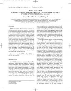
Fig. 1. (a–c) CLSM images showing PNA FISH of mixed planktonic bacteria labelled using three PNA probes. (a) Universal bacterial probe (Bac-Uni1-CY3) showing that all bacteria stain red. (b) P. aeruginosa probe (Psaer-FITC) showing that a proportion of the population stain green. (c) Multiplex PNA staining of P. aeruginosa (yellow bacteria showing hybridization with both universal and P. aeruginosa-specific probes), Staph. aureus (purple bacteria hybridized with both universal and Staph. aureus-specific probes), and Micrococcus luteus and Strep. oralis (red bacteria showing hybridization with the universal probe only). (d–g) Mixed bacterial CDFF biofilms labelled using three PNA probes: (d) universal bacterial probe (red); (e) P. aeruginosa-specific probe (green); (f) Staph. aureus-specific probe (blue); (g) multiplex PNA staining (overlay of d–f). 2606
Microbiology 155
Detection of wound bacteria by PNA FISH
http://mic.sgmjournals.org
2607
S. Malic and others
Fig. 2. Microscopical analysis of mixed-species biofilms formed in vitro on RHE tissue. (a) Intact RHE section showing Gramstained bacteria on the surface. (b) Artificially wounded RHE section showing Gram-stained bacteria within the tissue. (c) PNA FISH using the P. aeruginosa-specific (green bacteria) and the Staph. aureus specific probes (red bacteria) and with the epithelial cell nuclei counterstained with Hoechst dye (blue).
biofilm morphology or architecture due to the DNA/RNA extraction process. Traditional microscopical analyses and bacterial stains are useful, but limited, as they often rely on prior cultivation of the bacteria. Whilst Gram staining provides information on the type of bacterial cell wall it does not allow for species identification (Moter & Goebel, 2000). Since cultivation techniques tend to work for only a minority of species (only 2–3 % of bacteria are thought to be cultivable using currently available media), the development of alternative methods for the identification and visualization of bacteria has become a necessity (Amann et al., 2001; Moter & Goebel, 2000). The impact of micro-organisms on wound healing is poorly understood but there is strong evidence to suggest that bacteria within chronic wounds delay the healing process (Edwards & Harding, 2004; Percival & Bowler, 2004; Percival & Rogers, 2005). The production of 2608
destructive enzymes and toxins by bacteria results in tissue damage and indirectly promotes an inflammatory state (Percival & Rogers, 2005; Stephens et al., 2003). Moreover, removal of bacteria by host defence mechanisms and antimicrobial therapy is difficult in wound environments where biofilms have been established. In the present study, FISH in combination with CLSM was used to identify the presence of specific bacteria and to examine their spatial organization within in vitro biofilms established using the CDFF and RHE models, as well as in vivo in chronic wound biopsy sections. The PNA-FISH methodology was developed and validated for three PNA probes, to identify and visualize the polymicrobial population of fixed biofilms and clinical specimens. The bacteria used in the described in vitro models were all originally isolated from chronic wounds and included P. aeruginosa and Staph. aureus, which have Microbiology 155
Detection of wound bacteria by PNA FISH
invasiveness of bacteria in this intact tissue. However, in scratch-wounded RHE sections, it was apparent that bacteria started to penetrate the tissue and to migrate beneath the damaged surface layer. This suggests that once the integrity of the epithelial barrier has been disrupted (as would be encountered in a wounding situation) the bacteria can invade the tissue. A possible limitation of the RHE system was the lack of specific host immune responses, although proinflammatory cytokine responses by in vitro epithelial cell-lines in a similar reconstituted oral epithelium model have been reported (Schaller et al., 2002; Villar et al., 2005). Hence, although the RHE has some drawbacks, it is currently the most suitable in vitro model of human skin epithelium. Importantly, both models (CDFF and RHE) revealed heterogeneous biofilm structures with discrete clusters of bacterial species, with P. aeruginosa as the predominant organism. In the case of the former, this was confirmed using quantitative image analysis of the CLSM Z-stack dataset.
Fig. 3. Microscopical analysis of a section from a CVLU using PNA FISH and showing total bacteria labelled with the universal bacterial PNA probe (red bacteria) and Hoechst dye for epithelial cell nuclei (blue).
been shown by culture and molecular analysis to be predominant species in many wounds (Davies et al., 2004). Strep. oralis and M. luteus were also used. Both of these species had previously been shown to exhibit high growth rates and a propensity to produce biofilms in in vitro models (results not shown). These species, whilst not recognized as primary wound pathogens, are regularly isolated from acute and chronic wounds, and in immunocompromised patients can cause opportunistic infection.
It is likely that the bacterial species within the biofilms compete with each other for space, attachment and nutrients. Two possible factors for the predominance of P. aeruginosa in these mixed biofilms are its twitching and flagella-mediated motility, allowing the bacteria to migrate to optimal growth localities within the biofilm as it matures, as well as its faster growth rate. Clearly, these experiments do not exactly reflect the in vivo situation within a chronic wound. In vivo, different microbial species could well be expected to colonize at different times, with specific species acting as pioneer or primary colonizers. In our experiments, all four bacterial species were added to the RHE simultaneously. It would therefore be interesting to add the species in a successive manner. For instance, P. aeruginosa could be added to the model first to form a base for the biofilm, with other species added subsequently to determine if they were able to grow to a greater extent on the biofilm surface. Conversely, since it was observed in this study that P. aeruginosa appeared to ‘out-compete’ the other organisms, it might also be of interest to see whether staging of the infection times allows the other species to grow at higher levels, before P. aeruginosa is added to the system.
Initially, the use of PNA probes in this bacteria-specific FISH protocol permitted the simultaneous identification of mixed bacterial species in planktonic suspension. Hybridization was carried out under low-salt, high-temperature and highpH conditions. Surprisingly, the Staph. aureus-specific PNA probe showed very weak fluorescence compared to both the universal PNA bacterial and the P. aeruginosa-specific PNA probes. However, this has also previously been observed in other studies (Hartmann et al., 2005; Lefmann et al., 2006; Wellinghausen et al., 2007). To improve the fluorescence intensity of this probe, it was necessary to expose biofilms containing the Gram-positive organisms to lysozyme pretreatment for 15 min at 37 uC and to add 30 % formamide to the hybridization buffer. Thurnheer et al. (2004) found that a 1 h paraformaldehyde fixation treatment was sufficient to permeabilize Gram-positive cells, but this did not prove effective in the present study.
Further research could include incubating the infected RHE for longer than 24 h to see if this would result in greater infiltration of the bacterial cells within the keratinocyte wound model. The use of different culture media that promote bacterial growth as opposed to the tissue maintenance medium could also be assessed. In addition, detection of any extracellular polysaccharide matrix generated by the chronic wound bacterial biofilms may be possible, for example using calcofluor white, after completion of the FISH probing (Perry-O’Keefe et al., 2001b; Thurnheer et al., 2004).
In RHE sections, few bacteria were detected on the surface and only a sparse biofilm was evident, indicating poor
Analysis of chronic wound biopsy sections by FISH using the universal bacterial PNA probe did reveal the presence
http://mic.sgmjournals.org
2609
S. Malic and others
of colonizing bacteria. Routine haematoxylin and eosin staining (data not shown) as well as PNA FISH detection confirmed that bacteria were present in these biopsy sections. PNA FISH was shown to be a rapid and versatile tool for research purposes and potentially for clinical microbiology diagnostics when used in conjunction with CLSM and quantitative image analysis. CLSM has been established as a valuable method for obtaining high-resolution images and three-dimensional reconstructions of fluorescently labelled biofilms and biological samples (Lopez et al., 2005; Sunde et al., 2003; Thurnheer et al., 2004; Wagner et al., 2003). The continued application of this technique to clinical biofilms from infected tissues or indwelling medical devices could facilitate the identification and estimation of the relative proportions of bacteria within a biofilm. This approach could be used to assess biofilm management strategies or evaluate the effectiveness of antimicrobials against members of the biofilm consortium. Importantly, this technique could potentially be applied to clinical samples for both identifying and estimating the proportion of bacterial species present.
ACKNOWLEDGEMENTS We gratefully acknowledge financial support for this work from Cardiff University School of Dentistry (PhD studentship for S. M.). We are also grateful to Mrs Kath Allsopp for processing the tissue sections used in this study.
REFERENCES Amann, R., Fuchs, B. M. & Behrens, S. (2001). The identification of
microorganisms by fluorescence in situ hybridisation. Curr Opin Biotechnol 12, 231–236. Andersen, A., Hill, K. E., Stephens, P., Thomas, D. W., Jorgensen, B. & Krogfelt, K. A. (2007). Bacterial profiling using skin grafting,
predictive value of tissue biopsies and swabs. Wound Repair Regen 15, 17–22. Davis, S. C., Ricotti, C., Cazzaniga, A., Welsh, E., Eaglstein, W. H. & Mertz, P. M. (2008). Microscopic and physiologic evidence for
biofilm-associated wound colonization in vivo. Wound Repair Regen 16, 23–29. Edwards, R. & Harding, K. G. (2004). Bacteria and wound healing.
Curr Opin Infect Dis 17, 91–96. Gu, F., Lux, R., Du-Thumm, L., Stokes, I., Kreth, J., Anderson, M. H., Wong, D. T., Wolinsky, L., Sullivan, R. & Shi, W. (2005). In situ and
non-invasive detection of specific bacterial species in oral biofilms using fluorescently labeled monoclonal antibodies. J Microbiol Methods 62, 145–160. Hartmann, H., Stender, H., Schaefer, A., Autenrieth, I. B. & Kempf, V. A. J. (2005). Rapid identification of Staphylococcus aureus in blood
cultures by a combination of fluorescence in situ hybridisation using peptide nucleic acid probes and flow cytometry. J Clin Microbiol 43, 4855–4857. Hill, K. E., Davies, C. E., Wilson, M., Stephens, P., Harding, K. G. & Thomas, D. W. (2003). Molecular analysis of the microflora in chronic
venous leg ulceration. J Med Microbiol 52, 365–369. Howell-Jones, R. S., Price, P. E., Howard, A. J. & Thomas, D. W. (2006). Antibiotic prescribing for chronic skin wounds in primary
care. Wound Repair Regen 14, 387–393. James, G. A., Swogger, E., Wolcott, R., Pulcini, E. D., Secor, P., Sestrich, J., Costerton, J. W. & Stewart, P. S. (2008). Biofilms in
chronic wounds. Wound Repair Regen 16, 37–44. Lefmann, M., Schweickert, B., Buchholz, P., Goebel, U. B., Ulrichs, T., Seiler, P., Theegarten, D. & Moter, A. (2006). Evaluation of peptide
nucleic acid-fluorescence in situ hybridization for identification of clinically relevant mycobacteria in clinical specimens and tissue sections. J Clin Microbiol 44, 3760–3767. Leid, J. G., Willson, C. J., Shirtliff, M. E., Hassett, D. J., Parsek, M. R. & Jeffers, A. K. (2005). The exopolysaccharide alginate protects Pseudomonas aeruginosa biofilm bacteria from IFN-c-mediated
macrophage killing. J Immunol 175, 7512–7518. Lopez, C., Pons, M. N. & Morgenroth, E. (2005). Evaluation of
microscopic techniques (epifluorescence microscopy, CLSM, TPELSM) as a basis for the quantitative image analysis of activated sludge. Water Res 39, 456–468.
standard culture and molecular bacteriological methods. J Wound Care 16, 171–175.
Marsh, P. D. (2003). Are dental diseases examples of ecological
Bjarnsholt, T., Kirketerp-Moller, K., Jensen, P. O., Madsen, K. G., Phipps, R., Krogfelt, K., Hoiby, N. & Givskov, M. (2008). Why chronic
Mertz, P. M. (2003). Cutaneous biofilms: friend or foe? Wounds 15,
129–132.
wounds will not heal: a novel hypothesis. Wound Repair Regen 16, 2–10.
Moter, A. & Goebel, U. B. (2000). Fluorescence in situ hybridization
Coull, J. J. & Hyldig-Nielsen, J. J. (2003). US Patent 6664045. PNA
catastrophes? Microbiology 149, 279–294.
(FISH) for direct visualization of microorganisms. J Microbiol Methods 41, 85–112.
probes, probe sets, methods and kits pertaining to the detection of microorganisms.
Percival, S. L. & Bowler, P. G. (2004). Biofilms and their potential role
Davies, C. E., Wilson, M., Hill, K. E., Stephens, P., Hill, C. M., Harding, K. G. & Thomas, D. W. (2001). Use of molecular techniques to study
Percival, S. L. & Rogers, A. A. (2005). The significance and role of
in wound healing. Wounds 16, 234–240.
microbial diversity in the skin: chronic wounds reevaluated. Wound Repair Regen 9, 332–340.
biofilms in chronic wounds. In Biofilms: Persistence and Ubiquity. Edited by A. McBain, A. Allison, J. Pratten, D. Spratt, M. Upton & J. Verran. Manchester, UK: The Biofilm Club, University of Manchester.
Davies, C. E., Hill, K. E., Wilson, M., Stephens, P., Hill, C. M., Harding, K. G. & Thomas, D. W. (2004). Use of 16S ribosomal DNA PCR and
Perry-O’Keefe, H., Rigby, S., Oliveira, K., Sorensen, D., Stender, H., Coull, J. & Hyldig-Nielsen, J. J. (2001a). Identification of indicator
denaturing gradient gel electrophoresis for analysis of the microfloras of healing and nonhealing chronic venous leg ulcers. J Clin Microbiol 42, 3549–3557.
microorganisms using a standardized PNA FISH method. J Microbiol Methods 47, 281–292.
Davies, C. E., Hill, K. E., Newcombe, R. G., Stephens, P., Wilson, M. J., Harding, K. G. & Thomas, D. W. (2007). A prospective study of the
microbiology of chronic venous leg ulcers to reevaluate the clinical 2610
Perry-O’Keefe, H., Stender, H., Broomer, A., Oliveira, K., Coull, J. & Hyldig-Nielsen, J. J. (2001b). Filter-based PNA in situ hybridization
for rapid detection, identification and enumeration of specific microorganisms. J Appl Microbiol 90, 180–189. Microbiology 155
Detection of wound bacteria by PNA FISH Pratten, J. & Wilson, M. (1999). Antimicrobial susceptibility and
composition of microcosm dental plaques supplemented with sucrose. Antimicrob Agents Chemother 43, 1595–1599.
hybridization (FISH) for direct visualization of bacteria in periapical lesions of asymptomatic root-filled teeth. Microbiology 149, 1095– 1102.
Rhoads, D. D., Wolcott, R. W., Cutting, K. F. & Percival, S. L. (2007).
Thurnheer, T., Gmuer, R. & Guggenheim, B. (2004). Multiplex FISH
Evidence of biofilms in wounds and potential ramifications. In Biofilms: Coming of Age, pp. 131–143. Edited by P. Gilbert, D. Allison, M. Brading, J. Pratten, D. Spratt & M. Upton. Manchester, UK: The Biofilm Club, Manchester University.
analysis of a six-species bacterial biofilm. J Microbiol Methods 56, 37–47.
Schaller, M., Mailhammer, R., Grassl, G., Sander, C. A., Hube, B. & Korting, H. C. (2002). Infection of human oral epithelia with Candida
proinflammatory response to infection. Infect Immun 73, 4588–4595.
species induces cytokine expression correlated to the degree of virulence. J Invest Dermatol 118, 652–657. Serralta, V. W., Harrison-Balestra, C., Cazzaniga, A. L., Davis, S. C. & Mertz, P. M. (2001). Lifestyles of bacteria in wounds: presence of
biofilms? Wounds 13, 29–34. Socransky, S. S., Haffajee, A. D., Cugini, M. A., Smith, C. & Kent, R. L. (1998). Microbial complexes in subgingival plaque. J Clin Periodontol
25, 134–144. Stephens, P., Wall, I. B., Wilson, M., Hill, K. E., Davies, C. E., Hill, C. M., Harding, K. G. & Thomas, D. W. (2003). Anaerobic cocci populating
the deep tissues of chronic wound impair cellular wound healing responses in vitro. Br J Dermatol 148, 456–466. Sunde, P. T., Olsen, I., Goebe, U. B., Theegarten, D., Winter, S., Debelian, G. J., Tronstad, L. & Moter, A. (2003). Fluorescence in situ
http://mic.sgmjournals.org
Villar, C. C., Kashleva, H., Mitchell, A. P. & Dongari-Bagtzoglou, A. (2005). Invasive phenotype of Candida albicans affects the host Vroom, J. M., De Grauw, K. J., Gerritsen, H. C., Bradshaw, D. J., Marsh, P. D., Watson, G. K., Birmingham, J. J. & Allison, C. (1999).
Depth penetration and detection of pH gradients in biofilms by twophoton excitation microscopy. Appl Environ Microbiol 65, 3502–3511. Wagner, M., Horn, M. & Daims, H. (2003). Fluorescence in situ hybridisation for the identification and characterisation of prokaryotes. Curr Opin Microbiol 6, 302–309. Wall, I. B., Davies, C. E., Hill, K. E., Wilson, M., Stephens, P., Harding, K. G. & Thomas, D. W. (2002). Potential role of anaerobic cocci in
impaired human wound healing. Wound Repair Regen 10, 346–353. Wellinghausen, N., Bartel, M., Essig, A. & Poppert, S. (2007). Rapid
identification of clinically relevant Enterococcus species by fluorescence in situ hybridization. J Clin Microbiol 45, 3424–3426. Edited by: R. J. Lamont
2611