ANALYTICAL SCIENCES MAY 2015, VOL. 31
407
2015 © The Japan Society for Analytical Chemistry
Developing a New Micro Cloud Point Extraction Method for Simultaneous Preconcentration and Spectrophotometric Determination of Uranium and Vanadium in Brine Elham GHASEMI and Massoud KAYKHAII† Department of Chemistry, Faculty of Sciences, University of Sistan and Baluchestan, Zahedan 98135-674, Iran
A fast, simple, and economical method was developed for simultaneous spectrophotometric determination of uranium(VI) and vanadium(V) in water samples based on micro cloud point extraction (MCPE) at room temperature. This is the first report on the simultaneous extraction and determination of U(VI) and V(V). In this method, Triton X114 was employed as a non-ionic surfactant for the cloud point procedure and 4-(2-pyridylazo) resorcinol (PAR) was used as the chelating agent for both analytes. To reach the cloud point at room temperature, the MCPE procedure was carried out in brine. The factors influencing the extraction efficiency were investigated and optimized. Under the optimized condition, the linear calibration curve was found to be in the concentration range between100 – 750 and 50 – 600 μg L–1 for U(VI) and V(V), respectively, with a limit of detection of 17.03 μg L–1 (U) and 5.51 μg L–1 (V). Total analysis time including microextraction was less than 5 min. Keywords Micro cloud point extraction, uranium(VI), vanadium(V), water analysis, spectrophotometry (Received October 24, 2014; Accepted February 1, 2015; Published May 10, 2015)
Introduction Methods for the simultaneous determination of heavy metals have attracted a great deal of interest in recent years, especially techniques for determination when these cations are present in the same media. Usually, uranium ore contains large amounts of vanadium. One of the metallic impurities in uranium concentrates that is commonly known as “yellow-cake” is vanadium.1 Therefore, uranium mining and the nuclear energy cycle release uranium and vanadium to the environment at the same time. The presence of these elements in the environment means they can be ingested by humans; the simplest way is through contaminated water. In water the dissolved vanadium is normally present as vanadate and uranium is present as uranyl. Much research is being conducted on the simultaneous determination of uranium and vanadium in ores,2 and environmental3 and biological4 media. The methods based on conventional UV-Vis spectrophotometry analysis of vanadate and uranyl are some of the earliest detection methods adopted, mainly because of their low cost and simplicity of use;5,6 however, they suffer from poor sensitivity and are very time consuming. Moreover, due to the complexity of matrices of these target analytes, direct determination of them using this methodology is not very satisfactory. If coupled with some advanced preconcentration techniques, the analytical performance of spectrophotometry could be significantly improved. This would greatly expand its application, especially in less developed regions that lack more advanced analytical To whom correspondence should be addressed. E-mail:
[email protected]
†
instruments. Taking into account that U and V ions exist in very low concentrations in almost all samples and the volume of the samples available for analysis is very limited in some cases, applying a pretreatment separation/preconcentration step on real samples seems not only necessary, but also extraction methods need to be miniaturized. Since the introduction of micro extraction by Pawliszyn in the 1990s,7 there has been a lot of interest on minimizing the extraction methods to micro extractions. The volume of papers and reviews published on micro-extraction techniques indicates the importance of these techniques. Of the most important micro extraction techniques that have been developed for a variety of analytes, dispersive liquid liquid microextraction (DLLME),8 solid phase micro extraction (SPME),9,10 and headspace single drop micro extraction11 are most frequently used. One advantage of these techniques is that they use very little toxic, high-priced organic solvents. Besides, they are compatible with almost all analytical instruments and do not need a large sample volume. To make these techniques even more green, some research has been done on replacing the toxic organic solvents with more green solvents such as room temperature ionic liquids.12,13 Cloud point extraction (CPE) is a relatively green extraction method since it does not use toxic organic solvents.14 It is simple, inexpensive and safe procedure with high efficiency.15 CPE is compatible with many instruments, therefore it has been successfully applied as an efficient extraction procedure for the separation, preconcentration or purification of a variety of analytes, including organic,16–18 inorganic19,20 and even pharmaceutical21 and nano compounds.22 However, it suffers from some drawbacks that together with fast improvements in micro-extraction techniques made it less attractive. In CPE, micellar solution of non-ionic surfactants, which are
408 completely soluble in water, becomes turbid and separates into an enriched misellar phase when heated beyond a temperature called cloud point (CP). The hydrophobic analytes can transfer from the aqueous solution to this miscellar phase. It is obvious that the key point to achieving the cloudy solution ready for extraction is heating the sample solution (usually in water bath). This step is an energy and time consuming step and the time needed to reach the cloud point varies from 20 min to 4 h.23,24 Considering the time needed for the centrifugation and applying a cold bath to condensate the enriched surfactant phase, this could be a very long and time consuming method. CPE needs a large sample volume, usually up to 50 mL25 and in most cases, in coupling CPE with spectrophotometer, the enriched miscellar phase must be diluted with organic solvents (e.g. 2 mL methanol) because of its high density and the low volume of the enriched phase.26 However, because it is an environmentally friendly, inexpensive and easy to operate technique, we think CPE should not be neglected. In recent years there have been a few studies on improving the CPE procedure.27,28 To reach cloud point at room temperature, we can use the salting out effect instead of heating the mixture to the CP.29,30 This approach for reaching cloud point is applied here. For reducing the usage of organic solvents we decided to employ microcell for spectrophotometric determination instead of the conventional classic cells, which reduced the usage of the organic solvent as the diluting solvent from 2 to 0.05 mL. By carrying out the proposed micro-cloud point extraction (MCPE) procedure in brine, we not only succeeded in reducing the consumption of organic solvent, but we successfully minimized the volume of sample solution, eliminated the centrifuge step and reduced the time of the whole analysis to less than 5 min; as such we can claim that our proposed method is fast, green, easy to operate and economical. It is known that 4-(2-pyridylazo) resorcinol (PAR) is a very good compound for the spectrophotometric determination of heavy metals. Despite not being very selective, PAR is widely used as an analytical reagent for the sensitive spectrophotometric determination of uranyl31 and vanadate32 ions. Simultaneous spectrophotometric determination of U(IV) and V(V) in miscellar solution has been reported earlier.33 The proposed MCPE was successfully applied for simultaneous preconcentration and determination of U(VI) and V(V) ions in aqueous samples.
Experimental Instrument A Shimadzu UV/VIS spectrophotometer, UV-160 (Kyoto, Japan) equipped with two 10 μL microcells (Starna, UK) was used for measuring the absorbance and recording the spectra. Reagents and chemicals All reagents and solvents (i.e. acetone, methanol, ethanol, HCl and AcOH) were of analytical grade and were purchased from Merck KGaA (Germany) and used as received. Uranyl nitrate hexahydrate and vanadium standard solution (1000 ppm) were obtained from the same company. Solution of PAR (0.01 M), Na2SO4 (5% w/v) and Triton X114 (2% v/v) were prepared by dissolving appropriate amounts of each compound in doubly distilled water. The pH of the sample solution was adjusted using a suitable buffer prepared using AcOH/NaOAc or Na2HPO4/HCl. Doubly distilled water was used throughout all the procedures.
ANALYTICAL SCIENCES MAY 2015, VOL. 31
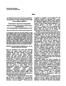
Fig. 1 Absorption spectra for the determination of 150 μg L–1 U(VI) (a), 250 μg L–1 V(V) (b), and mixture of U(VI) and V(V) (c); with PAR, 7 × 10⁻⁴ M; Triton X114, 0.2% (v/v); sample volume, 5 mL; pH, 5.5; Na2SO4 1% (w/v) diluting solvent, methanol, 50 μL; centrifugation time, 4 min at 4500 rpm.
Micro-cloud point extraction procedure An aliquot of the sample solution containing appropriate amounts of U(VI) and V(V) was transferred into a centrifuge test tube with a conical bottom and a mixture of 350 μL PAR (0.01 M), 500 μL Triton X114 (2% v/v) and 2 mL acetate buffer solution (pH = 5.5) was added to it. To reach cloud point and formation of a cloudy solution, 1 mL of 5% Na2SO4 solution was added to the mixture. Immediately after the addition of salt, the solution became cloudy and the complexes formed in the solution were extracted into miscelles of Triton. After that, the mixture was diluted to 5 mL with double distilled water. The obtained cloudy solution was centrifuged for 4 min at 4500 rpm. During the centrifugation the surfactant settled down at the bottom of the test tube to form a very high density liquid phase of which 10 μL was transferred to a vial and dissolved in 50 μL methanol. Finally, 25 μL of this mixture was transferred to a microcell for spectrophotometric determination.
Results and Discussion Absorption spectra of complex In order to find the maximum wavelength of absorption for the complex, the absorption spectra of U(VI)-PAR and V(V)PAR were determined in the wavelength range of 275 to 825 nm against the reagent blank, as shown in Fig. 1. The results indicate that the maximum absorption wavelengths were 545 for U(VI)-PAR and 568 nm for V(V)-PAR complexes. Accordingly, these wavelengths were selected as the chosen absorption wavelengths for further determinations. During all of the following experiments, the blank absorbance of all reagents was corrected. Effect of pH The level of pH plays an important role in the uranium and vanadium chelate formation because they exist in different species that depend largely on pH.34,35 Therefore, the effect of pH on the absorbance of U(VI)-PAR and V(V)-PAR was examined closely for the proposed MCPE method. In addition, PAR acts as a tridentate chelating agent that chelates with metal ions through the o-hydroxyl group. This species is dominant in pH ≥ 5.5.36 Also in basic pH, uranyl will be involved in polymeric reactions.37 Therefore, it was decided to study pH in
ANALYTICAL SCIENCES MAY 2015, VOL. 31
Fig. 2 Effect of pH of aqueous solution on MCPE for vanadium (♦) and uranium (■). MCPE conditions: U(VI)/V(V), 350 μg L–1; PAR, 7 × 10⁻⁴ M; Triton X114, 0.2% (v/v); sample volume, 5 mL; diluting solvent, methanol, 50 μL; centrifugation time, 5 min at 4500 rpm.
409
Fig. 4 Effect of concentration of PAR on MCPE for vanadium (♦) and uranium (■). MCPE conditions: U(VI)/V(V), 350 μg L–1; Triton X114, 0.2% (v/v); sample volume, 5 mL; pH, 5.5; diluting solvent, methanol, 50 μL; centrifugation time, 5 min at 4500 rpm.
a major effect on the extraction efficiency. In order to optimize the best concentration, different concentrations (0.1 – 0.5% v/v) of Triton X114 were subjected to the same procedure. Maximum peak responses for both complexes were obtained when 0.2% was added (Fig. 3). Effect of PAR concentration The effect of concentration of the chelating agent (PAR) on the absorbance of U(VI)-PAR and V(V)-PAR was also investigated (Fig. 4). The maximum absorbance was obtained at 7 × 10⁻⁴ M PAR for both complexes and remained almost constant for uranium and showed a decrease in absorbance for vanadium with increasing concentration up to 9 × 10⁻⁴ M. Fig. 3 Effect of concentration of Triton X114 on MCPE for vanadium (♦) and uranium (■). MCPE conditions: U(VI)/V(V), 350 μg L–1; PAR, 7 × 10⁻⁴ M; sample volume, 5 mL; pH, 5.5; diluting solvent, methanol, 50 μL; centrifugation time, 5 min at 4500 rpm.
the range of 4 to 7. It has been found that the absorbance of 350 μg L–1 U(VI) in the presence of 7 × 10⁻⁴ M PAR has its maximum value over the pH range 5 – 6. The absorbance of 350 μg L–1 V(V) under the same condition was found to have its maximum value at pH 5.5 (Fig. 2). Therefore, pH = 5.5 was chosen for as the optimal pH for the sample solution and further studies were performed at this pH. The effect of salt concentration Ionic strength of the sample solution is an important factor in reaching cloud point at room temperature. In order to find the best salt compound, different salts (NaCl, KCl, NaHSO4 and Na2SO4) were examined and among them Na2SO4 showed the best effect on forming the turbid solution at lower concentrations and without delay. Using the same concentrations, it was observed that in comparison to the other salts, the value of absorbance increases from 15% to more than 60% when Na2SO4 is used. By studying the effect of Na2SO4 concentration on the absorbance, it was found that the absorbance of both analytes reaches a maximum value at 1% (w/v) of Na2SO4 and the cloudy solution was formed immediately, therefore this concentration of sodium sulfate was used in all subsequent measurements. Optimization of the concentration of Triton The concentration of Triton X114 as the extractant solvent has
Selection of diluting solvent In the selection of the diluting agent, three important factors were considered: the ability of the chosen solvents to dissolve the sediment phase, its absorbance, and repeatability of the obtained data. For this purpose some conventional solvents including acetone, ethanol and methanol were investigated. Dissolution of the sediment phase in acetone took place with difficulty. Although absorbance of the complex using ethanol as the diluting solvent was high, its repeatability was poor. The results led us to select methanol as the diluting solvent. Moreover, the absorbance signals were relatively higher using this solvent. The effect of time of extraction and centrifugation We considered the extraction time as the time between the formation of a cloudy sample solution and the starting of centrifugation as is conventional in dispersive liquid liquid microextraction procedures.38 The extraction time was studied in the range of 0 – 8 min and it was observed that the extraction time has no significant effect on the extraction efficiency. Therefore, the samples were immediately subjected to centrifuge after the addition of salt. To obtain the desired spectra, it is necessary to separate two phases (aqueous and enriched surfactant phase). Some researchers prefer to wait until the aqueous and micellar phase separate on their own accord. However, this step requires considerable time. For the MCPE proposed here, this step takes about an hour; therefore, we decided to separate the enriched surfactant phase with the help of centrifuge. It was observed that at speeds less than 2000 at rpm, not only was the amount of the settled phase so small but also the absorbance was much lower than we expected. Therefore we decided to perform the
410
ANALYTICAL SCIENCES MAY 2015, VOL. 31
experiments at high speed of 4500 rpm centrifugation. Figure 5 shows the response of the analytical instrument as a function of centrifugation time. On the basis of the curve obtained, the best time of centrifugation was found to be 4 min at 4500 rpm, which was selected as the optimum time of centrifugation for all experiments. Linear range, limit of detection and precision Analytical figures of merit for the proposed method obtained under optimal conditions are shown in Table 1 in comparison with analytical data obtained in previous studies for simultaneous determination of uranium and vanadium. Limits of detection (LODs) were obtained based on a signal-to-noise ratio of 3. The repeatability of the method, expressed as relative standard deviation (RSD), was calculated for five replicates of the standard at an intermediate concentration of the calibration curve (350 μg L–1 for both analytes). The enrichment factor (EF), which was calculated as the ratio between the analyte concentration after MCPE (CMCPE) and the initial concentration of analyte (C0) within the sample (Eq. (1)),39 was found to be 11.8 fold for U(VI) and 15.0 fold for V(V). EF = CMCPE/C0
Simultaneous determination of U(VI) and V(V) in real samples In order to evaluate performance of the developed method in real matrices, it was applied for the analysis of a tap water sample. Since the absorption spectra of U(VI)-PAR and V(V)-PAR complexes are overlapping in some wavelengths, law of additivity was employed for the determination of both analytes in the same run. Two wavelengths, 545 and 568 nm (i.e. maximum wavelengths of U(VI)-PAR and V(V)-PAR), were selected and quantitative estimation of the analytes in real samples was carried out by solving Eqs. (2) and (3): A1 = AU1 + AV1 = εU1 bCU + εV1 bCV
(2)
A2 =AU2 + AV2 = εU2 bCU + εV2 bCV
(3)
where A1 and A2 are absorbances of the mixture at 545 and 568 nm, AU1 and AU2 are absorbances of U(VI)-PAR at 545 and 568 nm and similarly AV1 and AV2 are absorbances of V(V)-PAR at 545 and 568 nm; and CU and CV are the concentration of uranium and vanadium respectively. The molar absorptivity values, ε, of both complexes at both wavelengths were calculated to be:
(1) εU1 = 27047,
εU2 = 22137;
εV1 = 18457,
εV2 = 18407.
Table 2 Analytical results for simultaneous determination of U(VI) and V(V) in tap water and environmental calibration standard mix (n = 3) Matrix Tap water Tap water
Fig. 5 Effect of time of centrifuge on MCPE for vanadium (♦) and uranium (■). MCPE conditions: U, 500 μg L–1; PAR, 7 × 10⁻⁴ M; Triton X114, 0.2% (v/v); sample volume, 5 mL; pH, 5.5; diluting solvent, methanol, 50 μL.
Standard mix Standard mix
Analyte Uranium Vanadium Uranium Vanadium
added/ μg L–1
Recovery, %
150 350 700 100 350 600 0 0
108 97.42 103.0 112 96.2 96.8 97.9 97.4
Mean recovery, % 102.80 101.66 97.9 97.4
Experimental MCPE conditions: PAR, 7 × 10⁻⁴ M; Triton X114, 0.2% (w/v); Na2SO4 1% (w/v) dilution solvent, methanol, 50 μL; centrifugation time, 4 min at 4500 rpm; sample volume, 5 mL.
Table 1 Analytical figures of merit for MCPE extraction of U(VI)-PAR and V(V)-PAR in comparison with analytical data from earlier studies Technique used
analyte
Dynamic range of calibration curve/mg L–1
FI-ICP-AESc
Uranium Vanadium
0 – 0.005 0 – 0.005
ICP-AES
Uranium Vanadium
0 – 0.005 0 – 0.005
First derivative spectroscopy
Uranium Vanadium
0.4 – 4 4 – 18
MCPE
Uranium Vanadium
0.1 – 0.75 0.05 – 0.6
LODa/ μg L–1 0.9 0.08 10 1 250 300 17.03 5.51
RSDb, %
Recovery, %
EF
Ref.
0.5 – 4 0.5 – 4
101 96
8.4 8.8
40
0.5 – 4 0.5 – 4
101 96
NMd NM
40
73 – 102 97 – 102
0.4 – 0.9 0.4 – 1.2
NM NM
33
102.8 101.66
11.81 15.0
Present work
6.48 2.43
a. LOD, was based on 3 Sb/m criterion for 10 blank measurements for the present work. b. RSD, relative standard deviation, for 5 replicate measurements of 350 μg L–1 of each analyte for the present work. c. FI-ICP-AES, flow injection coupled to inductively coupled plasma atomic emission spectroscopy. d. NM, Not mentioned.
ANALYTICAL SCIENCES MAY 2015, VOL. 31
411
Subsequently, the mean absorptivity values were substituted to Eqs. (2) and (3), to form Eqs. (4) and (5): A1 = 27047CU + 18457CV
(4)
A2 = 22137CU + 18407CV
(5)
The concentrations of U(VI) and V(V) in the mixture were determined by solving Eqs. (4) and (5). No uranium and vanadium were detected in the tap water sample. To investigate the effect of sample matrices on extraction efficiency, the same sample was spiked with the target analytes on three levels. The results, which are shown in Table 2, indicate that the developed method can successfully be applied for the simultaneous determination of U(IV) and V(V) in water samples. The trueness of the method was evaluated by analyzing an environmental calibration standard mix obtained from AccuStandard, Inc. (CT, USA; Cat. No. AG-CAL-ASL-5) with certified U(IV) and V(V) concentrations. Results are again summarized in Table 2. Because of a ≥97.9% recovery, it is evident that the measured value is in good agreement with the certified value.
Conclusions In this study, a simple, inexpensive, green and effective method based on MCPE for preconcentration and determination of uranium in brine is developed. As a widely and commonly used detection method, spectrophotometry was coupled with the proposed MCPE, which largely minimized toxic organic solvent consumption and increased the sensitivity for the determination of the target analyte. The total analysis time was less than 5 min. The spectrophotometric instrumentation features such advantages as simplicity and low cost. The investigation carried out in this work shows the conventional spectrophotometer is expected to have broad prospects for simultaneous detection of trace amounts of metals and thus may expand its application.
References 1. D. A. Batistoni and P. N. Smichowski, Appl. Spectrosc., 1985, 39, 222. 2. A. N. Finn, J. Am. Chem. Soc., 1906, 28, 1443. 3. S. Sander, Anal. Chim. Acta, 1999, 394, 81. 4. U. Repinc and L. Benedik, J. Radioanal. Nucl. Chem., 2005, 264, 77. 5. J. Mendham, R. C. Denney, J. D. Barnes, and M. J. K. Thomas, “Vogel’s Quantitative Chemical Analysis”, 6th ed., 2000, Pearson Education Limited, Essex, England, 658. 6. T. M. Florence and Y. J. Farrar, Anal. Chem., 1970, 42, 271. 7. C. L. Arthur and J. Pawliszyn, Anal. Chem., 1990, 63, 2145. 8. L. Campone, A. L. Piccinelli, R. Celano, and L. Rastrelli, J. Chromatogr. A, 2011, 1218, 7648. 9. M. Kaykhaii, G. W. Dicinoski, and P. R. Haddad, Anal. Lett., 2010, 43, 1546. 10. J. Song, C. F. Forney, and M. A. Jordan, Food Chem., 2014, 160, 255.
11. M. Kaykhaii and M. H. Ghalehno, Anal. Methods, 2013, 5, 5622. 12. M. Kaykhaii and E. Ghasemi, Anal. Methods, 2013, 5, 5260. 13. M. Rahmani and M. Kaykhaii, Microchim. Acta, 2011, 174, 413. 14. K. Pytlakowska, V. Kozik, and M. Dabioch, Talanta, 2013, 110, 202. 15. C. B. Ojeda and F. S. Rojas, Anal. Bioanal. Chem., 2009, 394, 759. 16. E. Katsoyannos, O. Gortzi, A. Chatzilazarou, V. Athanasiadis, J. Tsaknis, and S. Lalas, J. Sep. Sci., 2012, 35, 2665. 17. H. Ma, F. Mu, S. Fan, X. Zhou, and Q. Jia, J. Sep. Sci., 2012, 35, 2484. 18. R. Carabias-Martínez, E. Rodríguez-Gonzalo, B. MorenoCordero, J. L. Pérez-Pavón, C. García-Pinto, and E. F. Laespada, J. Chromatogr. A, 2000, 902, 251. 19. S. Shariati and M. Golshekan, J. Anal. Chem., 2014, 69, 248. 20. N. N. Meeravali, M. A. Reddy, and S. J. Kumar, Anal. Sci., 2007, 23, 351. 21. H. Zhang and H. K. Choi, Anal. Bioanal. Chem., 2008, 392, 947. 22. S. M. Majedi, B. C. Kelly, and H. Lee, Anal. Chim. Acta, 2014, 814, 39. 23. H. Xu, W. Zhang, X. Zhang, J. Wang, and J. Wang, Procedia Environ. Sci., 2013, 18, 258. 24. S. Ameur, B. Haddou, Z. Derriche, J. P. Canselier, and C. Gourdon, Anal. Bioanal. Chem., 2013, 405, 3117. 25. A. Appusamy, I. John, K. Ponnusamy, and A. Ramalingam, Eng. Sci. Technol. Int. J., 2014, 17, 137. 26. A. T. Bis¸gin, M. Uçan, and I. Narin, J. Selçuk Univ. Nat. Appl. Sci., 2013, 517. 27. S. Gao, T. Sun, Q. Chen, and X. Shen, J. Hazard. Mater., 2013, 263, 562. 28. K. Seebunrueng, Y. Santaladchaiyakit, and S. Srijaranai, Anal. Bioanal. Chem., 2012, 404, 1539. 29. N. Sato, M. Mori, and H. Itabashi, Talanta, 2013, 117, 376. 30. K. Stamatopoulos, E. Katsoyannos, and A. Chatzilazarou, Antioxidants, 2014, 3, 229. 31. A. Ghosh, K. S. Patel, and R. K. Mishra, J. Radioanal. Nucl. Chem., 1991, 152, 243. 32. C. C. Goodno, Proc. Natl. Acad. Sci. U. S. A., 1979, 76, 2620. 33. M. N. Abbas, A. M. Homoda, and G. A. E. Mostafa, Anal. Chim. Acta, 2001, 436, 223. 34. R. Vochten, M. Deliens, and O. Medenbach, Can. Mineral., 2001, 39, 1685. 35. M. Sirokia and C. Djordjevicb, Anal. Chim. Acta, 1971, 57, 301. 36. R. M. Welch, Plant Physiol., 1973, 51, 828. 37. A. El-Sayed and M. M. Hamed, Eurasian J. Anal. Chem., 2009, 4, 36. 38. L. Fua, X. Liub, J. Hub, X. Zhaob, H. Wangc, and X. Wang, Anal. Chim. Acta, 2009, 632, 289. 39. Y. Assadi, M. A. Farjzadeh, and A. Bidari, “Comprehensive Sampling and Sample Preparation”, ed. J. Pawliszyn, 2011, Academic Press, Waltham, 183. 40. A. Sabarudin, O. Noguchi, M. Oshima, and S. Motomizu, Microchim. Acta, 2007, 159, 341.