[RNA Biology 3:2, 77-81, April/May/June 2006]; ©2006 Landes Bioscience
Development and Application of a High-Throughput Assay for glmS Riboswitch Activators Research Paper
ABSTRACT
Riboswitches are newly-discovered gene control elements that are promising targets for antibacterial drug development. To facilitate the rapid discovery and development of riboswitch-targeted compounds, modern drug discovery techniques such as structure-based design and high-throughput screening will need to be applied. One promising riboswitch drug target is the glmS riboswitch, which upon binding glucosamine-6-phosphate (GlcN6P) functions as a ribozyme and catalyzes self-cleavage. Herein we report the development of a high-throughput assay for glmS ribozyme cleavage that relies on fluorescence resonance energy transfer (FRET). This assay can be used to screen for compounds that bind to and activate glmS ribozyme cleavage. To validate the screen, we demonstrate that the assay can identify the active compounds from a library of GlcN6P analogs whose affinities for ribozyme were determined by commonly used electrophoretic methods with radiolabeled RNA. Furthermore, the primary screen of a library of 960 compounds previously approved for use in humans identified five active compounds, one of which is a GlcN6P analog known to stimulate ribozyme activity. These results demonstrate that modern high-throughput screening techniques can be applied to the discovery of riboswitch-targeted drug compounds.
Previously published online as a RNA Biology E-publication: http://www.landesbioscience.com/journals/rnabiology/abstract.php?id=3102
KEY WORDS antibiotics, fluorescence reporter, high-throughput screening, ribozyme, RNA drug targeting, RNA sensor
RIB
IST
INTRODUCTION
CE
ACKNOWLEDGEMENTS
OT D
Received 03/15/06; Accepted 06/14/06
ON
*Correspondence to: Ronald Breaker; Yale University; 266 Whitney Avenue; KBT 506; New Haven, Connecticut 06520 USA; Tel.: 203.432.9389; Fax: 203.432.0753; Email:
[email protected]
.D
1Department of Molecular, Cellular and Developmental Biology; 2Department of Molecular Biophysics and Biochemistry; 3Howard Hughes Medical Institute; Yale University; New Haven, Connecticut USA
UT E
.
Kenneth Blount1 Izabela Puskarz1 Robert Penchovsky1 Ronald Breaker1-3,*
IEN
The increasingly frequent emergence of pathogenic bacteria that are resistant to commonly prescribed antibiotics places the discovery of novel antibacterial therapeutics at the forefront of medical concerns.1 Most currently prescribed antibiotics target one of only four cellular processes—translation, cell wall formation, folate biosynthesis, and DNA replication.2 Since many pathogens already have well-developed mechanisms to circumvent the effects of drugs directed at these processes, new targets need to be discovered to revitalize our antibacterial arsenal.3 One group of RNA gene control elements termed riboswitches are promising candidates for novel and effective antibacterial drug targets.4,5 Riboswitches are structured RNA receptors found in the untranslated region (UTR) of messenger RNAs where they regulate the expression of the adjoining coding region or operon.6,7 Members of each known riboswitch class bind to a specific fundamental metabolite, which triggers structural changes in the adjoining mRNA that usually repress the expression of the protein(s) encoded in its open reading frame. One example is the 5'-untranslated region upstream of the glmS gene from Bacillus subtilis (Fig. 1A). The glmS gene codes for glucosamine-6-phosphate synthetase.5 At sufficiently high concentrations (the apparent KD is 200 µM, but concentrations as low as 1 µM activate cleavage), the product of this enzyme, glucosamine-6-phosphate (GlcN6P), binds to the glmS riboswitch and triggers self-cleavage at a specific nucleotide.8 Thus, the glmS riboswitch is a GlcN6P-dependent autocatalytic ribozyme. Several lines of reasoning suggest that the glmS ribozyme could be an excellent target for antibacterial drug development. Repression of the glmS gene would reduce the cellular concentration of GlcN6P, a precursor of uridine 5'-diphospho-N-acetyl-D-glucosamine, which is an essential substrate for cell wall formation.5 Accordingly, the GlmS protein is essential for normal cell growth.9 Thus, glmS repression by compounds that trigger ribozyme action would most likely inhibit growth. Moreover, since the glmS ribozyme is present and highly conserved in genomes of many high-priority bacterial pathogens, including Bacillus anthracis and Staphylococcus aureus, glmS ribozyme-targeted drugs could potentially inhibit a range of bacterial pathogens. To enable rapid riboswitch-targeted drug discovery, modern technologies such as high-throughput screening for ligands to these RNA receptors would be valuable. A recent
©
20
06
LA
ND
ES
BIO
SC
The authors thank Dr. Paul Fletcher and the Yale University Center for Chemical Genomics for assistance and technical advice for conducting high-throughput screening. The authors thank Dr. Jinsoo Lim for synthesizing the GlcN6P analogs. This work was supported by the National Institutes of Health and the Defense Advanced Research Projects Agency (DARPA). Riboswitch discovery science is also supported by the Howard Hughes Medical Institute.
www.landesbioscience.com
RNA Biology
77
Riboswitch High-Throughput Screening
report describes a high-throughputcompatible assay for detecting cleavage of a unimolecular B. subtilis glmS ribozyme.10 Herein we report the development of complimentary and fully automatable high-throughput assay to screen chemical libraries for compounds that bind to and activate a bimolecular glmS ribozyme from S. aureus. To validate the assay, we have screened a collection of rationally-designed GlcN6P analogs and a library of 960 bioactive compounds for those that bind to and activate the ribozyme. We find that the activity reported by this screen for the GlcN6P analogs accurately reflects the binding affinities measured by a commonly used assay (Lim J, Grove BC, Roth A and Beaker RR, manuscript submitted).
MATERIALS AND METHODS
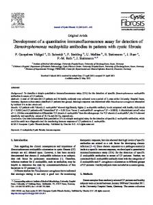
Chemicals and oligonucleotides. GlcN6P and glucosamine were purchased Figure 1. The sequences and secondary structures of two glmS ribozymes. (A) A unimolecular glmS ribozyme from B subtilis. Arrow identifies the site of ribozyme-mediate cleavage stimulated by GlcN6P from Sigma. The synthesis, purification, (1). (B) A bimolecular glmS ribozyme construct derived from S. aureus. This construct differs from the and full characterization of the remaining wild-type glmS ribozyme due to truncation of the P1 stem and the use of a 15-nucleotide substrate RNA GlcN6P analogs are reported elsewhere (shown in grey). The substrate is labeled with a Cy3™ acceptor at its 5’-terminus and a 5/6-FAM donor (Lim J, Grove BC, Roth A and Beaker at its 3’-terminus. The secondary structure model was adapted from reference 8 based on x-ray crystalloRR, manuscript submitted). Synthetic graphic data generated by A. Ferre’ D’Amare (personal communication).” DNAs were purchased and purified before use as described previously.11 The library of 960 bioactive High-throughput glmS assay. High-throughput fluorescence assay compounds was the Spectrum Collection™ purchased from validation and screening was performed in 384-well black MicroSource Discovery, Gaylordsville, CT. polypropylene plates (Corning part number 3677) in a 10 µL solution RNA preparation. The ribozyme domain representing the S. aureus of 50 mM HEPES (pH 7.5 at 25˚C), 10 mM MgCl2, and 200 mM glmS riboswitch (Fig. 1B) was prepared by in vitro transcription KCl, with 0.01% SDS where indicated in the text. A Tecan Aquarius using T7 RNA polymerase using methods similar to those multichannel pipettor (Tecan Group, LTD, Switzerland) was used described previously.12 The template for the transcription was gen- for automated liquid handling. In a typical experiment, a reaction erated from the corresponding region of the glmS leader sequence by mixture containing buffer and both RNA domains was transferred PCR amplification using genomic DNA from S. aureus subsp. aureus into the assay plates with the liquid handler, the fluorescence was Rosebach (ATCC 35556d). Transcribed RNA was purified by dena- measured, and the reaction was initiated by the addition of ~50 nl of turing PAGE. The substrate strand used for radiolabeled cleavage either GlcN6P, an analog of GlcN6P, or a library compound susanalysis, 5'-AAAGCGCCUGUGCAAAUA-3' was purchased from pended at 10 mM in DMSO using the Tecan pin tool (~50 µM final Dharmacon, Inc. (Lafayette, CO), deprotected according to the concentration of compound). Plates were sealed with nonbreathable manufacturer’s directions, and 5'-32P labeled as described previously.11 sealing tape and protected from light for the specified incubation The substrate strand 5'-AAGCGCCUGUGCAAA-3', labeled at the time, at which point the plate was briefly centrifuged and the fluo5'-end with a Cy3™ acceptor and at the 3'-end with a 5/6-FAM rescence measured. All multiplate fluorescence measurements were donor (isomeric mixture of 5- and 6-fluorescein) was purchased in conducted with a Wallach Envision (Perkin Elmer, Wellesly, MA) purified form from IBA GmbH (Göttingen, Germany). plate reader with a 480 nm excitation filter (30 nm bandwidth) and Low-throughput glmS kinetic analyses. All kinetic assays were a 535 nm emission filter (25 nm bandwidth). conducted in 50 mM HEPES (pH 7.5 at 25˚C), 10 mM MgCl2, and 200 mM KCl. To initiate the reaction, GlcN6P was added to give a final concentration of 200 µM. Kinetic assays with radiolabeled RESULTS AND DISCUSSION substrate were performed as described previously.13 Rate constants Although the glmS ribozyme sequence is highly conserved among were established by plotting the natural logarithm of the fraction of many bacterial species, the S. aureus sequence was chosen for screening uncleaved substrate remaining versus time and then determining development because of its clinical importance. To facilitate detection the negative slope of the resulting line. For the highest rate constants, of cleavage, the natural self-cleaving glmS ribozyme was converted to only the initial phase of the plots were used because ~20% of the a bimolecular ribozyme (Fig. 1B) in which separate substrate and RNA substrate remained uncleavable. Single reaction FRET-based ribozyme oligonucleotides associate via a truncated P1 stem. A kinetic assays were performed with λexcit = 488 nm (slit width = 1 nm), similar bimolecular glmS ribozyme from B. subtilis was recently λemit = 523 nm (slit width = 8 nm). characterized and found to have similar catalytic properties as the 78
RNA Biology
2006; Vol. 3 Issue 2
Riboswitch High-Throughput Screening
Figure 2. RNA cleavage activity of a bimolecular glmS ribozyme. (A) The apparent kobs for substrate cleavage (0.1 nM, 5'-32P-radiolabeled) was measured as a function of ribozyme concentration. (B) Cleavage of the bimolecular ribozyme-substrate complex in the presence of 40 µM GlcN6P (upper black trace) monitored by an increase in fluorescence of the FRET pair. No activity was observed in the absence of either the ribozyme (grey) or GlcN6P (black). (C) Change in fluorescence upon incubation of 10 nM each of the substrate and ribozyme RNAs in a 384-well plate for 68 hours in the presence of 200 µM GlcN6P. The error bars indicate the standard deviation. S and R denote substrate and ribozyme, respectively for each value from eight replicate reactions.
natural unimolecular ribozyme.13 The RNA substrate encompasses the 5'-half of the shortened P1 stem and the conserved nucleotides surrounding the cleavage site, whereas the ribozyme strand contains the 3'-half of P1 and the remainder of the glmS sequence from S. aureus. The cleavage activity of this bimolecular ribozyme was confirmed under multiple turnover conditions using a 5'-radiolabeled substrate (Fig. 2A). At 0.5 µM ribozyme, 200 µM GlcN6P, and saturating substrate concentrations, the bimolecular construct cleaves with an www.landesbioscience.com
apparent rate constant of 0.25 min-1 at 25˚C, comparable to the previously reported single-turnover rate constant of 1 min-1.8 Under these conditions, the apparent KD measured for the two strands was 0.2 µM, comparable to the value of 1 µM reported for a similar bimolecular construct.13 To enable FRET detection of cleavage, the substrate was synthesized with a 5-(and 6-)-carboxyfluorescein (5/6-FAM) donor at the 3' end and a Cy3™ acceptor at the 5' end. When excited at 485 nm, the 5/6-FAM donor normally emits at a maximum wavelength of 526 nm. When the substrate strand is intact, however, the fluorescence of 5/6-FAM is reduced presumably due to quenching by the proximal Cy3™ acceptor. After addition of GlcN6P and cleavage of the substrate strand, the two product strands dissociate, relieving the proximity of 5/6-FAM and Cy3™, with a concomitant increase of 5/6-FAM fluorescence. Similar fluorescence-based systems have previously been used to detect ribozyme cleavage in real time.14,15 Using this FRET detection strategy, the addition of 40 µM GlcN6P induces an increase in the fluorescence signal of nearly 6 fold within 20 minutes (Fig. 2B). We speculate that the modest burst in fluorescence increase during the first few seconds might be due to a conformational change in the substrate that occurs upon ligand binding. Tertiary contacts between ribozyme and substrate would constrain the substrate to a greater extent compared to that caused by the short base paired region, thus increasing the average distance between donor and acceptor. Importantly, no fluorescence increase was observed over this time course in the absence of either GlcN6P or the ribozyme. To facilitate rapid solution-based measurement of cleavage for large compound libraries, the assay conditions were optimized for automated liquid handling and fluorescence measurement in 384-well microplates. To reduce demand for enzyme and substrate RNAs, the concentrations of these molecules were varied to determine the minimal concentration of each that can be used to give a reproducible and statistically significant increase in the fluorescent signal. At 10 nM of each strand, a 4-fold increase in fluorescence was observed after 68 hours when 100 µM GlcN6P was present (Fig. 2C). At this low RNA concentration, the signal increase was dependent upon the inclusion of 0.01% SDS, perhaps because it minimizes adhesion of the RNA to the polypropylene plates. At incubation times less than 48 hours, the increase in fluorescence was noticeably less, most likely because the concentration of the two RNA strands is well below the measured dissociation constant of 0.2 µM, slowing the observed cleavage rate. Nevertheless, the fact that a signal increase is not observed in the absence of GlcN6P confirms that the signal increase reports ribozyme catalysis during this time course, rather than nonspecific RNA degradation. Furthermore, although ribozyme-substrate complex formation is most likely a rate limiting step in our high-throughput assays, the total fluorescence increase is a function of how well the ligand is able to induce ribozyme action once complex formation occurs. The signal change after incubation was nearly identical when the reactions were prepared either by manual pipetting or by the automated liquid handler (Fig. 2C). From these data, the Z' for the screen can be calculated: Z' = 1 - 3 * (SDpositive + SDnegative)/|Mpositive - Mnegative| where SDpositive, SDnegative, Mpositive, and Mnegative denote the standard deviation and mean for wells with or without GlcN6P, respectively. Z' is a relative indication of the separation between the positive and
RNA Biology
79
Riboswitch High-Throughput Screening
Figure 3. Identification of GlcN6P analogs that induce ribozyme activity. (A) The structures of the GlcN6P that were tested for their ability to activate glmS cleavage. The chemical differences from GlcN6P are highlighted in red. (B) The fluorescence at 21 or 44 hours, relative to time zero (left axis), after the addition of each analog identifies which compounds activate cleavage. For comparison, the relative fluorescence with no ligand (“NL”) is 0.8 and 0.9 at 21 and 44 hours, respectively, and the relative fluorescence after the addition of 200 µM GlcN6P (1) is 3.5 and 3.4 at 21 and 44 hours, respectively. For comparison, PAGE analysis revealed that each of the circled compounds activates the glmS ribozyme with an apparent affinity within 10-fold of GlcN6P.
Figure 4. A 960-compound pilot glmS activity screen. (A) Plate 1 of three shows the fluorescence response over 384 wells. Columns 1 and 2 contain only the substrate strand. Column 23 (green) contains the substrate and ribozyme strands, and column 24 (red) contains the ribozyme and substrate strands and 80 µM GlcN6P. Wells in which the fluorescence response is greater than 10-standard deviations above the no-ligand control are highlighted in blue. (B) The mean fluorescence response is shown for each control reaction and hit compound in Plate 1. Error bars indicate one standard deviation. S and R denote substrate and ribosome, respectively. (C) Five hits were identified from a screen of the 960-compound Spectrum Collection that show greater than 2.3-fold fluorescence increase after 44 hours. (D) Secondary analysis demonstrates that each of the five compounds identified in (C) exhibits a time-dependent increase in fluorescence.
80
RNA Biology
2006; Vol. 3 Issue 2
Riboswitch High-Throughput Screening
negative control populations and is widely accepted as an assessment of the statistical performance of a screen.16 The Z’ calculated for the data in (Fig. 2C) is 0.64, well above the industrial standard of >0.5 for a pharmaceutically useful screen, although this number was determined with fewer replicates than is typically used. This Z' also compares favorably with the previously published unimolecular glmS screen.10 This confirms the utility of the screen for high- throughput library screening. To evaluate how well the assay can distinguish active from inactive compounds, we screened a library of twelve GlcN6P analogs (Fig. 3) whose relative binding affinities for the glmS ribozyme had been previously determined by electrophoretic methods (the synthesis and ribozyme binding activity of these analogs will be reported elsewhere: Lim J, Grove BC, Roth A and Breaker RR, manuscript submitted). Each compound whose binding affinity for the ribozyme is within 10 fold of GlcN6P showed at least a 2.5-fold increase in fluorescence, compared to a 3.5-fold increase in fluorescence for GlcN6P. This cutoff for functional compounds (“hits”) of 2.5-fold increased fluorescence represents a statistically significant 18 standard deviations over the mean fluorescence value in the absence of ligand (“NL”). Conversely, each compound whose affinity is less than 10 fold of GlcN6P shows less than 1.5-fold increase in the fluorescence signal, except 5 and 13. The larger fluorescence increase observed for 5 and 13 most likely indicates that these compounds stimulate a slower rate of cleavage that is detectable after 44 hours but was undetectable by the previous gel analysis after a 30 minute incubation. Indeed, glucosamine induces a 3.0-fold increase in fluorescence after 44 hours, even though its measured affinity for the ribozyme is more than 100-fold lower than GlcN6P.17 Thus, the fluorescent report from the high-throughput screen accurately reflects binding activity for the glmS ribozyme. As a model screen to search for new compounds that might activate the glmS ribozyme, we screened a commercially available library of 960 bioactive compounds approved for use in humans. In control wells with only GlcN6P added, the fluorescence signal of the ribozyme- substrate mixture increased by 3.74-fold (± 0.28) after 34 hours (red in Fig. 4A and B), compared to only a 1.18-fold (± 0.09) increase in the absence of ligand (green in Fig. 4A and B), a difference of 28 standard deviations. For wells containing a library compound, an increase in fluorescence of greater than ten standard deviations above the mean relative fluorescence in the absence of ligand was considered to indicate an active compound. (This hit threshold was chosen in an effort to capture the maximum possible hit compounds). Using this parameter, five hit compounds were identified (Fig. 4C), most notably glucosamine. This hit rate of 0.06% compares favorably with other recently published screens,18-20 as does the average Z' factor of 0.57 calculated for each plate of the screen. Notably, although the previously described glmS screen showed no observable activity with glucosamine after 30 minutes,10 it is reasonable to assume that the longer incubation time used herein permits identification of this analog that binds with 100-fold weaker affinity than GlcN6P.17 Each of the five compounds identified was retested for glmS activation with either the fluorescence screen or by electrophoretic methods. Surprisingly, although each compound induces a reproducible and time-dependent increase in fluorescence (Fig. 4D), only glucosamine stimulates detectable cleavage by incubating for two hours (not shown). This result mirrors the previously- described glmS screen,10 in that novel glmS activating compounds have not yet www.landesbioscience.com
been identified, presumably due to the limited size and diversity of the library. Moreover, these types of infrequent false- positive hits have been observed for other fluorescence-based high-throughput screens18 and can readily be confirmed or refuted in our case with the secondary ribozyme cleavage assay.8 One possible explanation for the false positives is that they quench or otherwise modify the fluorescent label on the RNA over the 44 hour time course, or that the compounds become fluorescent over time. Nevertheless, the identification of glucosamine confirms that this screen can identify even low affinity glmS-activating compounds from complex compound libraries. In summary, we report the development of a fully automatable and statistically robust high-throughput screen for identifying compounds that bind to and activate the glmS ribozyme. This demonstrates that modern drug discovery technology can be applied to the discovery and development of riboswitch targeted drugs. Moreover, the technology described herein could be readily reconfigured for other riboswitches by engineering an allosteric fusion between an RNA-cleaving ribozyme and a riboswitch aptamer as previously described.22,23 These types of screens could significantly advance the discovery of novel anti- bacterial drugs directed at these promising classes of RNA targets. References 1. 2. 3. 4.
5. 6. 7. 8. 9. 10. 11. 12. 13. 14.
15.
16. 17. 18.
19. 20. 21. 22. 23.
Wolfson W. Holding back the tide of antibiotic resistance. Chem Biol 2006; 13:1-3. Walsh C. Where will new antibiotics come from? Nat Rev Microbiol 2003; 1:65-70. Nathan C. Antibiotics at the crossroads. Nature 2004; 431:899-902. Sudarsan N, Cohen-Chalamish S, Nakamura S, Emilsson Gail M, Breaker RR. Thiamine pyrophosphate riboswitches are targets for the antimicrobial compound pyrithiamine. Chem Biol 2005; 12:1253-358. Milewski S. Glucosamine-6-phosphate synthase: The multi-facets enzyme. Biochim Biophys Acta 2002; 1597:173-92. Winkler W, Nahvi A, Breaker RR. Thiamine derivatives bind messenger RNAs directly to regulate bacterial gene expression. Nature 2002; 419:952-6. Tucker BJ, Breaker RR. Riboswitches as versatile gene control elements. Curr Opin Struct Biol 2005; 15:342-8. Winkler WC, Nahvi A, Roth A, Collins JA, Breaker RR. Control of gene expression by a natural metabolite-responsive ribozyme. Nature 2004; 428:281-6. Kobayashi K, Ehrlich SD, Albertini A, Amati G, et al. Essential Bacillus subtilis genes. Proc Natl Acad Sci USA 2003; 100:4678-83. Mayer G, Famulok M. High-throughput-compatible assay for glmS riboswitch metabolite dependence. ChemBioChem 2006; 7:602-4. Seetharaman S, Zivarts M, Sudarsan N, Breaker RR. Immobilized RNA switches for the analysis of complex chemical and biological mixtures. Nature Biotechnol 2001; 19:336-41. Milligan JF, Uhlenbeck OC. Synthesis of small RNAs using T7 RNA polymerase. Methods Enzymol 1989; 180:51-62. Soukup GA. Core requirements for glmS ribozyme self-cleavage reveal a putative pseudoknot structure. Nucleic Acids Res 2006; 34:968-75. Singh K, Parwaresch R, Krupp G. Rapid kinetic characterization of hammerhead ribozymes by real-time monitoring of fluorescence resonance energy transfer (FRET). RNA 1999; 5:1348-1356. Hanne A, Ramanujam MV, Rücker T, Krupp T. Fluorescence resonance energy transfer (FRET) to follow ribozyme reactions in real time. Nucleosides Nucleotides 1998; 17:1835-50. Zhang JH, Chung TD, Oldenburg KR. A simple statistical parameter for use in evaluation and validation of high throughput screening assays. J Biomol Screen 1999; 1999:67-73. McCarthy TJ, Plog MA, Floy SA, Jansen JA, Soukup JK, Soukup GA. Ligand requirements for glmS ribozyme self-cleavage. Chem Biol 2005; 12:1221-6. Zhang JH, Wu X, Sills MA. Probing the primary screening efficiency by multiple replicate testing: A quantitative analysis of hit confirmation and false screening results of a biochemical assay. J Biomol Screen 2005; 10:695-704. Kroemer RT. Molecular modelling probes: Docking and scoring. Biochem Soc Trans 2003; 31:980-4. Macarrón R, Hertzberg RP. High-throughput screening: Methods and protocols. In: Janzen WP, ed. Totowa, NJ: Humana Press, 2002:1-29. Soukup GA, Breaker RR. Relationship between internucleotide linkage geometry and the stability of RNA. RNA 1999; 5:1308-25. Breaker RR. Engineered allosteric ribozymes as biosensor components. Curr Opin Biotechnol 2002; 13:31-9. Breaker RR. Natural and engineered nucleic acids as tools to explore biology. Nature 2004; 432:838-45.
RNA Biology
81